


 Close
Close
 4th June 2025
Hereditary Haemorrhagic Telangiectasia
Hereditary haemorrhagic telangiectasia (HHT) – sometimes known by its other name ‘Osler-Weber-Rendu syndrome’ – is a rare disorder of autosomal dominant inheritance. It is characterised by abnormal vasculature, including telangiectasia of the mucous membranes and skin, and arteriovenous malformations of the lung, liver and central nervous system. This produces characteristic skin signs and an increased tendency to bleed. This chapter will focus on the cutaneous signs of HHT.
Epidemiology
Worldwide, HHT is thought to affect between 1 in 5000 to 1 in 10,000 people. It is more commonly diagnosed in women, however this is likely due to men being less engaged with healthcare services. Given HHT is a multi-system disorder that can present in numerous different ways (e.g. epistaxis, gastrointestinal (GI) bleeds and intracerebral bleeds) medical professionals may fail to link the presentations to a unifying diagnosis; given all of this, HHT is probably under diagnosed.
Aetiology
The majority of HHT cases are caused by a mutation in one of two genes involved in blood vessel development: activin receptor-like kinase 1 gene (chromosome 12) or the endoglin gene (chromosome 9). This mutated gene (inherited in an autosomal dominant pattern) produces abnormal blood vessels without the usual capillaries between arteries and veins (arteriovenous malformations – AVM). When small these are described as telangiectasia (telangiectases – plural), when large arteries and veins are involved they are called AVMs. AVMs are weak and have an increased risk of bleeding.
In HHT, patients will often present with epistaxis due to AVMs within the nose. They can often be spontaneous or after light trauma, however the severity of bleeding may vary hugely between different patients.
Telangiectases on the mucous membranes, lips, and face produce the most characteristic skin signs of HHT.
Diagnosis
A diagnosis of HHT is made using the ‘Curacao’ diagnostic criteria:
- Epistaxis – must be recurrent and spontaneous
- Telangiectases – multiple, on the mucous membranes, lips, face and hands
- AVMs – internal organ involvement
- First-degree relative with a diagnosis of HHT
For a definite diagnosis of HHT, a patient must meet at least three of the above criteria.
Clinical signs of HHT:
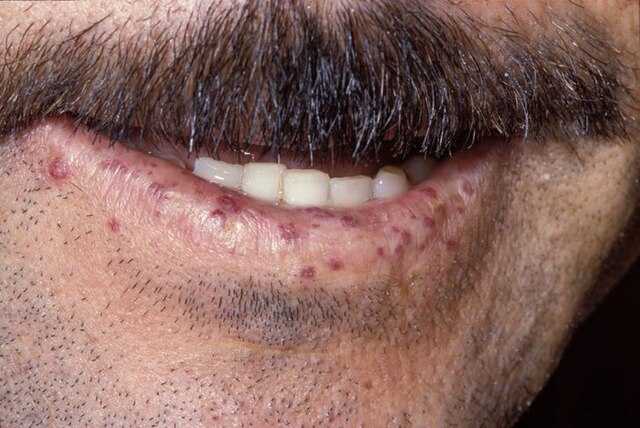
https://upload.wikimedia.org/wikipedia/commons/thumb/2/2dCase_115.jpg/640px-Case_115.jpg
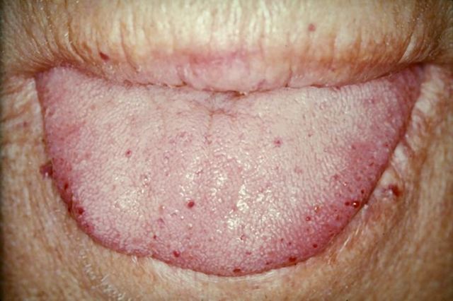
https://upload.wikimedia.org/wikipedia/commons/thumb/7/77/TongueTelang.JPG/640px-TongueTelang.JPG
Medical management
It is not possible to prevent or cure HHT, however the symptoms can be managed. Mild HHT (up to 1/3 of those diagnosed) may not require any treatment.
- Topical intranasal treatment with timolol or sirolimus can reduce the frequency and severity of nosebleeds.
- Tranexamic acid can reduce the severity of bleeds.
- Bevacizumab and thalidomide inhibit angiogenesis and are being investigated for their use in the treatment of HHT.
Surgical management
- Laser therapy for the treatment of skin telangiectases.
- Laser therapy can also be used to manage mild-moderate epistaxes. Severe epistaxis may require skin transplants to the septum (septal dermoplasty).
- Internal organ AVMs often require surgical management.
Complications
- Anaemia
- Epistaxis
- Pulmonary AVMs and pulmonary haemorrhage
- Central nervous system AVMs and stroke or spinal cord bleed
- GI AVMs and GI bleed
- Hepatic AVMs and hepatic bleed – can also lead to hepatic failure or cardiac failure
Complete Guide to Clinical Genetics ST3 Application and Recruitment (2027)
Written by Medibuddy. Last updated: October 2026. Clinical Genetics is

Complete Guide to Clinical Radiology ST3 Application and Recruitment (2027)
Recruitment paused: NHS England is not running Clinical Radiology ST3+ recru

Complete Guide to Paediatric Surgery ST3 Application and Recruitment in United Kingdom (2026)
Written by Medibuddy. Last updated: October 2026. If you’re planning


Take your subscriptions with you
Our mobile app allows you to access your interview and exam question banks wherever you are.

