


 Close
Close
 5th June 2025
Scleroderma (Systemic Sclerosis)
Classification
Scleroderma is a term that translates to hardened skin. However, the general term ‘scleroderma’ is often used for both systemic sclerosis (systemic scleroderma) and morphoea (localised scleroderma). Though the skin changes in the two conditions are virtually indistinguishable, on both a clinical and histopathological level, morphoea does not result in damage and fibrosis of the lungs, heart, kidneys and/or gastrointestinal tract.
Systemic sclerosis (SSc) is an autoimmune inflammatory condition that causes fibrosis, inflammation and vasculopathy and involves multiple systems, such as skin, lungs, heart, gastrointestinal tract and kidneys. There are two subtypes of systemic sclerosis according to distribution of skin involvement – limited cutaneous SSc (lcSSc) and diffuse cutaneous SSc (dcSSc).
Epidemiology
Different world populations have different prevalence, with higher rates in the US compared to Europe or Asia. The prevalence is 88 per million seen in England compared to 250 per million adults in the US. Women are more often affected, with a ratio of 5:1 female to male. SSc is rare in childhood, more common in young adults and has a peak incidence in patients’ 50s.
Aetiology
Aetiology is unknown but genetic, immunological and environmental factors have been suggested:
- Genetic: <5% of monozygotic and dizygotic twins are clinically concordant for systemic sclerosis; however, most monozygotic twins are concordant for antinuclear antibodies
- Immunology: 90% of patients have positive ANA on indirect immunofluorescence; this group of patients shows subsets of mutually exclusive auto-antibodies that are associated with distinct clinical phenotypes
- Environmental: exposure to silica dust and multiple solvents have been proposed, but no clear link has been identified
Pathophysiology
The precise mechanism is unknown, but many models have been proposed. At the start of the disease, the immune system, endothelial cells and fibroblasts are all activated. A sub-population of hyper-proliferative fibroblasts is selected for, which are confirmed to be over-represented in scleroderma. These cells produce high levels of collagen. Activated T-cells contribute to disease by producing pro-fibrotic cytokines and downregulating cytokines that inhibit growth or synthesis.
Diagnosis
LcSSc and dcSSc both show the same signs and symptoms, but the skin distribution and course of the disease is different. Two thirds of patients with scleroderma have dcSSc, which has widespread skin involvement including proximal limbs. This is often rapidly progressive, has significant internal organ involvement and has a worse prognosis. One third of patients with scleroderma have lcSSc, where the sclerosis is limited to the digits, face and distal limbs (does not spread further than elbows or knees). It has a slower progression and less organ involvement, but carries a risk of pulmonary artery hypertension. Up to 20% of patients have an overlap syndrome with another connective tissue disease.
Systemic features
- Gastrointestinal: dysmotility, gastroesophageal reflux (GORD), dysphagia, bloating, nausea and vomiting, alternating diarrhoea/constipation
- Cardiopulmonary: interstitial lung disease (ILD), pulmonary artery hypertension, cardiomyopathy
- Renal dysfunction: patients may present with abrupt onset of moderate/marked hypertension, which is a sign of scleroderma renal crisis
- Musculoskeletal: friction rubs over joints and tendons, synovitis, weakness and limited movement causing contractures
- Ophthalmological: tight eyelids, reduced tear secretion, retinopathy
- Fatigue, arthralgia, myalgia
- Sicca symptoms and Sjogren syndrome
Cutaneous features
- Skin sclerosis, commonly sclerodactyly (hardening of the skin causing the fingers to curl inward) or microstomia
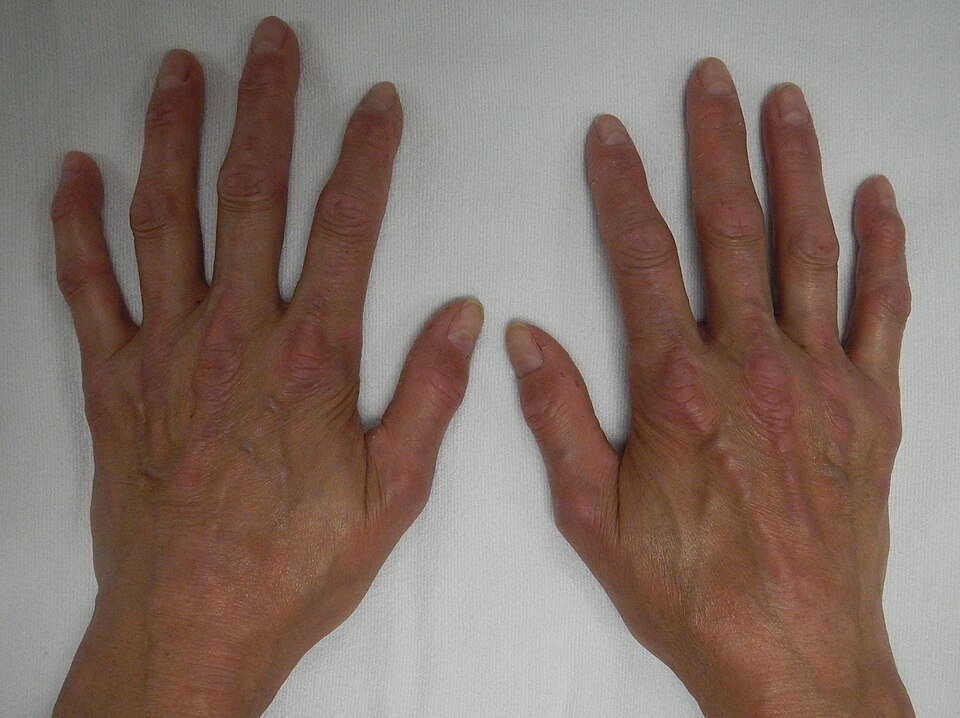 Image Source: Wikimedia Commons (2015). Crest 1, Licensed under a creative commons licence. Available for free at: https://commons.wikimedia.org/wiki/File:CREST1.JPG (Accessed 19 May 2025).
 - Puffy hands, often one of the first symptoms
- Raynaud’s phenomenon
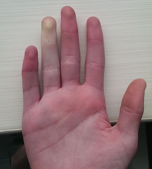 Image Source: Wikimedia Commons (2014). Raynaud Phenomenon, Licensed under a creative commons licence. Available for free at: https://commons.wikimedia.org/w/index.php?search=Raynaud%E2%80%99s+phenomenon&title=Special:MediaSearch&go=Go&type=image (Accessed 19 May 2025).
 - Digital pits and/or ulceration
- Abnormal nail fold capillaries
- Telangiectases – often located on the face, hands and mucous membranes
- Subcutaneous calcinosis – small, hard masses on pressure points (eg. Fingers, forearms)
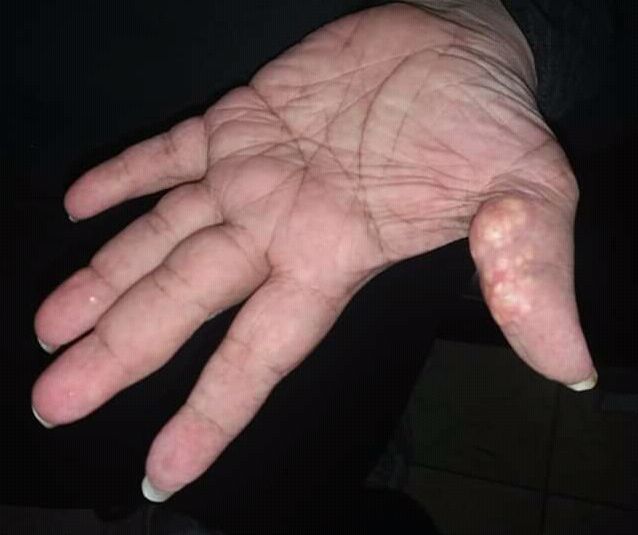 Image Source: Wikimedia Commons (2018). Calcinosis Cutis, Licensed under a creative commons licence. Available for free at: https://commons.wikimedia.org/wiki/File:Calcinosis_cutis_new_image.jpg (Accessed 19 May 2025).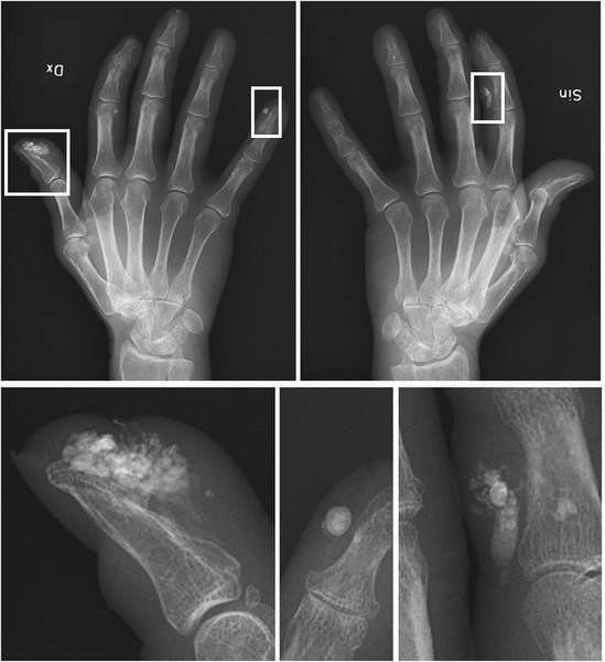
Image Source: Wikimedia Commons (2018). Calcinosis of Crest Syndrome, Licensed under a creative commons licence. Available for free at: https://commons.wikimedia.org/wiki/File:Calcinosis_of_CREST_syndrome.jpg (Accessed 19 May 2025).
- Salt and pepper dyspigmentation – hyperpigmentation with vitiligo-like depigmentation
Though the previous name, CREST syndrome, of lcSSc is no longer used, it remains a useful mnemonic to remember the key features:
- Calcinosis
- Raynaud’s phenomenon
- (O)esophageal dysmotility
- Sclerodactyly
- Telangiectasia
Investigations
Initial investigations look at auto-antibodies, with over 90% of patients having a positive ANA. Within this group there are subsets of mutually exclusive antibodies, which are associated with distinct clinical pictures:
- Anti-topoisomerase I (anti-Scl70)
- Seen in 20% of patients
- Associated with an increased risk of ILD and diffuse skin involvement
- Anti-RNA polymerase III antibody
- Seen in 20% of patients
- Associated with renal crisis
- Anti-centromere antibodies
- Seen in 20-25% of patients
- Associated with limited skin involvement and overall better prognosis
- In the remaining 40% no specific auto-antibody has been identified yet
Further investigations to assess for systemic involvement are required. Pulmonary involvement can be investigated with pulmonary function tests and high-resolution CT of the lungs. Cardiac involvement requires ECG and echocardiogram (ECHO). If the ECHO finds elevated right ventricular systolic pressure, the patient should be referred for a right-heart catheterisation and full evaluation, as ECHO findings may not be indicative of the true pulmonary artery pressures. For gastrointestinal symptoms such as dysmotility and reflux, a barium swallow can be a useful test. Anaemia or melaena can indicate chronic GI blood loss from gastric antral vascular ectasia syndrome (GAVE) and required upper GI endoscopy. If inflammatory myositis develops, patients will require blood tests for serum muscle enzymes, possibly followed by a nerve conduction study and a muscle biopsy.
Complications
The greatest complications of systemic sclerosis are interstitial lung disease (ILD) and renal crisis. Development of ILD will need specialist referral and can ultimately require lung transplantation.
Severe and life-threatening renal disease occurs in 10-15% of patients and was the largest mortality cause before ACE inhibitors (ACEi) were introduced. Early dcSSc patients are at highest risk of developing a renal crisis. The signs of a renal crisis are onset of acute renal failure, abrupt onset of moderate or marked hypertension, urinalysis indicating proteinuria with few cells or casts and micro-angiopathic haemolytic anaemia. ACEi must be titrated to the maximal tolerated dose and if required, further anti-hypertensives can be added (usually calcium channel blockers).
Small pericardial effusions can occur when cardiac involvement is noted, which can progress to cardiac tamponade. Oral prednisolone is given for pericardial effusions, but strict monitoring is required, as corticosteroids can precipitate a renal crisis. Tamponade is managed with a pericardial window and systemic corticosteroids.
Patients should be monitored with regular ECHOs to look for the development of pulmonary hypertension, with referral for right heart catheterisation if needed.
Raynaud’s phenomenon can lead to digital ulceration and skin sclerosis can cause fixed contractures. Patients may develop gastric antral vascular ectasia (GAVE). Heartburn can lead to strictures and Barrett’s metaplasia and needs early intervention.
Management
Treatment options are according to the organ system affected. As patients with systemic sclerosis often have multiple systems affected, multi-modal therapies may be required. Involvement of multiple specialists may also be required to manage the complications, such as respiratory, renal and gastroenterology input.
Prognosis
DcSSc has a poorer prognosis compared to lcSSc, though both can develop significant visceral involvement as detailed above. Mean survival is approximately 12 years after diagnosis, but with a wide variation in disease severity. At the milder end of the disease, survival is close to the general population.
Complete Guide to Clinical Genetics ST3 Application and Recruitment (2027)
Written by Medibuddy. Last updated: October 2026. Clinical Genetics is

Complete Guide to Clinical Radiology ST3 Application and Recruitment (2027)
Recruitment paused: NHS England is not running Clinical Radiology ST3+ recru

Complete Guide to Paediatric Surgery ST3 Application and Recruitment in United Kingdom (2026)
Written by Medibuddy. Last updated: October 2026. If you’re planning


Take your subscriptions with you
Our mobile app allows you to access your interview and exam question banks wherever you are.

