


 Close
Close
 5th June 2025
Dermatomyositis
Dermatomyositis is an idiopathic inflammatory autoimmune disorder with characteristic myopathy and cutaneous signs.
Epidemiology
Dermatomyositis is a rare disease that can occur at any age. Two peaks of incidence have been associated – between 5-15 years old and between 40-60 years old.
Data on incidence and prevalence is available from a US-population study. The estimated annual incidence of all subtypes is 9.63 per million people, with 13.98 per million in females and 4.68 per million in males. The overall prevalence in all subtypes was estimated at 21.42 in 100,000 people, with 33.09 in 100,000 females and 8.35 in 100,000 in males.
Juvenile-onset dermatomyositis has an annual incidence of 2-3 cases per million. Females are more commonly affected with the mean age of onset being 10 years old. Adult-onset dermatomyositis affects females more than males, with a ratio of 2.5:1. It is more commonly seen in the black population compared to the white population. Malignancy-associated dermatomyositis affects males and females equally but is more common in over 50s, with risk increasing with age.
Aetiology and Pathophysiology
The aetiology of dermatomyositis is unknown – the current opinion is that it is due to an interaction between environmental factors and genetic susceptibility.
- Genetic: a genetic predisposition can be inferred from the familial associations investigated
- Immunological: certain HLA subtypes appear to be connected to an increased risk of developing dermatomyositis
- Environmental: overall only weak associations found but the strongest evidence is available for ultraviolet radiation intensity
- Infection: some reports are available of patients with antibodies to Borrelia burgdorferi and Toxoplasma gondii developing myositis; other reports suggest this with viral antibodies, particles, nucleic acids
- Drug-induced: D-penicillamine, hydroxyurea, statins, phenylbutazone, terbinafine
The pathogenesis is unclear, but it is widely accepted to be an autoimmune condition.
Diagnosis
Clinical signs and symptoms
Muscle and skin signs develop simultaneously in approximately 60% of patients. In 30%, skin lesions precede muscle weakness, though the time in between can be up to several months. Between 10-20% of patients develop skin signs, but never develop any apparent muscle disease (known as amyopathic dermatomyositis). As it is considered an autoimmune disorder, dermatomyositis may also present with systemic manifestations.
Myopathy
This often manifests as a symmetrical weakness of the proximal muscles, though the severity varies between patients. Oftentimes, this manifests as difficulty carrying out certain activities – climbing stairs, rising from a sitting or crouching position, lifting objects, washing or combing hair, difficulty swallowing. Myalgia is present in less than 30% of patients. On examination, muscle tenderness is seen in 50% of patients. Muscle atrophy can be seen in long-standing disease, but is not common and so an alternative diagnosis should be considered in these cases.
Cutaneous signs
- Heliotrope rash – bilateral violaceous to erythematous discolouration of eyelids with swelling of the eyelids and surrounding skin; pathognomonic sign of dermatomyositis
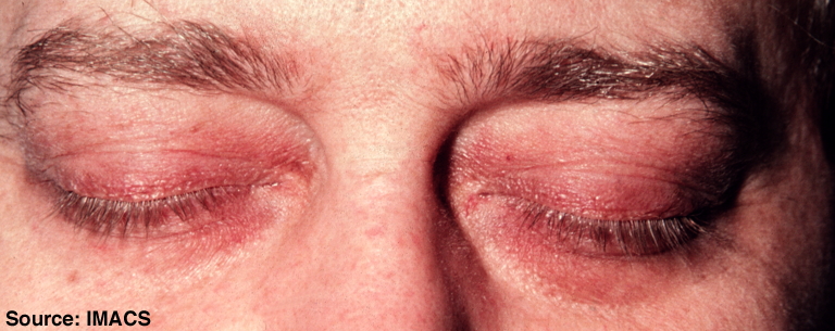 Image Source: Wikimedia Commons (2010). Dermatomyositis, Licensed under a creative commons licence. Available for free at: https://commons.wikimedia.org/wiki/File:Dermatomyositis10.jpg (Accessed 19 May 2025)
 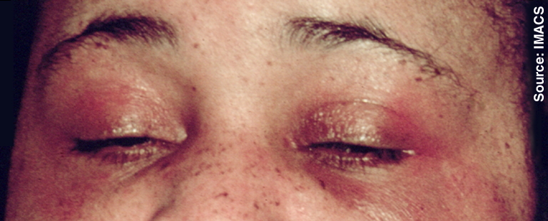 Image Source: Wikimedia Commons (2010). Dermatomyositis, Licensed under a creative commons licence. Available for free at: https://commons.wikimedia.org/wiki/File:Dermatomyositis11.jpg (Accessed 19 May 2025).
 - Shawl sign – fixed redness of the back, shoulders, chest and neck
- V-sign – fixed redness of the face, neck and upper chest
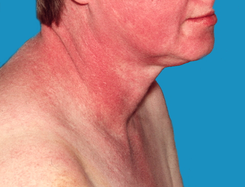 Image Source: Wikimedia Commons (2011). Dermatomyositis 18, Licensed under a creative commons licence. Available for free at: https://commons.wikimedia.org/wiki/File:Dermatomyositis18.jpg (Accessed 19 May 2025).
 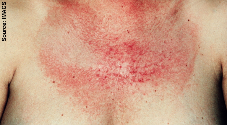 Image Source: Wikimedia Commons (2010). Dermatomyositis 17, Licensed under a creative commons licence. Available for free at: https://commons.wikimedia.org/wiki/File:Dermatomyositis17.jpg (Accessed 19 May 2025).
 - Photosensitivity
- Gottron’s papules – pathognomonic of dermatomyositis; violaceous papules and plaques on dorsal knuckles, and more rarely on wrists, elbows, knees, malleoli
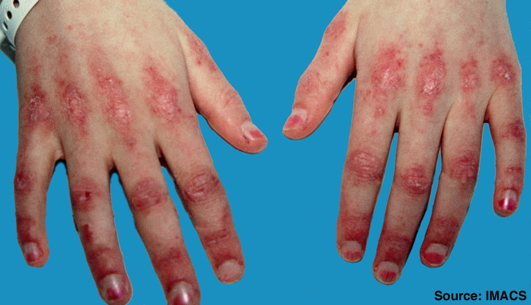 Image Source: Wikimedia Commons (2010). Stomach blood supply, Licensed under a creative commons licence. Available for free at: https://commons.wikimedia.org/wiki/File:Dermatomyositis2.jpg (Accessed 19 May 2025).
 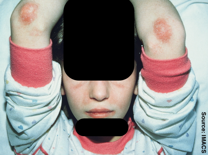 Image Source: Wikimedia Commons (2010). Dermatomyositis 4, Licensed under a creative commons licence. Available for free at: Https://commons.wikimedia.org/wiki/File:Dermatomyositis4.jpg (Accessed 19 May 2025).
 - Itchy scalp with inflammation and diffuse, non-scarring hair thinning
- Nailfold changes – periungal erythema, cuticular overgrowth (ragged cuticles), dilated capillaries of the nailfold
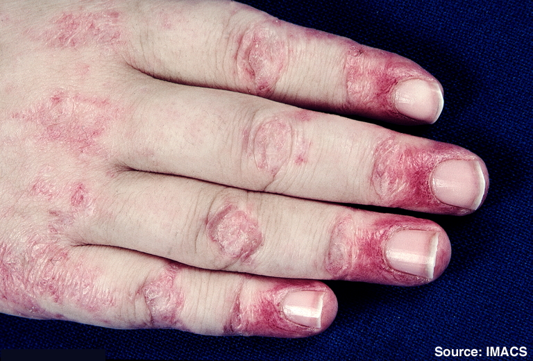 Image Source: Wikimedia Commons (2010). Dermatomyositis 7, Licensed under a creative commons licence. Available for free at: https://commons.wikimedia.org/wiki/File:Dermatomyositis7.jpg (Accessed 19 May 2025).
 - Mechanic’s hands – hyperkeratotic changes in hands seen in antisynthetase syndrome
- Holster sign – erythema over buttocks and lateral thighs
- Calcinosis cutis (calcium deposits)
Systemic manifestations
- Inflammatory polyarthritis – when present, has a similar distribution to rheumatoid arthritis
- Interstitial lung disease
- Cardiac involvement
- Raynaud’s phenomenon
Investigations
The following investigations should be considered in dermatomyositis:
- Serum creatinine kinase (CK) – the most sensitive muscle enzyme test
- Serum aldolase – glycolytic pathway enzyme found mainly in skeletal muscle, brain and liver; not as specific or sensitive as CK, but can be elevated in dermatomyositis when CK is normal
- Muscle biopsy – should be performed in all suspected dermatomyositis cases
- Electromyography (EMG) – recommended if raised CK and typical muscle biopsy findings are present
- Skin biopsy – required in patients that only present with skin manifestations to differentiate from other connective tissue diseases, biopsy appearance is similar to cutaneous lupus
- Antinuclear antibody (ANA) – positive in 80% of patients
- Anti-double-stranded DNA, anti-Ro, anti-La, anti-Sm, anti-ribonucleoprotein (RNP) – useful for differentiation from other connective tissue diseases
- Myositis-specific antibodies (MSAs) and myositis-associated antibodies (MAAs)
MSAs and MAAs are found in most dermatomyositis patients and are associated with different clinical subsets. They are not widely available, but can offer information regarding clinical courses and therapeutic responses.
- Anti-Mi-2 – directed against a helicase involved in transcriptional activation
- Strongly associated with the classical presentation of dermatomyositis, but found in only 10% of patients
- Anti-U1 ribonucleoprotein (RNP) and anti-Ku found in overlap syndromes that present similarly to anti-Mi-2 positive dermatomyositis
- Anti-transcription intermediary factor 1 gamma (TIF1-gamma)
- Most commonly isolated antibody
- Strongly associated with malignancy
- Anti-signal recognition particle (SRP)
- Found almost exclusively in polymyositis and only occasionally in dermatomyositis
- When present, associated with acute severe, treatment-resistant necrotising myositis
- Anti-Jo-1 and other anti-amioacyl-tRNA synthetase antibodies (including anti-PL-7, anti-PL-12, anti-EJ, anti-OJ, anti-KS) -> anti-synthetase syndrome
- Patients positive for the above present with a distinct clinical picture called anti-synthetase syndrome
- Main clinical features include interstitial lung disease, fever, myositis, polyarthritis, mechanic’s hands, Raynaud’s phenomenon
- Anti-Jo-1 associated with a higher frequency of myositis
- The other are associated with a higher frequency of interstitial lung disease
- Anti-melanoma differentiation-associated gene 5 (MDA5, also known as CADM-140) – specific to clinically amyopathic dermatomyositis
- Strongly associated with a rapidly progressive interstitial lung disease with poor prognosis
- May present with tender palm papules and diffuse alopecia
- Anti-NPX2
- Strongly associated with juvenile-onset dermatomyositis; a high proportion of these patients suffer from cutaneous calcinosis
- Anti-PM-Scl
- Found in patients with overlapping myositis and scleroderma
Management
Treatment depends on the presentation of dermatomyositis – though there can be some overlap in management, muscle and skin manifestations are treated with different agents. Dermatomyositis is considered to be the most responsive idiopathic inflammatory myopathy, but even with good control of myopathy, skin lesions may remain treatment-resistant.
In severe disease or an acute flare, intravenous corticosteroids are used in the short-term until better control is achieved and the patient can be switched to oral agents. Patients with anti-synthetase syndrome may be corticoid-resistant and may require concomitant immunosuppressives. If anti-SRP positive, patients often require immunosuppressives, IVIG or rituximab early in the disease due to the association with acute severe recalcitrant necrotising myositis. If the patient is MDA5 positive, which is associated with rapidly progressive ILD, they often also require high-dose systemic corticosteroids from an early stage.
Management of myopathy
- First-line: high doses of oral corticosteroids
- Second-line:
- Methotrexate – often preferred over azathioprine as it acts quicker; a low dose is useful for both muscle and skin involvement; effective corticosteroid-sparing agent
- Azathioprine – appears to take longer to have an effect (6 months or more)
- Third-line – can be used as a monotherapy or combined with corticosteroids or other immunosuppressants
- Ciclosporin – corticosteroid-sparing agent in some patients
- Tacrolimus and mycophenolate
- Cyclophosphamide – useful in refractory disease associated with vasculitis, ILD or respiratory muscle involvement
- Chlorambucil – successful in a few studies of refractory disease
- In some cases, the disease remains active regardless of any immunosuppressive therapy; Adjunctive IVIG in patients with significant muscle weakness has demonstrated some benefit in small studies and case reports
Management of skin manifestations
- First-line: topical therapy
- Patient education regarding adequate sun protection due to photosensitive nature of skin lesions
- Topical corticosteroids – beneficial even in patient who are already on systemic corticosteroids; help control erythema and pruritus
- Topical antipruritic treatments as adjuncts to topical corticosteroids – emollients with added menthol, phenol or camphor or topical antihistamines (e.g., Doxepin)
- If severe pruritus unresponsive, trial of oral antihistamine
- Second-line: antimalarials
- Hydroxychloroquine – only effective for skin lesions and scalp pruritus with no effect on muscle symptoms
- Chloroquine – risk of retinopathy greater than with hydroxychloroquine
- Topical tacrolimus – only anecdotal evidence exists for this agent
- Third-line: immunosuppressives
- Methotrexate – can be used in combination with topical treatment and antimalarials; useful for both muscle and skin manifestations
- Azathioprine – only anecdotal evidence exists of benefit in methotrexate-resistant dermatomyositis
- Mycophenolate – shown to be beneficial in recalcitrant skin disease
- Ciclosporin – beneficial in resistant skin disease and early ILD; contra-indicated in renal impairment and hypertension
- Anti-inflammatory agents such as thalidomide and dapsone are alternatives to immunosuppressives – case reports and anecdotal evidence exist
- Adjunctive IVIG can be used in refractory skin involvement.
Prognosis
Classic dermatomyositis has a 5-year survival of 95% and 10-year survival of 84%. Despite improved survival rates with newer treatments, one third of patients will suffer from permanent disability. Classic dermatomyositis has a relapsing-remitting course and patients will require long-term treatment.
Amyopathic dermatomyositis have a good response to treatment of myopathy, but cutaneous disease is often more resistant to treatment. Follow-up is required to check for any myopathy, but prognosis is overall good.
Poor prognostic factors include
- Delay in treatment of more than 6 months after symptom onset
- Presenting with greater muscle weakness
- Respiratory muscle involvement
- ILD
- Dysphagia
- Malignancy
- Cardiac involvement
Complete Guide to Clinical Genetics ST3 Application and Recruitment (2027)
Written by Medibuddy. Last updated: October 2026. Clinical Genetics is

Complete Guide to Clinical Radiology ST3 Application and Recruitment (2027)
Recruitment paused: NHS England is not running Clinical Radiology ST3+ recru

Complete Guide to Paediatric Surgery ST3 Application and Recruitment in United Kingdom (2026)
Written by Medibuddy. Last updated: October 2026. If you’re planning


Take your subscriptions with you
Our mobile app allows you to access your interview and exam question banks wherever you are.

