


 Close
Close
 5th June 2025
Lupus
Lupus erythematosus (LE) encompasses a group of auto-inflammatory conditions. Systemic lupus erythematosus (SLE) is a generalised disorder that can affect any body system. Cutaneous lupus, or skin involvement, is seen in 80% of patients with SLE.
Epidemiology
Incidence and prevalence have been difficult to assess worldwide – studies have mainly focused on white populations in small geographical areas. The incidence in the UK is estimated to be 4.91 per 100,000 person-years, with the highest reported incidence seen in North America (23.2 per 100,000 person-years). Incidence is higher in women compared to men, with reported ratios ranging from 2:1 to 15:1. Peak age of onset is 30-70 for women and 50-70 for men. Prevalence of SLE in the US is estimated to be 72.8 per 100,000 person-years. This prevalence estimate is 9 times higher among women compared to men. There is also a racial and ethnic component, with the highest prevalence seen in African-Americans (230.9 per 100,000 in women, 26.7 per 100,000 in men), followed by Hispanics (120.7 per 100,000 in women, 18 per 100,000 in men), and lowest in white populations (84.7 per 100,000 in women, 8.9 per 100,000 in men). Global data appears to confirm these differences noted in the US data.
Aetiology
The aetiology of SLE is not understood, but it is generally agreed that an environmental factor affecting a genetically susceptible individual is fundamental in its pathogenesis. The high female preponderance also suggests hormonal factors may play a role.
The importance of genetic factors is confirmed by familial aggregation and higher-than-expected rates of concordance in twin studies, with the heritability of SLE estimated at 43.9%. Genome-wide association studies have identified more than 60 genetic risk loci, with the one most consistently associated with SLE shown below:
- IRF5 (interferon regulatory factor 5) codes a transcription factor involved in the regulation of pro-inflammatory cytokines
- STAT4 (signal transducer and activator of transcription 4) acts as a transcription activator and helps mediate responses to interleukin-12 in lymphocytes and regulate T helper cell differentiation
- BANK1 (B-cell scaffold protein with ankyrin repeats 1) codes for a B-cell-specific scaffold protein and may contribute to SLE by altering B-cell signalling
- ITGAM (integrin alpha M) – polymorphism of this gene has a significant association with SLE in multiple ethnic populations
- PTPN22 (protein tyrosine phosphatase, non-receptor type 22) inhibits T-cell receptor signalling and selectively promotes type I interferon responses; a missense single-nucleotide polymorphism of this is associated with an increased risk for SLE
Related environmental factors may be non-infectious or infectious.
- Infectious agents include Epstein-Barr virus (EBV), parvovirus B19, cytomegalovirus (CMV) and human immunodeficiency virus 1 (HIV1).
- Non-infectious agents can include exposure to sunlight, drugs, hormones, toxins such as cigarette smoke or stress and emotional upset.
- Drugs can either be an environmental factor triggering a flare or unmasking of latent LE, or can specifically cause drug-induced lupus erythematosus, which is a lupus-like syndrome different from idiopathic LE.
Drug-induced LE has a time relationship with the causative drug, with symptoms beginning after starting treatment with the causative drug. However, the latent time between starting the medication and the symptoms beginning can range between 1 month and 10 years, which can make it very difficult to establish a causative relationship. Unlike idiopathic LE, drug-induced LE resolves once the causative agent is stopped. Some of the drugs commonly associated with LE are procainamide, minocycline, terbinafine, sulfasalazine, isoniazid, phenytoin and carbamazepine.
Pathophysiology
SLE is an auto-immune disease where high-affinity immunoglobulin G antibodies target double-stranded DNA as well as nuclear proteins. When there is rapid clearance of cells via apoptosis, the nuclear antigens do not have time to be exposed to the immune system and recognised as native. Failure of this process, along with other processes that ensure immune tolerance for nuclear antigens, results in antinuclear antibodies being produced. The complement system is activated, causing inflammation and tissue injury.
Diagnosis
Systemic signs
SLE is a systemic disease so presenting signs and symptoms may involve a range of organ systems.
- Constitutional: fatigue, fever and weight loss are common symptoms; unexplained fever is common and characteristic of SLE
- Lymphadenopathy: peripheral lymphadenopathy more often regional than generalised; nodes are usually non-tender, vary in size from a few mm to 3-4cm, in cervical and axillary regions; histology of a lymph node frequently shows reactive hyperplasia
- Mucocutaneous signs and symptoms are covered in more detail below, commonly seen
- Musculoskeletal: joint pain symptoms are common; correctable ulnar deviation and joint subluxations in the hands in the absence of radiological damage are characteristic; may also develop myositis leading to muscle weakness and pain
- Raynaud’s phenomenon: can develop secondary to SLE; recent onset of triphasic colour change (white – ischaemia, blue – deoxygenation and red – reperfusion) in hands and feet in a young woman due to exposure to cold or emotional stress should prompt further investigation into SLE.
- Renal: involvement of this system is seen in 50-70% of patients, though more common in male patients; this can manifest as lupus nephritis (more common in Hispanic and black patients), glomerulonephritis (more common in patients positive for anti-dsDNA antibodies), hypertension, nephrotic syndrome, renal failure; most patients are asymptomatic
- Central nervous system (CNS): major CNS involvement is uncommon; when it is seen, it usually manifests as seizures, cranial nerve abnormalities (visual field defects, blindness, papilloedema, nystagmus, ptosis, facial palsy) and psychiatric illnesses (psychosis, depressive disorders, organic brain syndromes); cerebral infarcts may occur secondary to co-existing positive antiphospholipid antibodies
- Cardiac: pericarditis, myocarditis, endocarditis, arterial and venous thrombosis, premature atherosclerotic coronary artery disease; the risk of cardiovascular events such as MI and stroke is 2-3x higher in SLE patients
- Pulmonary: pleuritis, pleural effusions (usually unilateral and exudative), diffuse interstitial lung disease, pulmonary hypertension, rarely pulmonary haemorrhage; pleural embolisms may occur particularly if antiphospholipid antibodies are positive; shrinking lung syndrome is a rare respiratory manifestation characterised by dyspnoea, chest pain, a raised hemidiaphragm and a restrictive pattern on pulmonary function tests
- Haematological: anaemia, leukopenia and thrombocytopenia are commonly seen; anaemia is often due to chronic disease and improves as disease activity becomes controlled; haemolytic anaemia can rarely occur but it is severe when it presents
- Gastrointestinal tract: oral ulcers are common; oesophageal hypomobility can less commonly cause dysphagia; lupus peritonitis or mesenteric artery occlusion can cause abdominal pain, vomiting and diarrhoea; pancreatitis may occur due to SLE; chronic active hepatitis can occur
- Serositis: pleuritis and pericarditis is more common than peritonitis
Mucocutaneous signs
Skin involvement is the first sign of SLE in 25% of patients but up to 80% will develop cutaneous LE. Skin involvement can present as LE-specific or LE-non-specific manifestations. LE-specific lesions are aggravated by UV exposure and are localised in sun-exposed sites such as the face, neck, upper chest and upper back. Cutaneous lupus (CLE) has specific acute, subacute, intermittent and chronic manifestations. Typically, SLE will present with acute CLE whereas only 5% of patients with chronic CLE have SLE, due to only the skin being involved.
Acute CLE: typically, SLE patients present with acute CLE manifestations
- Malar or ‘butterfly’ face rash – violaceous erythema extending from the cheeks over the bridge of the nose and sparing the nasolabial folds; usually lasts for a few days and resolves without scarring; often recurs after sun exposure; seen in 30-40% of patients and may be more common in females
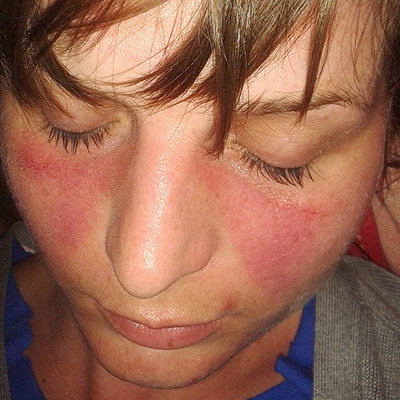 Image Source: Wikimedia Commons (2013). Lupus, Licensed under a creative commons licence. Available for free at: https://commons.wikimedia.org/wiki/File:Lupusfoto.jpg (Accessed 19 May 2025).
 - Bullous systemic lupus erythematosus – presenting sign in one third of SLE patients;
- Tense vesicles, bullae and erosions on a background of normal or erythematous skin usually in a sun-exposed area (often the face is the area affected);
- Blisters heal without scarring and the pruritus is absent or mild;
- Often associated with a flare of lupus nephritis
- Three types which can only be distinguished by immunohistochemistry; type I is associated with auto-antibodies to type VII collagen
- Treatment of choice is dapsone, which leads to a rapid clinical improvement and this also differentiates bullous SLE from SLE with a blistering rash
- Maculopapular rash resembling a morbilliform drug eruption – morbilliform rash can be seen in SLE
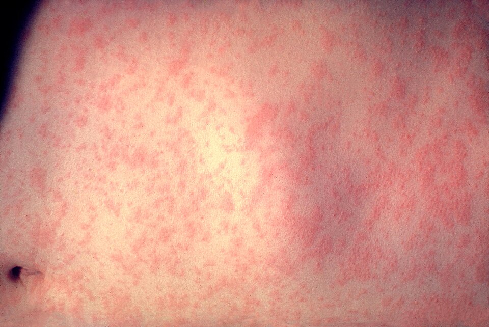 Image Source: Wikimedia Commons (2008). Morbillivirus Measles Infection, Licensed under a creative commons licence. Available for free at: https://commons.wikimedia.org/wiki/File:Morbillivirus_measles_infection.jpg (Accessed 19 May 2025).
 - Mucosal erosions and ulcerations (mouth, genitals, lips, nose) – mouth ulcers, and less commonly nose ulcers, occur in up to 45% of patients; usually large and painless
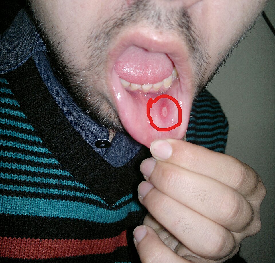 Image Source: Wikimedia Commons (2014). Aphthous Stomatitis on the Labial Mucosa, Licensed under a creative commons licence. Available for free at: https://commons.wikimedia.org/wiki/File:Aphthous_stomatitis_on_the_labial_mucosa.jpg (Accessed 19 May 2025).
 - Photosensitivity, which can be mild or severe
- Diffuse hair loss (nonscarring alopecia) with brittle hair shafts
Subacute CLE: less commonly associated with SLE (50% have a mild form of SLE); 20-40% are thought to have drug-induced subacute CLE; comprises 10-15% of CLE presentations
- Psoriasiform papulosquamous rash on upper back, chest and upper arms
- Annular, polycyclic plaques with central clearing
- Lesions seen on trunk and arms resolve to leave dyspigmentation and telangiectases, but no scarring
- Signs precipitated or aggravated by sun exposure
Intermittent (lupus tumidus)
- Affects sun-exposed areas – chest, back, upper arms, face
- Erythematous patches and plaques with a smooth surface, with round or annular shapes
- Signs resolve with no scarring left in the winter
Chronic CLE: affects 25% of SLE patients
- Classic discoid lupus is the most common manifestation – scaly plaques with follicular prominence (carpet tack sign) that cause post-inflammatory dyspigmentation and/or scarring
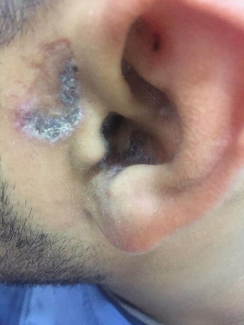 Image Source: Wikimedia Commons (2018). Stomach blood supply, Licensed under a creative commons licence. Available for free at: https://commons.wikimedia.org/wiki/File:Shuster_sign_of_discoid_lupus_ertherymatouses.jpg (Accessed 19 May 2025).
 - Hypertrophic (warty) lupus – variant of discoid LE with thickened warty plaques resembling viral warts
- Mucosal lupus – plaques, ulcers and scaling of the lips, nose, mouth and sometimes genitals
- Lupus profundus/panniculitis – inflammation of the subcutaneous without vasculitis; presents as firm, deep, tender nodules which leave dents in the skin
- Chilblain lupus – rare variant involving mainly the fingers and toes of smokers; triggered by exposure to a moist cold environment
Non-specific cutaneous SLE – features related to underlying illness rather than an autoimmune attack; can occur in other connective tissue or autoimmune diseases
- Nail fold capillary telangiectasia
- Raynaud phenomenon
- Diffuse hair thinning
- Cutaneous vasculitis – palpable purpura or urticarial vasculitis
- Livedo reticularis – in 20-30% of SLE patients
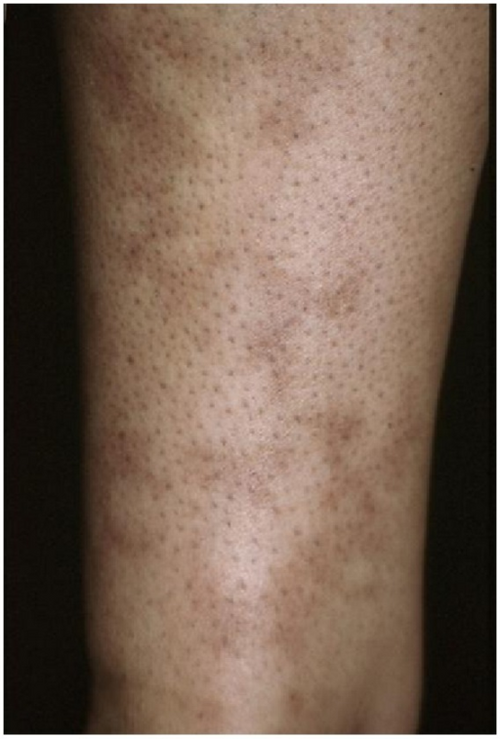 Image Source: Wikimedia Commons (2012). Livedo Reticularis of an Arm, Licensed under a creative commons licence. Available for free at: https://commons.wikimedia.org/wiki/File:Livedo_reticularis_of_an_arm.png (Accessed 19 May 2025).
 - Papular mucinosis (mucin deposits)
- Calinosis cutis (calcium deposits)
Investigations
Initial investigations should include:
- Antinuclear antibody (ANA) – a positive ANA does not diagnose SLE, but a diagnosis of SLE requires a positive ANA
- Anti-Ro antibody – rarely, ANA can be negative in anti-Ro-antibody positive lupus
- Double-stranded DNA, Smith antigen – highly specific for SLE and often confirmatory of diagnosis if present
- Full blood count – often leukopenia caused by neutropenia, anaemia
- Clotting screen – activated partial thromboplastin time may be prolonged if antiphospholipid antibodies present
- Kidney function and urinalysis – deranged if renal system affected
- ESR and CRP – elevated due to systemic inflammation
Subsequent investigations which can be considered:
- Coombs’ test to assess for haemolysis
- Complement levels – often raised in SLE
- Antiphospholipid antibodies
- Skin or renal biopsy
Management
Treatment is divided into preventative measures, topical therapy and systemic therapy.
Preventative measures to minimise flares and organ damage include:
- Diligent minimisation of sun exposure with clothes, accessories, sunscreen
- Smoking cessation
- Psychological interventions
Topical therapy includes potent topical corticosteroids and calcineurin inhibitors (tacrolimus ointment and pimecrolimus cream).
Systemic therapy choice depends on which organ systems are most affected. The medications generally used are as follows:
- NSAIDs – first line for musculoskeletal stiffness or pain
- Hydroxychloroquine – recommended in all SLE patients; response rates are about 80% in CLE; risk of retinal toxicity
- Corticosteroids – mainstay of treatment in the ill SLE patient; can give intravenous methylprednisolone for immediate therapeutic effect before switching to oral
- Immunosuppressive agents include methotrexate, azathioprine, mycophenolate
- Biological agents – B-cell targeting agents such as belimumab and rituximab
Prognosis
SLE often presents as flares with periods of remission. In some patients, all signs of active disease resolve. The 5-year survival is 95% and 10-year survival is 92%, with the most common causes of mortality being cardiovascular disease, infection and severe disease activity.
Complete Guide to Clinical Genetics ST3 Application and Recruitment (2027)
Written by Medibuddy. Last updated: October 2026. Clinical Genetics is

Complete Guide to Clinical Radiology ST3 Application and Recruitment (2027)
Recruitment paused: NHS England is not running Clinical Radiology ST3+ recru

Complete Guide to Paediatric Surgery ST3 Application and Recruitment in United Kingdom (2026)
Written by Medibuddy. Last updated: October 2026. If you’re planning


Take your subscriptions with you
Our mobile app allows you to access your interview and exam question banks wherever you are.

