


 Close
Close
 5th June 2025
Rheumatoid arthritis
Rheumatoid arthritis (RA) is a chronic autoimmune systemic inflammatory disorder that most often symmetrically affects the small joints of the hands and feet.
Epidemiology
RA is a common condition, affecting about 1% of the UK population. It is 2-4 times more common in women compared to men and the peak age of onset is 30-50 years old with incidence increasing with age. The peak age of incidence in the UK for both men and women is around 70 years of age. Extra-articular involvement is seen in 40% of cases and skin changes are seen in 30%.
Aetiology
The aetiology of RA is multifactorial with no clear cause found. RA is an autoimmune condition with antibodies seen against rheumatoid factor or anti-citrullinated protein antibodies. It is also associated with genetic factors, most notably HLA-DR4 which may be involved in the pathogenesis. Some genetic variants of PTPN22 and other genes have been identified as risk factors. It has also been proposed that an infection may be the trigger RA in a susceptible individual, but no specific causative factor has been identified.
Pathophysiology
Central to the pathogenesis is the formation of inflamed synovium in joints. Increased inflammation in the synovium causes the synovial lining to become hyperplastic with immune cell infiltration in the sublining. Increased cytokines are seen, with tumour necrosis factor (TNF), interleukin-1 (IL-1) and interleukin-6 (IL-6) most abundantly seen. Angiogenesis serves the hypertrophic synovium. The inflammatory setting, along with increased levels of metalloproteinase activity is thought to drive the joint destruction.
Diagnosis
Clinical features
Patients often present with symmetrical synovitis of the small joints of the hands and feet, though any joint can be affected. This consists of pain often worse at rest, swelling of the joint, heat and early morning stiffness lasting over 1 hour. Extra-articular changes are often skin-related and can be numerous:
- Atrophic, dry, friable skin
- Raynaud phenomenon
- Nail changes e.g., onycholysis, nail ridging and splitting, clubbing
- Rheumatoid nodules – only seen in RA
- solitary or multiple skin-coloured nodules on sites of recurrent pressure or trauma, generally asymptomatic
- Rheumatoid vasculitis – only seen in RA
- An unusual complication of long-standing severe rheumatoid arthritis involving small to medium-sized vessels
- Can present as palpable purpura, nailfold infarcts, digital necrosis, ulcers, and urticarial vasculitis
- A specific manifestation of this vasculitis is a Bywaters lesion – painless purpuric papules of <1mm diameter found on the fingertip or around the nail
- Rheumatoid neutrophilic dermatitis – only seen in RA
- Rare severe skin manifestation of severe long-standing RA
- Symmetrical asymptomatic eruption of erythematous papules, nodules, and plaques which may show an annular distribution at the extremities
- Associated with a disease flare and resolves with treatment
There are three classical deformities seen in RA, though they are no longer commonly seen in presenting patients due to the advent of disease-modifying anti-rheumatic drugs (DMARDs).
- Swan neck deformity – DIP hyperflexion, PIP hyperextension
- Boutonniere deformity – PIP flexion, DIP hyperextension
 Image Source: Wikimedia Commons (2021). Swan Neck Deformation Finger, Licensed under a creative commons licence. Available for free at: https://commons.wikimedia.org/wiki/File:Swan_neck_deformation_finger.jpg (Accessed 19 May 2025).
 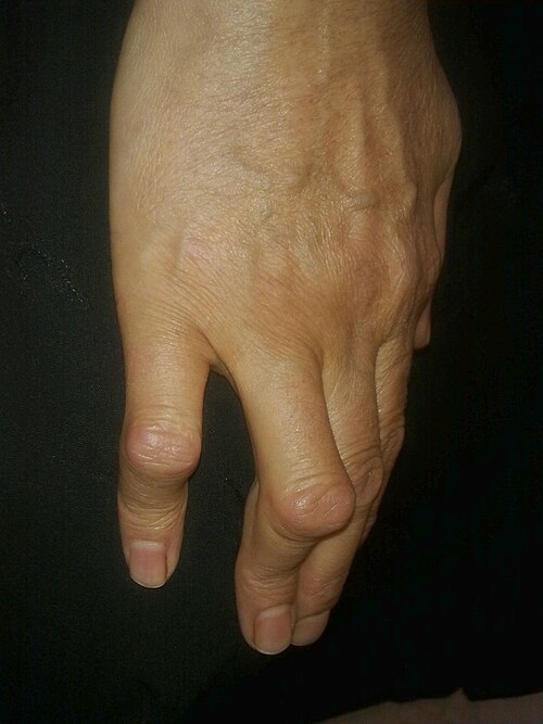 Image Source: Wikimedia Commons (2013). Boutonniare Deformity, Licensed under a creative commons licence. Available for free at: https://commons.wikimedia.org/wiki/File:Boutonni%C3%A8re_deformity.jpg (Accesed 19th May 2025).
 - Ulnar deviation – inflammation of MCP joints causing finger joint dislocation and deviation to the ulnar side
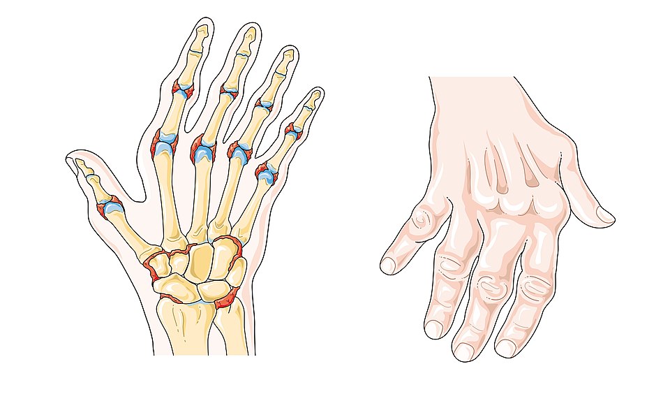 Image Source: Wikimedia Commons (2019). Rheumatoid Arthritis – Smart Servier, Licensed under a creative commons licence. Available for free at: https://commons.wikimedia.org/wiki/File:Rheumatoid_arthritis_–_Smart-Servier_(cropped).jpg (Accessed 19 May 2025).
RA is a systemic disease and in more severe forms can be associated with pleuritis, interstitial lung disease, pericarditis, and inflammatory eye disease.
Investigations
Diagnosis is clinical however investigations can speed up the process. The first line investigations include:
- Rheumatoid factor (RF) – positive in 60-70% of RA patients, found in 90% of patients with rheumatoid nodules
- Anti-cyclic citrullinated peptide (anti-CCP) antibodies – found in 80% of RA patients
- X-rays of hands and feet – can aid diagnosis but often helpful to monitor progress of disease
- Ultrasonography – may detect synovitis of the wrist and fingers at the initial presentation
All people suspected to have RA should have a rheumatology appointment within 3 weeks to confirm this. If the small joints of the hands and feet are affected, more than one joint is affected or there has been a delay of 3 months or more between onset of symptoms and patient presenting, a referral should be made within 3 working days.
Complications
- Joint destruction causing work disability and requiring joint replacement surgeries
- Shortened life span – 8-10 years on average
- Increased risk for coronary artery disease
- Interstitial lung disease (ILD)
- Felty syndrome – defines as the presence of RA, splenomegaly, and an abnormally low white blood cell count; seen in less than 1% of patients
- Carpal tunnel syndrome – seen in 10-20%
- Secondary to immunosuppressive effects of treatment – infection, malignancy, methotrexate-related liver toxicity
Management
Management recommendations depend on the disease severity at presentation.
For mild disease at initial presentation:
- 1st line: DMARD monotherapy
- 1st option: Hydroxychloroquine
- 2nd options: Sulfasalazine, Methotrexate or Leflunomide
- Consider corticosteroids and NSAIDs in acute flares
For moderate-to-severe disease at initial presentation.
- 1st line: Methotrexate monotherapy
- 2nd options: Sulfasalazine, Hydroxychloroquine, Leflunomide
- 2nd line: Add biological agent or targeted DMARD
- Biological agent options: etanercept, infliximab, adalimumab, certolizumab pegol, golimumab, abatacept, rituximab, tocilizumab, sarilumab
- DMARD options: tofacitinib, baricitinib, upadacitinib, filgotinib
- Consider corticosteroids and NSAIDs in acute flares
For pregnant or planning pregnancy patients:
- 1st line: Corticosteroids
- 2nd line: Hydroxychloroquine or Sulfasalazine
Complete Guide to Clinical Genetics ST3 Application and Recruitment (2027)
Written by Medibuddy. Last updated: October 2026. Clinical Genetics is

Complete Guide to Clinical Radiology ST3 Application and Recruitment (2027)
Recruitment paused: NHS England is not running Clinical Radiology ST3+ recru

Complete Guide to Paediatric Surgery ST3 Application and Recruitment in United Kingdom (2026)
Written by Medibuddy. Last updated: October 2026. If you’re planning


Take your subscriptions with you
Our mobile app allows you to access your interview and exam question banks wherever you are.

