


 Close
Close
 3rd June 2026
Atherosclerosis
Atherosclerosis is a chronic inflammatory process caused by an accumulation of plaque materials made up of lipids and connective tissue matrix proteins. These lipoproteins are deposited and retained in the subintimal space of blood vessels, secondary to loss of endothelial structure. As these retained plaques harden over time, they cause narrowing of the opening of the arteries and restricted blood flow. As the plaques rupture, there can be formation of thrombosis, leading to possible complications such as myocardial infarction (MI), unstable angina or cardiac death.
Atherosclerosis is the underpinning mechanism of coronary artery disease (CAD), peripheral arterial disease (PAD) and cerebrovascular disease, depending on which arteries are affected.
Cardiovascular disease is the leading cause of death in the UK, with atherosclerosis as the main underlying cause, specifically accounting for roughly 50% of all cardiovascular events that occur. Atherosclerotic cardiovascular disease (ASCVD) had a prevalence of 6.58% in England in 2022 and 8.26% in Northern Ireland.
Epidemiology
Modifiable factors include:
- Hypertension
- Hyperlipidaemia (specifically high LDL, low HLD)
- Smoking
- Diabetes
- Obesity
- Excess alcohol
- Chronic Kidney Disease (CKD)
- Presence of implantable cardiac devices
- Chronic haemodialysis
Non-modifiable factors include:
- Male sex
- Age
- Genetics (e.g familial hypercholesterolaemia)
- Inflammatory conditions
- Autoimmune conditions
Aetiology and Pathophysiology
The underlying mechanism of atherosclerosis is one of endothelial dysfunction. Damage is undertaken on the intima, which may be caused by stress at vessel bifurcations. This endothelial damage leads to a decrease in the bioavailability of nitric oxide and an increase in the amount of reactive oxygen species that are produced. This is made worse by factors such as dyslipidaemia and hypertension.
LDL cholesterol moves into the arterial wall at the site of the damage, leading to formation of a fatty streak as macrophages cross the endothelium to enter the subintimal space. Smooth muscle cells follow and collagen proliferates, leading to a plaque/ fibrous cap developing. A necrotic core develops within the plaque, containing oxidised cholesterol, secondary to the foam cell accumulation.
The plaque growth not only narrows the arterial lumen, leading to restricted blood flow and ischaemia, but also risks rupture, in turn leading to blood clot formation. These clots can then lead to life-threatening conditions such as MIs, Transient ischemic attacks (TIAs) and ischaemic strokes.
Table 1. The 3 stages of atherosclerosis summarised
| Stage of plaque formation | |
|---|---|
| 1. Lipid-streak formation | Lipids are retained in the arterial wall. Macrophages will move into the intimal wall and bring in excess amounts of lipids. This leads to foam cell formation. |
| 2. Fibrous plaque formation | Smooth muscle cells move into the intimal arterial wall and lead to the fibrous cap being built. Further accumulation of foam cells initiate necrosis inside the plaque. |
| 3. Thrombosis and further lesions | As the core of necrosis grows, the cap will disappear. The substances making up the core will interact with coagulation cells and can lead to thrombosis. |
Key features of an atherosclerotic plaque are:
- Proliferation of macrophages, fibroblasts and smooth muscle cells
- Deposition of lipids
- High levels of collagen
- Necrosis
Once a certain vascular territory is damaged by atherosclerosis, the total risk for any CV event is much higher, as well as in the organ that is damaged by the compromised blood flow.
Diagnosis
The diagnosis of atherosclerosis can be made based on a thorough medical history, including risk factors (family history and lifestyle, etc.), physical examination, blood tests, and imaging.
History
- Any previous atherosclerotic conditions, e.g., cerebral vascular disease?
- Any risk factors for atherosclerotic disease, e.g., hypertension, smoking
- Family history of any coronary events, especially at a young age.
Clinical examination
- Xanthomata – Yellow raised deposits on the skin, usually around the tendons, knuckles, elbows, and ankles. These are an indication of familial hypercholesterolaemia.
- Xanthelasma – Yellow raised deposits that appear around the eyelids
- Corneal arcus – Grey ring that forms around the cornea edge. Associated with ageing, however, if present in younger patients may be a sign of familial hypercholesteremia.
- Buerger’s test – Tests for arterial insufficiency, which may be present secondary to atherosclerosis. It involves raising the feet of the patient and supporting them at 45-degrees for 2 to 3 minutes. Look for developing pallor to indicate draining of the blood from the peripheries. Then sit the patient up with their legs over the side of the bed, watching for reactive hyperaemia (positive test). The feet will have a delay in return of colour and will be a dusky red.
- Diminished pulses over the posterior tibial pulse and dorsalis pedis pulse — may indicate PAD.
- Bruits on auscultation of pulses (peripherally, carotids, renal arteries) – may indicate stenosis of arteries
- Colour changes to peripheries.
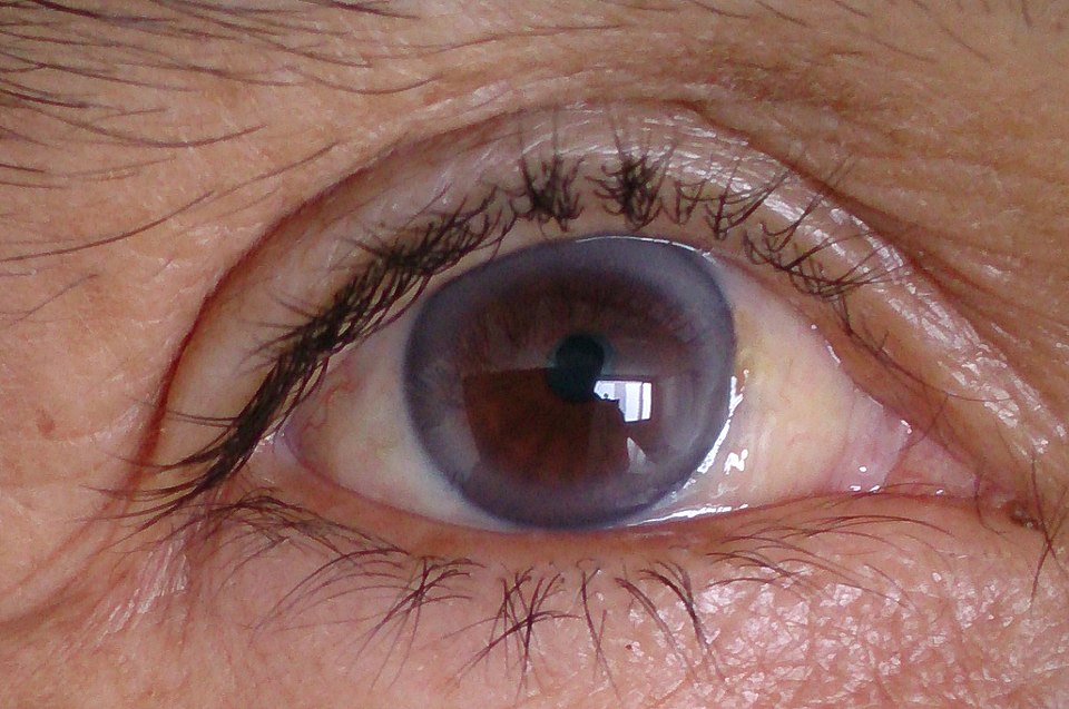
Corneal arcus
Image source: Wikimedia Commons (2020), ECG AVNRT, Licenced under a creative commons licence. Available for free at: https://commons.wikimedia.org/wiki/File:Arcus_Senilis_(cropped).jpg (Accessed: 27 April 2026)
Clinical Manifestations
Atherosclerosis can manifest in different clinical conditions/signs depending on which vessels the deposits are situated in, and how much blood flow is restricted.
- Carotid artery disease – Altered mental status, bruits over carotid arteries, may be asymptomatic until a stroke or TIA occurs.
- Stroke/ TIA – Sudden onset of neurological symptoms, e.g., hemiparesis or hemisensory symptoms, ataxia, vertigo, altered mental status, vomiting, dysarthria, dysphagia. TIA symptoms completely resolve in 24 hours.
- Coronary heart disease – chest pain at rest or activity, neck/shoulder/arms/ jaw radiation, dyspnoea, syncope, palpitations
- Peripheral arterial disease – pain on movement in legs and buttocks (usually bilateral but can be unilateral), parasetheisa in legs, hair loss on legs and feet, non-healing ulcers, blue discolouration of feet
- Mesenteric artery ischaemia – abdominal pain that is acute and severe, usually starting about 30 minutes after eating, nausea, vomiting, diarrhoea.
- Erectile Dysfunction
- Renal artery stenosis – Hypertension that can be difficult to treat, worsening kidney function, and flash pulmonary oedema.
Investigations
Risk Assessment:
- Assessment of CVD risk is undertaken depending on previous risk factors. This is followed by a full formal risk assessment if the patient’s estimated 10-year risk of CVD is 10% or more.
- The QRISK3 tool is used to calculate the estimated CVD risk for people aged between 25 and 84 without existing CVD over the following 10 years.
- You cannot use QRISK scores for patients with T1DM, CKD (eGFR <60) or familial hypercholesteremia.
Blood tests:
- Lipid profile (Total cholesterol, HDL- C, LDL-C, triglycerides) – a non-fasting sample is required
- Non-HDL cholesterol – This can give a more accurate estimate of cardiovascular risk with non-fasting samples
- LFTS – To assess liver function before statin initiation
- Glucose and HbA1c
- Renal function (eGFR, creatinine, us&es) – to assess for reduced renal function secondary to atherosclerosis, for dosing of statins and risk stratification
- TSH – To exclude hypothyroidism as a secondary cause of hyperlipidaemia
- Genetic testing of Lipoprotein (a) – may be undertaken if there is a history of familial hypercholesteremia
Imaging:
- Coronary Artery Disease
- Coronary angiography (Gold standard) – especially if intervention (PCI/CABG) is likely
- CT Coronary Angiography – Has the benefit of being non-invasive, and is the first-line investigation in outpatient cardiology or chest pain clinics.
- CT calcium scoring – A non-invasive test that provides a calcium score regarding the exact location and degree of plaque that is calcified on the coronary arteries.
- Stress testing – myocardial perfusion scan, stress echocardiograms
- ECG – To look for evidence of a prior MI
- Cerebrovascular Disease
- Carotid duplex ultrasound – to assess for stenosis in stroke/TIA patients
- Transcranial Doppler (TCD) — non-invasive and portable, uses a Doppler transducer to evaluate blood flow within major cranial arteries
- CT angiography of carotids for pre-operative planning
- Non-contrast CT
- MRI
- Peripheral Arterial Disease
- Ankle-Brachial Pressure Index (ABPI)
- Normal: 1-1.4; PAD <0.9 : Critical limb ischaemia: <0.5
- Duplex ultrasound – first line when assessing stenosis
- CT angiogram or conventional angiography – when considering revascularisation
- Ankle-Brachial Pressure Index (ABPI)
Medical management
Lifestyle interventions
Lifestyle change support, such as weight management and smoking cessation should be offered before commencement of medication in primary prevention.
If managing secondary prevention, lifestyle measures should be discussed alongside initiation of treatment.
Risk factor control
Blood pressure: Target BP <140/90mmHg in low risk. Target BP should be 130/80mmHg in high-risk patients (established atherosclerosis, CKD, diabetes).
Glycaemic control: Aiming for a fasting glucose <6mmol/L and HbA1c of less than 6.5%
Antiplatelet therapy: If patients have known peripheral or coronary disease caused by atherosclerosis, then aspirin 75mg OD should be considered, with clopidogrel as a second-line choice. Aspirin should not be considered in primary prevention, as the risk of GI bleeding outweighs the benefits.
Pharmacological Management
Lipid-lowering agents such as Atorvastatin 20mg are used for primary prevention if QRISK3 > or equal to 10%. The latest guidance from NICE advises to ‘not rule out’ those with a QRISK score of less than 10% for treatment. Atorvastatin 80mg is used for secondary prevention regardless of cholesterol level.
Repeat lipid levels and LFTS must be checked in 3 months of starting a lipid-lowering agent, then annually. Non-HDL cholesterol targeted with greater than or equal to 40% reduction for primary prevention. Non-HDL target is ≤ 2.6mmol/L or LDL target is ≤ 2.0 mmol/L for secondary prevention.
Clinicians should increase the dose of the statin if these are not met, and the patient agrees. If the target level is still not achieved post an increase in dose, Ezetimibe 10mg OD can be added in.
If target is still not achieved, newer lipid-lowering agents (PCSK9 inhibitors or inclisiran) may be added in, however, these may need to be discussed with specialist care.
Please see (Williams et al., 2024) for a summary of the NICE guidelines regarding primary and secondary prevention.
Surgical/ Interventional management
Coronary Artery Disease
- Percutaneous Coronary Intervention (PCI) – Utilised in acute coronary syndrome (ACS) or for angina not controlled by medical therapy
- Coronary Artery Bypass Grafting (CABG) – Utilised when plaques block coronary arteries, usually in complex blockages, unstable or uncontrolled angina. It involves bypassing the blood flow with arterial or venous conduits. The most commonly used vessel for a conduit is the left internal thoracic artery (ITA) due to its patency over long time periods. The radial artery and saphenous vein are also used frequently, however may be less patent in the long term.
Carotid disease
- Carotid Endarterectomy (CEA) – This involves the removal of cholesterol deposits and clots in the artery wall. Utilised for symptomatic carotid stenosis above 70%, however may be beneficial for patients with 50-69% stenosis. When performed in asymptomatic stenosis, the risk of any stroke is reduced by 30% over the next 3 years.
- Carotid Stenting – Utilised if there is severe stenosis plus co-morbidities, high surgical risk or contraindications to endarterectomy.
Peripheral Arterial Disease
- Treatment depends on severity.
- Angioplasty or bypass surgery – if there are persistent symptoms or critical limb ischaemia
- Amputation – In non-reconstructable ischaemia or severe infection
Complications
As discussed above, complications of atherosclerosis are dependent on which vessels the plaques are found in.
- Neurological – Ischaemic stroke, TIA, vascular dementia
- Cardiac – Myocardial infarction, heart failure, arrhythmias, angina
- Peripheral – Ulcers, claudication, critical limb ischaemia, gangrene, limb loss
- Renal – CKD, Renovascular hypertension
- Abdominal – Abdominal aortic aneurysms, mesenteric ischaemia
Complete Guide to Clinical Genetics ST3 Application and Recruitment (2027)
Written by Medibuddy. Last updated: October 2026. Clinical Genetics is

Complete Guide to Clinical Radiology ST3 Application and Recruitment (2027)
Recruitment paused: NHS England is not running Clinical Radiology ST3+ recru

Complete Guide to Paediatric Surgery ST3 Application and Recruitment in United Kingdom (2026)
Written by Medibuddy. Last updated: October 2026. If you’re planning


Take your subscriptions with you
Our mobile app allows you to access your interview and exam question banks wherever you are.

