


 Close
Close
 6th July 2026
Deep vein thrombosis
Diagnosis
Suspicion for DVT is based on history and clinical presentation. Signs and symptoms include pain and tenderness over the affected area. NICE guidelines suggest the following pathway in the initial diagnosis and management of DVT:
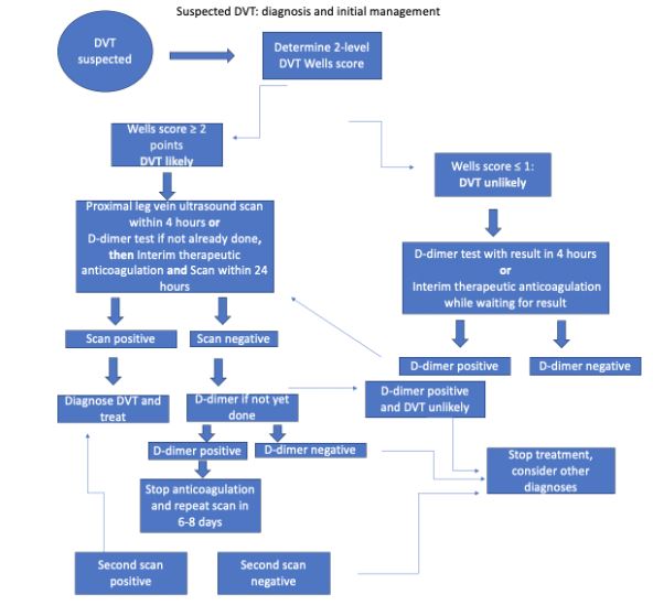
Figure 1: Suspected DVT diagnosis and initial management, adapted from NICE guidelines: Venous thromboembolic diseases: diagnosis, management and thrombophilia testing
Image Source: Wikimedia Commons (2023). Visual Summary, Licensed under a creative commons licence. Available for free at: https://www.nice.org.uk/guidance/ng158/resources/visual-summary-pdf-11193380893 (Accessed 8 May 2025).
The components of the Wells score have been summarised into the table below:
| Clinical feature | Points |
|---|---|
| Active cancer (patient either receiving treatment within the last 6 months, or is palliative) | 1 |
| Paralysis, paresis, or (recent) plaster immobilisation of the lower limbs | 1 |
| 1 | |
| Localised tenderness along the distribution of the deep venous system | 1 |
| Swelling of the entire leg | 1 |
| Swelling of the calf at least 3 cm larger than on the asymptomatic side (to be measured 10 cm below the tibial tuberosity) | 1 |
| Pitting oedema of only the symptomatic side | 1 |
| Collateral superficial veins (non-varicose) | 1 |
| History of previous DVT | 1 |
| Alternative diagnosis at least as likely as DVT | -2 |
| DVT likely | 2 points or more |
| DVT unlikely | 1 point or less |
Table 5- 2 Level DVT Wells Score (adapted from: https://www.nejm.org/doi/full/10.1056/NEJMoa023153)
Investigation
Patients will require different investigations depending on which leg they follow on the flow chart. All patients being investigated for a DVT should have baseline blood tests done – full blood count, renal and hepatic function, prothrombin time (PT), and activated partial thromboplastin time (APTT). If starting interim therapeutic anticoagulation, do not wait for blood results. Other mainstay investigations may include: proximal leg vein ultrasound, D-dimer test, CTPA, and V/Q scan. If suspecting a PE, patients will need a chest X-ray and ECG.
Management
Whilst investigating a potential DVT, patients may already have been started on interim therapeutic anticoagulation whilst awaiting further results. If confirmed, this remains the first line treatment for both DVTs and PEs. The standard anticoagulation used is a direct oral anticoagulant (DOAC), or low molecular weight heparin (LMWH) if DOACs are not suitable. There are several contraindications to DOACs. These are summarised below:
- Mechanical heart valves
- Renal impairment* (check local guidelines)
- Triple positive antiphospholipid syndrome
- Severe active bleeding or risk factors for major bleeding
- Pregnancy and breastfeeding
- Liver disease
- Active malignancy
- Allergy/hypersensitivity to drug or its excipients
*A patient with renal impairment is not automatically unsuitable for a DOAC. You need to check the creatinine clearance of the patient which will determine suitability. As per NICE guidelines:
CrCl of 15-50 ml/min can be given:
- Apixaban
- Rivaroxaban
- LMWH fLMor a minimum of 5 days then
-
- Edoxaban or
- Dabigatran if CrCl ≥ 30 ml/min.
- LMWH or UFH and a Vitamin K antagonist (VKA) for a minimum of 5 days or until INR is at least 2.0 on 2 consecutive measurements, then a VKA alone.
CrCl < 15ml/min can be given:
- LMWH
- UFH
- LMWH or UFH and a VKA for a minimum of 5 days or until INR is at least 2.0 on 2 consecutive measurements, then a VKA alone.
Thrombolytic therapy may be used in symptomatic iliofemoral DVT. Treatment is continued for at least 3 months if DVT/PE is confirmed and may be continued for up to 6 months if the VTE was unprovoked. Blood results will guide further treatment regimens.
In a patient presenting with an unprovoked VTE, patients should have a review of their medical history and baseline bloods, including FBC, renal and hepatic profiles, and a clotting screen. If they do not have any signs or symptoms of cancer, further investigation for any malignancy is not suggested. If there is no clear underlying cause, thrombophilia testing is appropriate. Tests for thrombophilia may be affected by anticoagulation, so specialist advice may be required.
As always, it is important to consider relevant comorbidities, contraindications, and patient preference. Important factors to consider include patient bodyweight, haemodynamic instability, any renal impairment, including renal failure, active cancer, pregnancy/breastfeeding, and patients with triple antiphospholipid syndrome. Results of blood tests will guide further treatment regimens.
If anticoagulation is contraindicated for whatever reason, alternative treatments include mechanical interventions – inferior vena cava (IVC) filters, graduated compression stockings, and percutaneous mechanical thrombectomy. To give a brief explanation of these methods:
- IVC filters are small devices made from metal that are inserted into the IVC. It then traps the clot.
- Graduated compression stockings apply pressure, or, as the name implies, compress your legs to encourage blood to flow back up to the heart, rather than the blood pooling in your legs.
- Percutaneous mechanical thrombectomy is similar to IVC filters in that a device is passed through your veins. Rather than trapping the clot, however, it dissolves the clot into small pieces.
Complete Guide to Clinical Genetics ST3 Application and Recruitment (2027)
Written by Medibuddy. Last updated: October 2026. Clinical Genetics is

Complete Guide to Clinical Radiology ST3 Application and Recruitment (2027)
Recruitment paused: NHS England is not running Clinical Radiology ST3+ recru

Complete Guide to Paediatric Surgery ST3 Application and Recruitment in United Kingdom (2026)
Written by Medibuddy. Last updated: October 2026. If you’re planning


Take your subscriptions with you
Our mobile app allows you to access your interview and exam question banks wherever you are.

