


 Close
Close
 6th July 2026
Investigation and management of VTE in pregnancy
Pregnancy, in and of itself, is a risk factor for thromboembolism. In preparation for delivery, the body goes into a procoagulant state, to combat the risk of any haemorrhage during or after delivery.
If DVT is suspected, compression duplex ultrasound of the leg should be done. If this is negative AND there is low clinical suspicion, anticoagulation can be stopped.
In all women presenting with signs and symptoms of PE, ECG and CXR should be performed, as well as baseline bloods. If there is concurrent presentation of DVT, duplex ultrasound should be performed. Otherwise, V/Q scan or CTPA should be done. If the CXR is abnormal, CTPA is preferential to a V/Q scan. Both CTPA and V/Q scans will expose the patient and foetus to ionising radiation with V/Q scans causing a slight increase in the risk of cancer developing in childhood but producing a lower dose of radiation to breast tissue (which is more sensitive to radiation during pregnancy). The absolute risk for both is minimal, however patients should be counselled on the risks and consented:
V/Q scans produce a lower dose of radiation to breast tissue (breast tissue is more sensitive to radiation during pregnancy) but carries a slightly increased risk of childhood cancer
CTPA scans will give a smaller dose of radiation to the foetus
The first line management for VTE is low molecular weight heparin (LMWH), and should be started if DVT/PE is clinically suspected, even if imaging has not yet been performed (unless there is a strong contraindication to treatment). Continue to treat with therapeutic doses of subcutaneous LMWH throughout the rest of the pregnancy, and for 6 weeks postnatally – a minimum total of 3 months. In the haemodynamically unstable patient, rapid assessment by the on-call team (including the consultant obstetrician) is crucial. Patients with a massive PE who are haemodynamically unstable should be treated with IV unfractionated heparin.
The same mechanical treatments are available for use in pregnancy under certain circumstances, and would require specialist input.
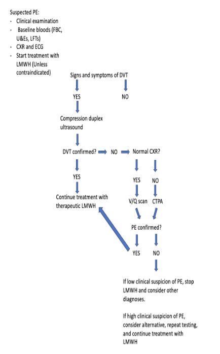
Figure 4 – Algorithm for the investigation and initial management of suspected PE in pregnancy and puerperium. (Adapted from the RCOG guidelines for Thromboembolic Disease in Pregnancy and Puerperium: Acute Management: https://www.rcog.org.uk/media/wj2lpco5/gtg-37b-1.pdf
Complete Guide to Clinical Genetics ST3 Application and Recruitment (2027)
Written by Medibuddy. Last updated: October 2026. Clinical Genetics is

Complete Guide to Clinical Radiology ST3 Application and Recruitment (2027)
Recruitment paused: NHS England is not running Clinical Radiology ST3+ recru

Complete Guide to Paediatric Surgery ST3 Application and Recruitment in United Kingdom (2026)
Written by Medibuddy. Last updated: October 2026. If you’re planning


Take your subscriptions with you
Our mobile app allows you to access your interview and exam question banks wherever you are.

