


 Close
Close
 15th July 2026
Dermatitis
Allergic contact dermatitis
Clinical features
Allergic contact dermatitis is associated with Type IV hypersensitivity reaction. It requires previous exposure to the substance (sensitisation). Rash usually takes around 48-72 hours after the exposure to appear. Usually confined to contacted area, but can sometimes extend outside the contacted area or may be generalised. Reaction usually produces erythema, oedema and blisters. Diagnosis usually can be made with detailed and careful history taking on exposure to common allergens.
Common sources of allergic contact dermatitis include
- Nickel, Chromate
- Hair dyes(paraphenylenediamine), tights, shoes
- Creams, ointments, cosmetics
Investigations
- Patch testing
Management
- Avoidance of allergens
- Emollient creams
- Topical steroids
- Oral antibiotics if secondary infection
- Topical calcineurin inhibitor
Irritant contact dermatitis
Clinical features
Irritant contact dermatitis is a non-allergic reaction which commonly affects the hands. It is due to repetitive exposure to irritants on the skin. Erythema is normally confined to the area of exposure to the irritant. In acute phase, it can present with erythema, swelling, papules and blisters. It usually occurs soon after the exposure, and severity is dependent on a few factors such as length of exposure, concentration and quantity of irritants. Frequent hand scrubbing, exposure to temperature extremes, and humidity can cause irritant contact dermatitis. It can happen because of the cumulative impact of multiple irritants.
Common sources of allergic contact dermatitis include
- Detergent
- Cement
- Organic solvent
- Friction
- Hand sanitisers
Investigations
- Diagnosis is mainly from history taking and clinical examination.
- Patch testing may be used to distinguish between allergic and contact dermatitis.
Management
- Avoid all potential irritants
- Emollient
- Barrier creams +/- topical steroids.
Atopic Dermatitis
Clinical features
Atopic dermatitis is also known as atopic eczema. It is the most common inflammatory dermatitis. It tends to affect people with positive family history or personal history of atopy. Usually starts from infancy and may settle in late childhood or adolescence. All ages can be affected. Risk factors for developing atopic dermatitis can be broken down into genetic and environmental risk factors. Genetic: positive family history, filaggrin gene defects (FLG), which cause a skin barrier defect, and the immune system (Th2-associated cytokines contribute to loss of skin barrier function). Environmental factors include dry skin associated with low humidity, frequent hand washing, winter weather, and irritants such as detergents.
It commonly affects the flexural aspect of the skin; however, it can affect all parts of the skin depending on age. In darker skin types, it may also affect the extensor surfaces more commonly than in lighter skin types. In chronic cases, skin may become thickened (lichenified) and less erythematous compared to the acute phase.
Investigations
- Normally, a clinical diagnosis
- Patch testing may be utilised in treatment-resistant cases.
Management
- Avoidance of irritants or exacerbating factors
- Topical therapy such as regular emollients, topical steroids, topical antibiotics (if only secondary infection), and topical calcineurin inhibitors
- Phototherapy: UVB or PUVA
- Systemic treatment: Prednisolone (severe flare), ciclosporin, azathioprine, antihistamines
- Janus Kinase Inhibitor: Abrocitinib, bariticinib
- Biologics: Dupilumab, Tralokinumab
Steroid Potencies
| Mild | Moderate | Potent | Very Potent |
|---|---|---|---|
| Hydrocortisone 0.5%-2.5% | Betamethasone valerate 0.025% ClobetaSONE butyrate 0.05% (Eumovate) | Betametasone valerate 0.1% (Betnovate) | ClobetaSOL propionate 0.05% (Dermovate) |
Complications
- Secondary infection:
- Bacterial – Staphylococcus and streptococcal infection
- Viral – Eczema herpeticum, Eczema coxsackium, Molluscum contagiosum
- Fungal- Dermatophyte (Trichophyton rubrum / Epidermophyton skin infection)
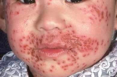
Child with facial eczema herpeticum.
Image Source: Wikimedia Commons (2020), ECG AVNRT, Licenced under a creative commons licence. Available for free at: https://commons.wikimedia.org/wiki/File:Eczema_herpitcum.jpg (Accessed: 24 May 2026).
Clinical features
Eczema Herpeticum is commonly seen as a complication of atopic dermatitis. It is characterised by monomorphic painful and itchy blisters with punched out lesions, and new blisters have a central dimple (umbilication). It is caused by Herpes Simplex Virus(HSV)Type 1 or 2. It can also happen in non-eczematous conditions such as thermal burns and psoriasis. The patient can be very unwell and present with fever and swollen lymph nodes. Eye involvement will require urgent ophthalmology attention.
Investigations
- Viral swabs
- Bacterial swabs
- Rule out secondary infection.
Management
- Oral antiviral, e.g., Aciclovir 400mg, five times a day
- Oral antibiotics if secondary bacterial infection
- Topical steroids generally NOT recommended unless there is active underlying atopic dermatitis that requires treatment
Dyshidrotic eczema (Pompholyx)
Clinical features
Pompholyx is characterised by vesicles and can be as large as bullae, usually deep-seated on the palms and soles. It is often associated with intense itchiness and a burning sensation. Once the blisters healed, the skin can become red, dry and cracked. However, most of the time, the vesicles will gradually subside or resolve spontaneously. Up to 50% of patients will have a family or personal history of atopic dermatitis. Females are more commonly affected than males. Cause of the rash is unknown and thought to be aggravated by hot and humid weather, emotional stressors, exposure to irritants or allergens.
Investigations
- Usually diagnosed clinically
- Skin biopsy can be performed if diagnosis is uncertain
- Patch testing can be used if a component of allergic dermatitis is suspected
Management
- General measures include wet dressings to dry up blisters, such as diluted potassium permanganate, emollient, antiperspirant applied to palms and soles at night, and wear gloves if working with irritants or allergens
- Topical steroids- Greater potency steroid is normally used, given thick skin on palms and soles
- Antibiotics if infection present
Seborrhoeic dermatitis
Clinical features
Seborrheic dermatitis is a self-limiting, chronic and relapsing skin condition. It has a biphasic model, which affects infants and peaks in adults. It mainly affects areas with rich sebaceous glands, such as the face (eyebrow, eyelid, cheeks), scalp and trunk. It can affect the eyelid, causing blepharitis or scaly patches or scales on the scalp. The lesion has a defined margin, dull red or yellowish, covered with greasy scale. In adults, Malassezia furfur growth plays a role in the pathogenesis of seborrhoeic dermatitis. There are a few conditions known to be associated with severe seborrhoeic, such as HIV and Parkinson’s disease.
Seborrhoeic dermatitis is usually associated with flares during the winter months and improves during the summer. Itchiness is not a common feature. Salmon pink scaly rash can be seen on the facial folds. On the scalp, the presentation ranges from mild dandruff to scaling of the scalp.
Investigations
- Usually, a clinical diagnosis
- Consider investigations for associated conditions, such as HIV in refractory cases
Management
- Scalp: first line: Shampoo with zinc pyrithionine, tar shampoos, ketoconazole shampoos
- Other parts of the body: Topical antifungal such as ketoconazole 2% cream for facial involvement, Ketoconazole 2% bodywash can be used on trunk involvement
- Mild topical corticosteroid can be considered if there is active inflammation
Discoid Dermatitis (Nummular Eczema)
Clinical features
Discoid dermatitis is characterised as single or multiple, itchy, well-defined, coin-shaped, erythematous plaques with +/- vesiculation. It usually involves the lower legs, the forearm and the dorsum of the hand. It commonly affects young women and middle-aged adults. The cause of this is unknown; it is associated with contact allergy manifestation of atopy or localised injury.
Investigations
- Skin swab – usually shows Staphylococcus aureus colonisation
- Skin scraping for mycology is useful to rule out fungal infection
- Patch testing for the suspected trigger as a contact allergy
Management
- Emollient
- Avoid allergens
- Topical steroid
- Antibiotics if lesion is infected
References
- Calonje, E., Brenn, T., Lazar, A., & Billings, S. D. (Eds.). (2012). Spongiotic, psoriasiform and pustular dermatoses: Eczematous dermatitis (Chapter 6, pp. 180–186). In McKee’s Pathology of the Skin (4th ed.). Elsevier
- Johnston, G. A., Exton, L. S., Mohd Mustapa, M. F., Slack, J. A., Coulson, I. H., English, J. S., & Bourke, J. F. (2017). British Association of Dermatologists’ guidelines for the management of contact dermatitis 2017. The British journal of dermatology, 176(2), 317–329. https://doi.org/10.1111/bjd.15239
- Oakley, A. (2012). Contact dermatitis. DermNet NZ. Retrieved October 26, 2025, from https://dermnetnz.org/topics/contact-dermatitis
- Fransway, A. F., & Reeder, M. J. (2023). Irritant contact dermatitis in adults. UpToDate. https://www.uptodate.com/contents/irritant-contact-dermatitis-in-adults?search=contact%20dermatitis&source=search_result&selectedTitle=1~150&usage_type=default&display_rank=1
- Oakley, A. (2012). Irritant contact dermatitis. DermNet NZ. Retrieved October 26, 2025, from https://dermnetnz.org/topics/irritant-contact-dermatitis
- Oakley, A. (1997). Allergic contact dermatitis. DermNet NZ. Updated by Post, R. (2016). Retrieved October 26, 2025, from https://dermnetnz.org/topics/allergic-contact-dermatitis
- Sasseville, D., & Houle, M.-C. (2024). Seborrheic dermatitis in adolescents and adults. UpToDate. Retrieved October 26, 2025, from https://www.uptodate.com/contents/seborrheic-dermatitis-in-adolescents-and-adults?search=seborrheic%20dermatitis&source=search_result&selectedTitle=1~150&usage_type=default&display_rank=1
- Oakley, A. (2008). Seborrhoeic dermatitis [CME module]. DermNet NZ. Retrieved October 26, 2025, from https://dermnetnz.org/cme/dermatitis/seborrhoeic-dermatitis
- Jain, M. A., & Jarrett, P. (2023, February). Dyshidrotic eczema (pompholyx). DermNet NZ. Retrieved October 26, 2025, from https://dermnetnz.org/topics/dyshidrotic-eczema
- Sasseville, D., & Houle, M.-C. (2023). Acute palmoplantar eczema (dyshidrotic eczema). UpToDate. Retrieved October 26, 2025, from https://www.uptodate.com/contents/acute-palmoplantar-eczema-(dyshidrotic-eczema)?search=pompholyx&source=search_result&selectedTitle=1~21&usage_type=default&display_rank=1
- Oakley, A. (n.d.). Atopic dermatitis. DermNet NZ. Retrieved October 26, 2025, from https://dermnetnz.org/topics/atopic-dermatitis
- Weston, W. L., & Howe, W. (2020). Atopic dermatitis (eczema): Pathogenesis, clinical manifestations, and diagnosis. UpToDate. Retrieved October 26, 2025, from https://www.uptodate.com/contents/atopic-dermatitis-eczema-pathogenesis-clinical-manifestations-and-diagnosis?search=atopic%20dermatitis&source=search_result&selectedTitle=2~150&usage_type=default&display_rank=2
- Lin, C.-Y. (2010). Eczema herpeticum. DermNet NZ. Retrieved October 26, 2025, from https://dermnetnz.org/topics/eczema-herpeticum
- M. S. Hirsch & J. Mitty (2025, July). Epidemiology, clinical manifestations, and diagnosis of herpes simplex virus type 1 infection. UpToDate. Retrieved October 26, 2025, from https://www.uptodate.com/contents/epidemiology-clinical-manifestations-and-diagnosis-of-herpes-simplex-virus-type-1-infection?search=eczema%20herpeticum&source=search_result&selectedTitle=1~22&usage_type=default&display_rank=1
Complete Guide to Clinical Genetics ST3 Application and Recruitment (2027)
Written by Medibuddy. Last updated: October 2026. Clinical Genetics is

Complete Guide to Clinical Radiology ST3 Application and Recruitment (2027)
Recruitment paused: NHS England is not running Clinical Radiology ST3+ recru

Complete Guide to Paediatric Surgery ST3 Application and Recruitment in United Kingdom (2026)
Written by Medibuddy. Last updated: October 2026. If you’re planning


Take your subscriptions with you
Our mobile app allows you to access your interview and exam question banks wherever you are.

