


 Close
Close
 26th January 2024
Osteoporosis
Background
Osteoporosis is a skeletal disease characterised by low bone mass and abnormal bone architecture, which results in compromised bone strength, an increase in bone fragility and subsequent susceptibility to fracture. Osteoporotic (fragility) fractures are defined as those which result from mechanical forces, such as falling from standing height or less, or routine tasks, such as bending and lifting, that would not normally result in fractures. They are associated with a low BMD and most commonly occur at the spine, wrist, hip and shoulder.
Osteoporosis is defined by the World Health Organisation (WHO) as a bone mineral density (BMD) of 2.5 standard deviations below the BMD mean for young, healthy adults. BMD is measured by dual-energy X-ray absorptiometry (DEXA) at the femoral neck and reported as a T-Score. The four categories of T-scores are as follows:
- Normal: T-score ≥-1 (within 1 standard deviation of young, healthy white women)
- Osteopenia (low bone mass): T-score <-1 but >-2.5
- Osteoporosis: T-score of ≤-2.5
- Severe (or established) osteoporosis: T-score ≤-2.5 with presence of one or more fragility fractures)
Bone strength is not only determined by BMD but can also be associated with factors such as size and shape of the bone, bone turnover, quality of bone micro-architecture and bone mineralisation.
Osteoporosis itself is asymptomatic; therefore, the condition usually presents only after a bone fracture. The most common sites for fragility fractures are spine (vertebrae), hip (proximal femur) and wrist (distal radius); however, they may also occur in the arm, pelvis, ribs and other bones. Clinical signs and examination findings depend on the site of the fracture.
Epidemiology
Women are at greater risk of osteoporosis than men due to a decrease in oestrogen production after the menopause. It is estimated that more than 2 million women have osteoporosis in England and Wales. The prevalence of osteoporosis increases markedly with age, to more than 25% at 80 years in women. White men and women are predominantly affected, but people of any ethnicity can develop this condition. 1 in 3 women and 1 in 5 men are thought to suffer with an osteoporotic fracture in their lifetime.
Pathophysiology
Osteoporosis results from an imbalance in the normal process of bone remodelling by osteoclasts and osteoclasts. Through the process of normal ageing, the breakdown of bone via osteoclasts increases and is not balanced by the formation of new bone by osteoblasts. This culminates in reduced BMD and changes in the composition, architecture, size and structure of the bone. The remodelling of bone is regulated by various cytokines, including interleukins 1, 6, and 11, colony-stimulating factors, and hormones such as parathyroid hormone, 1,25-dihydroxy vitamin D, calcitonin, and oestrogen. Factors such as tumour necrosis factor (TNF), receptor activator of nuclear factor- kappa B (RANK), RANK ligand (RANKL), and osteoprotegerin (OPG) contribute to the resorption of bone in postmenopausal osteoporosis. RANKL secreted by osteoblasts interacts with RANK receptors on osteoclasts to promote the formation, function and differentiation of osteoclasts, thereby triggering bone resorption. OPG is expressed by osteoblasts and plays a role in the inhibition of the activation of RANK.
Causes and risk factors
Osteoporosis manifests when there is inadequate peak bone mass and/or ongoing bone loss. Peak bone mass is achieved in early adulthood. It depends on genetics, sex hormone status, levels of physical activity and nutritional intake of calcium and vitamin D. The rate of bone loss is accelerated in women around the time of the menopause due to oestrogen deficiency, decreased testosterone in elderly men and hyperparathyroidism.
The risk of sustaining a fragility fracture is influenced by factors, including bone strength (determined by BMD), the individual patient’s risk of falls and the presence of certain risk factors. As the BMD decreases, the risk of fractures increases, but BMD alone cannot be used to predict fracture risk in the absence of consideration of other risk factors. The risk factors affecting bone strength can be subdivided into those that reduce BMD, those which do not reduce BMD and those with unestablished mechanisms as shown in the table below.
| BMD dependent | BMD independent | Unestablished mechanisms |
|---|---|---|
| Female sex | Increasing age (above 50 for women and above 65 for men) | Drugs such as:
|
| Premature menopause | Oral corticosteroids (dependent on dose and duration of treatment). | |
| Caucasian/Asian ethnicity | Smoking | |
| Endocrine disorders including diabetes, hyperthyroidism, hyperparathyroidism, hypogonadism | Alcohol (3 or more units daily) | |
| Gastrointestinal disorders that cause malabsorption, such as inflammatory bowel disease, coeliac disease and chronic pancreatitis | Previous fragility fracture (risk increases with increasing number of fractures). Highest risk for previous hip fractures and lowest for previous vertebral fractures | |
| Chronic kidney disease | Parental history of hip fracture. | |
| Chronic liver disease | Rheumatological disorders such as rheumatoid arthritis | |
| Chronic obstructive pulmonary disease | ||
| Immobility | ||
| Body mass index of less than 18.5 kg/m² | ||
| Low dietary calcium intake and vitamin D insufficiency | ||
| Other disorders such as mastocytosis, multiple myeloma, osteogenesis imperfecta, homocystinuria |
Table 4: Risk factors affecting bone strength
Adapted from Kumar and Clark’s Clinical Medicine. 2017 Kumar P, Clark ML. Kumar and Clark’s Clinical Medicine E-Book. Elsevier Health Sciences; 2012 Jun 4.
Falls are often multifactorial; however, some common risk factors are impaired vision, impaired gait, lower limb weakness, use of alcohol and drugs, and polypharmacy.
References
- Johansen A, Harding K, Evans R, Stone M. Trauma in elderly people: what proportion of fractures are a consequence of bone fragility? Archives of gerontology and geriatrics. 2000 Feb 1;29(3):215-21.
- Wilkinson I, Wilkinson IB, Raine T, Wiles K, Goodhart A, Hall C, O’Neill H. Oxford handbook of clinical medicine. Oxford University Press; 2017.
- Kumar P, Clark ML. Kumar and Clark’s Clinical Medicine E-Book. Elsevier Health Sciences; 2012 Jun 4.
- Manolagas SC. Birth and death of bone cells: basic regulatory mechanisms and implications for the pathogenesis and treatment of osteoporosis. Endocrine reviews. 2000 Apr 1;21(2):115-37.
- Conaghan PG, Dickson J, Grant RL. Care and management of osteoarthritis in adults: summary of NICE guidance. Bmj. 2008 Feb 28;336(7642):502-3.
- National Institute for Health and Care Excellence (Great Britain). Osteoporosis: assessing the risk of fragility fracture. National Institute for Health and Care Excellence (NICE); 2017
- Christodoulou C, Cooper C. What is osteoporosis? Postgraduate medical journal 2003 Mar 1;79(929):133-8.
Diagnosis
The assessment of patients with osteoporosis consists of three main stages: assessment of fragility fracture risk, ruling out secondary causes for fragility fractures and osteoporosis, and the diagnosis of osteoporosis via DEXA scan. The overall aim is to assess patients depending on their risk factors and initiate treatment in order to prevent fragility fractures. However, unfortunately, in many patients, the occurrence of a fragility fracture may be the first indicator of an underlying diagnosis of osteoporosis, which is otherwise an asymptomatic condition.
Assessment of fragility fracture risk
NICE guidelines recommend the following algorithm for the identification of high-risk patients to assess for risk of developing a fragility fracture:
- All women over the age of 65 and all men over the age of 75
- Women between the ages of 50 and 64 and men between the ages of 50-74 who have at least one of the following risk factors:
- Personal or family history of osteoporotic fracture
- Use of oral glucocorticoids
- History of falls
- Causes of secondary osteoporosis
- Low BMI
- Smoking
- Alcohol intake more than 14 units per week for women and more than 21 units per week for men
- Secondary cause of osteoporosis, e.g. rheumatological or endocrine conditions
- Men and women under the age of 50 years with any of the following risk factors:
- Use of oral glucocorticoids
- Untreated premature menopause
- Previous fragility fracture
- Men and women under the age of 40 years with any of the following risk factors:
- Use of oral glucocorticoids equivalent to, or more than, 7.5 mg prednisolone daily for 3 months or more
- Previous fragility fracture
- Consider assessing fracture risk for patients taking certain medications, e.g. SSRIs, antiepileptics, aromatase inhibitors, PPIs, and thiazolidinediones.
Ruling out secondary causes of fragility fractures and osteoporosis
In patients who are diagnosed with osteoporosis or sustain a fragility fracture, further investigations are recommended by the National Osteoporosis Guideline Group (NOGG) to exclude disorders that present like osteoporosis, identify causes and risk factors for osteoporosis, assess risk of further fractures and enable appropriate selection of treatment.
The following investigations are recommended by NOGG, in addition to a full history and clinical examination:
- FBC
- ESR or CRP
- Serum calcium – low in osteomalacia, elevated in hyperparathyroidism
- Serum albumin – may be decreased in osteoporosis
- Serum creatinine – to rule out chronic kidney disease-associated bone disorders
- Serum phosphate – low in osteomalacia
- Serum alkaline phosphatase – raised in osteomalacia
- Liver function tests
- Thyroid function tests – to rule out hyperthyroidism as a cause of osteoporosis
Additional procedures are also recommended if there is a suspicion of alternative diagnoses:
- Lateral radiographs of lumbar and thoracic spine – to investigate for fractures
- Protein immunoelectrophoresis and urinary Bence-Jones proteins – in elderly patients with multiple spontaneous vertebral fractures to rule out multiple myeloma
- 25-hydroxy vitamin D – to rule out vitamin D deficiency
- Serum parathyroid hormone – to rule out hyperparathyroidism
- Serum testosterone – should be considered in all young men with osteoporosis – low levels may indicate hypogonadism
- 24-hour urinary cortisol/dexamethasone suppression test – in patients with features suggestive of Cushing’s syndrome
- Endomysial and/or tissue transglutaminase antibodies – if symptoms suggestive of coeliac disease
- Isotope bone scan – if concerned about malignancy
- Biochemical markers of bone resorption and formation can mostly be used to monitor response to treatment rather than diagnosis of osteoporosis.
- Urinary calcium excretion – to rule out chronic kidney disease
| Markers of bone formation
(low levels in osteoporosis) |
Markers of bone resorption
(elevated levels in osteoporosis) |
|---|---|
| serum procollagen type I N propeptide (PINP) | serum C-terminal telopeptide of type I collagen (CTX), |
| urinary deoxypyridinoline | |
| N-telopeptides |
Table 5: Biochemical markers of bone formation and resorption
Adapted from Vasikaran S, Eastell R, Bruyere O, Foldes AJ, Garnero P, Griesmacher A, McClung M, Morris HA, Silverman S, Trenti T, Wahl DA. Markers of bone turnover for the prediction of fracture risk and monitoring of osteoporosis treatment: a need for international reference standards. Osteoporosis international. 2011 Feb;22(2):391-420.
Calculating fracture risk
The diagnosis of osteoporosis is mainly based on an assessment of BMD. However, most osteoporotic fractures occur in patients who do not have a T score of less than -2.5; therefore, an assessment of clinical risk factors and the extent to which these can contribute towards risk of fall should be carried out, independently of BMD. According to NICE guidelines, patients who are at high risk of developing osteoporosis should have an assessment of fracture risk prior to arranging a DEXA scan – this can be done by using a validated online risk calculator such as QFracture or FRAX, which predict the absolute risk of hip and major osteoporotic fracture over a 10 year period. The FRAX calculator calculates the probability of people between the ages of 40 and 90 having a major fragility fracture. The following risk factors are taken into account:
- Sex
- BMI
- History of previous fracture
- Parental history of fracture
- Current smoking status
- Use of glucocorticoids
- History of rheumatoid arthritis
- Secondary osteoporosis
- Alcohol intake of 3 or more units per day
- Femoral neck BMD (if result available)
Patients are then classified as being low, intermediate or high risk according to their results, and subsequent NOGG advice is given regarding the need for treatment. The QFracture score is based on UK data and can be calculated for patients aged 30-99 without a value for BMD. In addition to the risk factors calculated in the FRAX score, the QFracture score also includes ethnicity, residence in care home, history of falls, dementia, cerebrovascular disease, chronic liver disease, and whether the patient is on high-risk medications other than glucocorticoids. Those patients who are identified as having a 10-year fracture risk of 10% or more should then be considered for undergoing a DEXA scan. Fragility fracture risk scores should ideally be carried out prior to DEXA scan; however, in patients over the age of 50 years with a history of fragility fracture or patients younger than 40 years with major risk factors for fracture, DEXA can be carried out without the use of FRAX or QFracture scores.
DEXA scan
The gold standard method of diagnosis for osteoporosis is a DEXA scan. Although DEXA carries an accuracy of more than 90% at the hip, it is important to note that false-positive results can be caused by conditions such as osteomalacia, osteoarthritis or soft-tissue calcification. A T-score of less than -2.5 on the DEXA indicates osteoporosis; a score of less than -2.5 with the presence of fragility fractures indicates severe osteoporosis. DEXA is the preferred method of diagnosis of osteoporosis; however, NICE guidelines states that for women aged 75 years or older who have one or more independent risk factors, DEXA may not be required if the clinician deems it to be clinically inappropriate or impractical to carry out. In addition, for some patients with vertebral fractures, it may be suitable to commence drug treatment without carrying out a DEXA scan. Other investigations, such as ultrasound measurement of bone at the heel or single-energy x-ray absorptiometry, can be used for the assessment of fracture risk; however, have not been validated for the diagnosis of osteoporosis. Plain x-rays can detect fractures and osteopenia (low bone density but not severe enough to cause osteoporosis) but not osteoporosis. CT scan can be used if DEXA is not available.
References
- Vasikaran S, Eastell R, Bruyere O, Foldes AJ, Garnero P, Griesmacher A, McClung M, Morris HA, Silverman S, Trenti T, Wahl DA. Markers of bone turnover for the prediction of fracture risk and monitoring of osteoporosis treatment: a need for international reference standards. Osteoporosis international. 2011 Feb;22(2):391-420.
- Conaghan PG, Dickson J, Grant RL. Care and management of osteoarthritis in adults: summary of NICE guidance. Bmj. 2008 Feb 28;336(7642):502-3.
- Compston J, Bowring C, Cooper A, Cooper C, Davies C, Francis R, Kanis JA, Marsh D, McCloskey EV, Reid DM, Selby P. Diagnosis and management of osteoporosis in postmenopausal women and older men in the UK: National Osteoporosis Guideline Group (NOGG) update 2013. Maturitas. 2013 Aug 1;75(4):392-6.
- National Institute for Health and Care Excellence (Great Britain). Osteoporosis: assessing the risk of fragility fracture. National Institute for Health and Care Excellence (NICE); 2017
Differential diagnoses
The following conditions can be difficult to distinguish clinically from osteoporosis. However, a thorough history, clinical examination and some investigations can be used in order to rule out conditions which can also either lead to bone pain or increase the risk of fractures.
- Multiple myeloma – can present with bone pain, but patients would most commonly have other associated features such as renal failure or anaemia. This can be ruled out by the presence of Bence Jones proteins on urine electrophoresis or monoclonal gammopathy on serum electrophoresis.
- Osteomalacia – in osteomalacia, there is a normal amount of bone with a low mineral content (in contrast to osteoporosis, where mineralisation levels are unchanged, but there is overall bone loss). It is associated with vitamin D deficiency or resistance. Blood tests in patients with osteomalacia would normally reveal low vitamin D, calcium and phosphate levels and raised alkaline phosphatase and parathyroid hormone (PTH) levels. It can be distinguished from osteoporosis using bone biopsy.
- Chronic renal disease and associated bone and mineral disorders – patients often have poor renal function with elevated serum creatinine and PTH levels.
- Primary hyperparathyroidism – this can present with features of hypercalcaemia such as constipation, abdominal pain, and psychiatric disturbances in addition to bone pain. Investigations in patients with primary hyperparathyroidism would normally reveal high calcium and alkaline phosphatase levels and low phosphate levels. PTH would be elevated or inappropriately normal. 24-hour urinary calcium levels would be raised.
- Primary or metastatic bone malignancy – patients may have a known history of cancer or presence of symptoms indicating primary malignancy. DEXA scan may be normal, but CT or radio nuclear bone scans could reveal presence of a tumour. Bone biopsy is the definitive test of differentiation between malignancy and osteoporosis.
- Vertebral deformities – conditions such as osteoarthritis or scoliosis may be mistaken for osteoporotic vertebral fractures. X-ray and other imaging modalities can be used in cases of diagnostic uncertainty.
References
- Wilkinson I, Wilkinson IB, Raine T, Wiles K, Goodhart A, Hall C, O’Neill H. Oxford handbook of clinical medicine. Oxford University Press; 2017.
- Compston J, Bowring C, Cooper A, Cooper C, Davies C, Francis R, Kanis JA, Marsh D, McCloskey EV, Reid DM, Selby P. Diagnosis and management of osteoporosis in postmenopausal women and older men in the UK: National Osteoporosis Guideline Group (NOGG) update 2013. Maturitas. 2013 Aug 1;75(4):392-6.
Management
The main aim of management of osteoporosis is prevention of fragility fractures, particularly hip, vertebral and radial.
General measures
All patients at risk of osteoporosis or with established disease should receive adequate supplementation with calcium and vitamin D. Vitamin D replacement should be prescribed to all patients; calcium should be prescribed in addition to this if oral intake of calcium is deemed inadequate. Eating a balanced diet and engaging in regular exercise is recommended to maintain bone strength. Smoking cessation and alcohol reduction advice should be given.
Pharmacological management
Post-menopausal women
Bisphosphonates
Bisphosphonates, e.g. alendronic acid, ibandronic acid, risedronate, and zoledronic acid, are used as first-line therapy for the prevention and treatment of osteoporosis in women. They are indicated for postmenopausal women with a history of an osteoporotic fracture and/or a T score of less than -2.5. Patients who have had a fragility fracture and are aged 75 years or over are presumed to have underlying osteoporosis and should be commenced on first-line therapy with an oral bisphosphonate, without the need for DEXA. Bisphosphonate acts by inhibiting osteoclast activity and, therefore, reducing the rate of bone resorption. They have been shown to reduce the risk of both vertebral and non-vertebral fractures, aside from ibandronic acid, which only reduces the risk of femoral fractures. Alendronate and risedronate can be given as once daily or once weekly oral preparations, whereas ibandronic acid is given orally once a month. Zoledronic acid is given by a single intravenous infusion once a year. The main adverse effects of oral bisphosphonates are related to gastrointestinal disturbance, including dysphagia, oesophagitis and gastric ulcers. Rare cases of atypical femoral fractures and osteonecrosis of the jaw have also been reported.
Patients in whom treatment with oral bisphosphonates is not tolerated or contraindicated, an intravenous preparation such as ibandronic acid or zoledronic acid can be tried. If this is not tolerated, NICE recommends referral to a specialist. Further treatment options include strontium ranelate, raloxifene, denosumab, and teriparatide. The exact choice of drug depends on numerous factors, including cost, patient choice, T score and presence of certain risk factors. Hormonal replacement therapy (HRT) can be considered for younger postmenopausal women to reduce the risk of osteoporotic fractures and alleviation of menopausal symptoms.
Denosumab
Denosumab should be considered as second-line therapy for postmenopausal women with osteoporosis. It is a human monoclonal antibody which works by inhibiting the receptor activator of nuclear factor-kappa B ligand (RANKL), to inhibit the formation and maturation of osteoclasts. It is delivered as a single subcutaneous injection every 6 months and is given in large doses to adults with bone metastases from solid tumours. Caution should be taken in prescribing denosumab to patients with pre-existing hypocalcaemia or those with severe renal disease. Potential adverse effects include increased risk of serious infection, osteonecrosis of the jaw, atypical femoral fractures and impaired fracture healing.
Raloxifene
Raloxifene is a selective oestrogen receptor modulator (SERM) which reduces the risk of vertebral fractures. It may increase the risk of thromboembolic events and exacerbate menopausal symptoms; however, unlike HRT, it decreases the risk of breast cancer.
Teriparatide
Teriparatide is a recombinant form of parathyroid hormone, given by subcutaneous injections daily for a maximum of two years. It is very effective at increasing BMD and has been shown to reduce the risk of vertebral fractures; however, its use is restricted due to cost. Possible adverse effects include nausea, dizziness and leg cramps.
Strontium ranelate
Strontium ranelate acts by promoting the production of osteoblasts and inhibiting osteoclasts. However, this was withdrawn by the manufacturer worldwide due to a number of adverse effects, including risk of cardiovascular and thromboembolic events and Steven Johnson syndrome.
Men
First-line treatment for men with osteoporosis is with oral bisphosphonates Alendronic acid or Risedronate. Second-line treatment includes teriparatide or denosumab, although specific guidelines are lacking.
Glucocorticoid-induced osteoporosis
Patients on long-term steroids, in particular a dose of 7.5mg daily for 3 or more months, are at a significant risk of osteoporosis. These patients should all be offered calcium and vitamin D supplementation, together with lifestyle advice. In addition, the Royal College of Physicians recommends that patients on steroids over the age of 65 or those who have a history of previous fragility fracture should be offered bone protection with alendronate. Those who are under the age of 65 years should be offered a DEXA scan with further management dependant on the result:
- T score greater than 0 = reassure
- T score between 0 and -1.5 = repeat bone density scan in 1-3 years
- T score less than -1.5 = offer bone protection.
Follow up
Patients at risk of fragility fracture should be followed up regularly to check adherence to medication or development of any adverse effects. For patients taking oral glucocorticoids, pharmacological treatment with bisphosphonates and/or calcium and vitamin D should continue until the course of treatment is complete and then reassessed to determine whether this needs to be continued in high-risk patients. For all other patients, the need for treatment with bisphosphonates should be reviewed after 3-5 years. Patients who remain at high risk should be offered Alendronic acid for up to 10 years and risedronate for up to 7 years. For other patients, a repeat DEXA scan should be performed – if the T score is less than -2.5, treatment should be continued, and BMD should be rechecked every 3-5 years. If T score is greater than -2.5, treatment should be stopped and BMD re-measured after 2 years.
Summary of management of osteoporosis
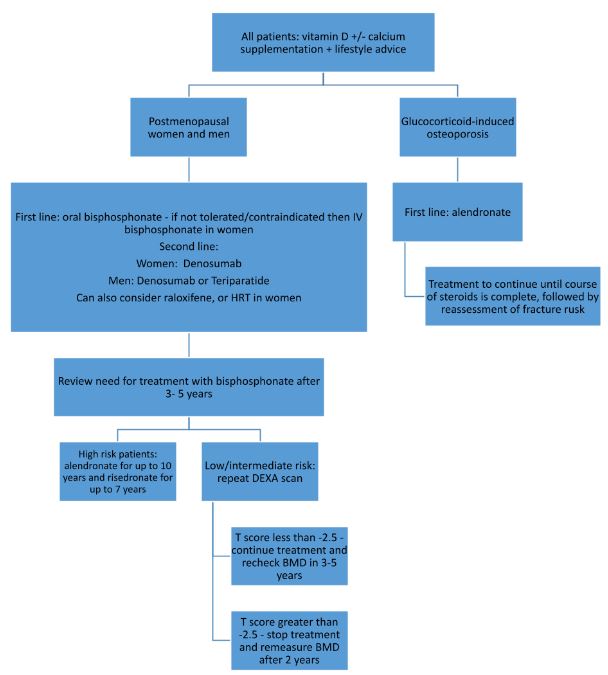
Adapted from NICE guidelines, Osteoporosis: assessing the risk of fragility fracture. National Institute for Health and Care Excellence (NICE); 2017.
References
- Mauck KF, Clarke BL. Diagnosis, screening, prevention, and treatment of osteoporosis. InMayo Clinic Proceedings 2006 May 1 (Vol. 81, No. 5, pp. 662-672). Elsevier.
- Chesnut III CH, Skag A, Christiansen C, Recker R, Stakkestad JA, Hoiseth A, Felsenberg D, Huss H, Gilbride J, Schimmer RC, Delmas PD. Effects of oral ibandronate administered daily or intermittently on fracture risk in postmenopausal osteoporosis. Journal of bone and mineral research. 2004 Aug;19(8):1241-9.
- National Institute for Health and Care Excellence (Great Britain). Osteoporosis: assessing the risk of fragility fracture. National Institute for Health and Care Excellence (NICE); 2017.
Complete Guide to Clinical Genetics ST3 Application and Recruitment (2027)
Written by Medibuddy. Last updated: October 2026. Clinical Genetics is

Complete Guide to Clinical Radiology ST3 Application and Recruitment (2027)
Recruitment paused: NHS England is not running Clinical Radiology ST3+ recru

Complete Guide to Paediatric Surgery ST3 Application and Recruitment in United Kingdom (2026)
Written by Medibuddy. Last updated: October 2026. If you’re planning


Take your subscriptions with you
Our mobile app allows you to access your interview and exam question banks wherever you are.

