


 Close
Close
 22nd May 2023
Anaemia
Classification of anaemia
Aetiology of anaemia
The World Health Organisation defines anaemia as a haemoglobin level <120g/L for women and <130g/L for men. Due to variations in normal haemoglobin ranges with age and sex, as well as between laboratory instruments, anaemia is widely defined as a haemoglobin below the reference range provided by the testing laboratory.
Haematopoeitic stem cells in the bone marrow differentiate into reticulocytes, which in turn are released into the circulation to develop into mature red blood cells. The average lifespan of a red blood cell is 120 days before it is removed from the circulation by macrophages.
Anaemia can present clinically with pallor of skin and mucous membranes, tiredness, exertional dyspnoea, palpitations, dizziness, exacerbation of existing angina or heart failure, or symptoms of the underlying cause of anaemia.
Anaemia can be classified by mechanism or by morphology. Mechanisms of anaemia include:
- Decreased production of red blood cells
- Increased loss of red blood cells
- Dilutional
Classification of anaemia by morphology is more commonly used clinically and is defined by the size of red blood cells, measured by mean corpuscular volume (MCV):
- Macrocytic (MCV>100fL)
- Normocytic (MCV 80-100fL)
- Microcytic (MCV<80fL)
Macrocytic anaemia
Macrocytosis is defined as an MCV>100fL, and when associated with anaemia is termed macrocytic anaemia. It can be subdivided into megaloblastic and non-megaloblastic anaemia.
Megaloblastic anaemia
Megaloblastic anaemia is named due to the presence of enlarged, immature red blood cells (megaloblasts) and hypersegmented neutrophils on blood film. This results from impaired DNA synthesis or maturation during red blood cell production, which delays the progression of the cell cycle from the G2 growth stage to mitosis, leading to continuing cell growth and enlarged red blood cells.
Causes of megaloblastic anaemia can be divided into those resulting in vitamin B12 deficiency and folate deficiency.
Common causes of vitamin B12 deficiency include:
- Pernicious anaemia – autoimmune destruction of gastric parietal cells causing reduced production of intrinsic factor, a glycoprotein required for B12 absorption in the terminal ileum.
- Post-gastrectomy or post-terminal ileum resection e.g. due to Crohn’s disease
- Proton-pump inhibitors e.g. omeprazole
- Bacterial overgrowth
- Decreased intake e.g. vegan diet without vitamin B12 supplementation
Common causes of folate deficiency include:
- Decreased intake e.g. alcohol dependence, elderly, anorexia nervosa
- Decreased absorption e.g. coeliac disease
- Increased demand e.g. pregnancy, malignancy, chronic inflammation
- Excessive urination e.g. in congestive heart failure or dialysis
- Drugs e.g. phenytoin, methotrexate, trimethoprim, sulfasalazine, cholestyramine
Non-megaloblastic anaemia
Non-megaloblastic anaemias (also known as normoblastic anaemias) comprise other causes of macrocytosis without impairment of DNA synthesis. Therefore, megaloblasts and hypersegmented neutrophils do not feature on the blood film in non-megaloblastic anaemia.
Causes of non-megaloblastic anaemia include:
- Alcohol excess
- Reticulocytosis – reticulocytes are large, immature red cells. Reticulocytosis can be seen when there is increased bone marrow activity to replace red blood cell loss, such as in haemolysis or haemorrhage.
- Liver disease
- Hypothyroidism
- Tyrosine-kinase inhibitors e.g. imatinib
- Myelodysplasia
- Multiple myeloma
Normocytic anaemia
Anaemia associated with an MCV between 80fL and 100fL is termed normocytic.
Causes of normocytic anaemia include:
- Large volume acute blood loss
- Anaemia of chronic disease e.g. chronic kidney disease, rheumatoid arthritis
- Pregnancy
- Bone marrow failure e.g. aplastic anaemia
- A combination of conditions producing both a microcytic and macrocytic anaemia
Microcytic anaemia
Microcytosis is defined as an MCV<80fL, and when associated with anaemia is termed microcytic anaemia.
Causes of microcytic anaemia include:
- Iron deficiency
- Anaemia of chronic disease
- Thalassaemia
- Sideroblastic anaemia – results from impaired incorporation of iron into haemoglobin molecules. This can be congenital or secondary to alcohol excess, drugs (isoniazid, chloramphenicol), or myelodysplasias. The MCV is low or normal in congenital forms but can sometimes be raised in acquired forms.
- Lead poisoning
Erythrocyte morphology
Normal erythrocyte morphology
Erythrocytes (i.e. red blood cells) are the commonest type of blood cell. They lack a nucleus and other organelles but are rich in haemoglobin, which is responsible for transporting oxygen and carbon dioxide in the circulation. Normal erythrocytes are flexible, biconcave discs. This structure creates a large surface area to volume ratio for optimal gas exchange within tissues, whilst facilitating easy movement through narrow blood vessels.
Abnormalities in size, shape, colour, or distribution of erythrocytes can indicate an underlying disease process. Poikilocytosis refers to abnormalities in the shape of erythrocytes, whereas anisocytosis refers to the presence of varying sizes of erythrocytes.
Poikilocytosis
| Poikilocytosis | Morphology | Appearance on blood film | Associations |
|---|---|---|---|
| Teardrop cells | Teardrop shaped | 
Image Source: Wikimedia Commons (2009). Teardrop Cells smear, Licensed under a creative commons licence. Available for free at: https://commons.wikimedia.org/wiki/File:Teardrop_Cells_smear_2009-09-22.JPG (Accessed on: 10 January 2023) |
Myelofibrosis Thalassaemia |
| Sickle cells | Sickle shaped | 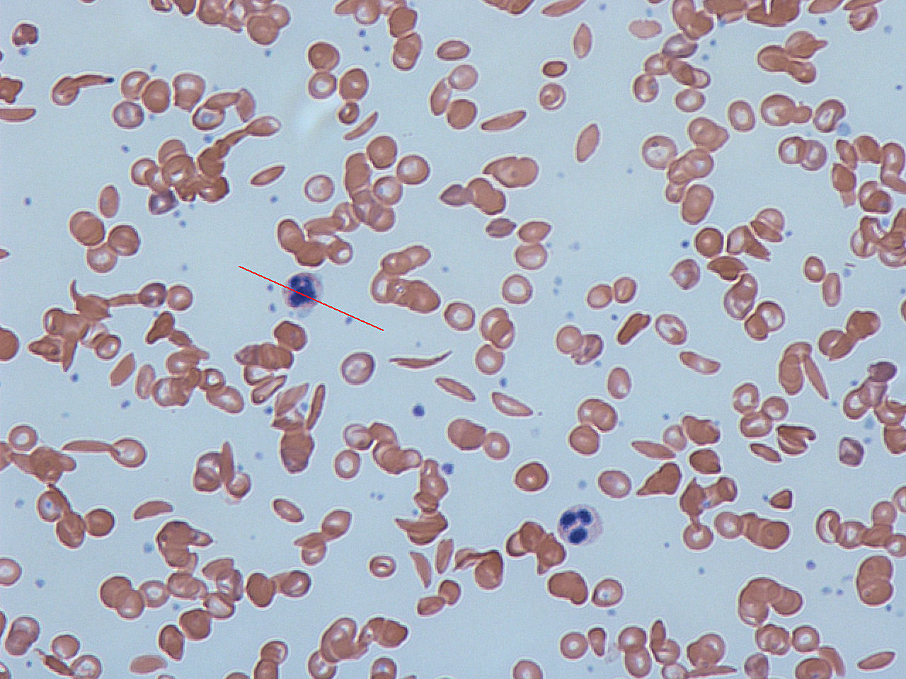
Image Source: Wikimedia Commons (2012). Sickle cells, Licensed under a creative commons licence. Available for free at: https://commons.wikimedia.org/wiki/File:Sickle_cells.jpg (Accessed on: 10 January 2023). |
Sickle cell diseases |
| Helmet cells (schistocytes) | Fragmented | 
Image Source: Wikimedia Commons (2013). Blood smear with typical schistocytes in TTP marked in blue, Licensed under a creative commons licence. Available for free at: https://commons.wikimedia.org/wiki/File:Blood_smear_with_typical_schistocytes_in_TTP_marked_in_blue_1.tif (Accessed on: 10 January 2023). |
Microangiopathic haemolytic anaemia (e.g. DIC, TTP, HUS)
Mechanical damage (e.g. metallic heart valves) |
| Pencil cells | Pencil shaped, hypochromic and microcytic. A variant of elliptocyte associated with iron deficiency. | 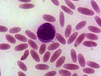
Image Source: Wikimedia Commons (2006). Elliptocytosis, Licensed under a creative commons licence. Available for free at: https://upload.wikimedia.org/wikipedia/commons/0/01/Elliptocytosis.jpg (Accessed on: 10 January 2023). |
Iron deficiency anaemia |
| Elliptocytes | Oval, elongated. | 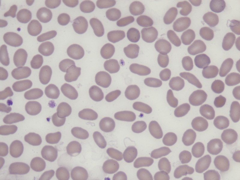
Image Source: Wikimedia Commons (2015). Hereditary Elliptocytosis in a 70-year-old man, Licensed under a creative commons licence. Available for free at: https://commons.wikimedia.org/wiki/File:Hereditary_Elliptocytosis_in_a_70-year-old_man.tif (Accessed on: 10 January 2023). |
Hereditary elliptocytosis Iron deficiency Thalassaemia Myelofibrosis |
| Spherocytes | Sphere shaped | 
Image Source: Wikimedia Commons (2010). Hereditary Spherocytosis smear, Licensed under a creative commons licence. Available for free at: https://commons.wikimedia.org/wiki/File:Hereditary_Spherocytosis_smear_2010-03-17.JPG (Accessed on: 10 January 2023). |
Hereditary spherocytosis Autoimmune haemolytic anaemia. |
| Target cells | Bullseye | 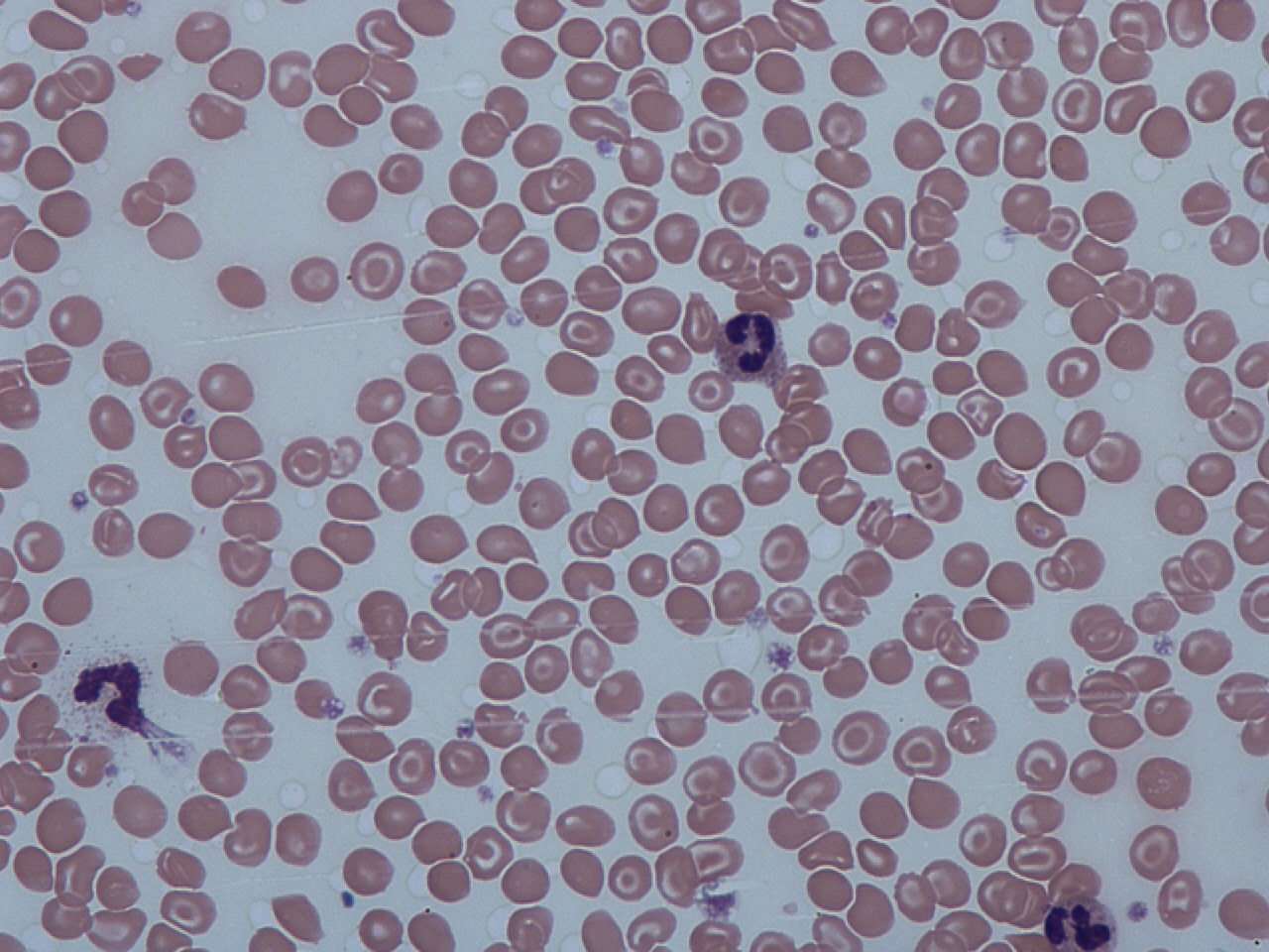
Image Source: Wikimedia Commons (2012). Target cells and spherocytes, Licensed under a creative commons licence. Available for free at: https://commons.wikimedia.org/wiki/File:Target_cells_and_spherocytes.jpg (Accessed on: 10 January 2023). |
Thalassaemia Haemoglobin C and S Liver disease Post-splenectomy Iron deficiency |
| Burr cells (echinocytes) | Smooth cytoplasmic projections | 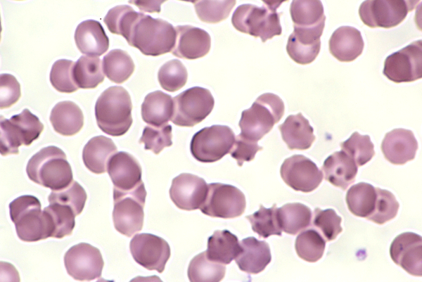
Image Source: Wikimedia Commons (2016). Echinocytes-12, Licensed under a creative commons licence. Available for free at: https://commons.wikimedia.org/wiki/File:Echinocytes-12.JPG (Accessed on: 10 January 2023). |
Liver disease Uraemia Pyruvate kinase deficiency |
| Bite cells | Bite marks | 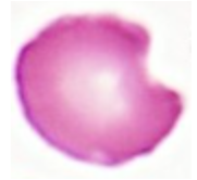
Image Source: Wikimedia Commons (2020). Bite Cells, Licensed under a creative commons licence. Available for free at: https://commons.wikimedia.org/wiki/File:BITE_CELLS.jpg (Accessed on: 10 January 2023). |
G6PD deficiency |
Some conditions result in erythrocytes with inclusion bodies.
| Inclusion bodies | Contents | Associations |
|---|---|---|
| Heinz bodies | Denatured haemoglobin | G6PD deficiency Thalassaemia |
| Howell-Jolly bodies | DNA | Hyposplenism |
| Basophilic stippling | RNA | Lead toxicity Sideroblastic anaemia Thalassaemia |
| Ringed sideroblasts | Iron | Sideroblastic anaemia |
| Pappenheimer bodies | Iron | Sideroblastic anaemia Haemolytic anaemia Hyposplenism |
Iron metabolism
Pathophysiology of iron metabolism
Iron is an essential component of the normal production and regulation of red blood cells, particularly due to its role in haemoglobin formation.
The average human has 3-6g of iron in their body, 80% of which is functional iron contained within haemoglobin and myoglobin. 20% is stored as ferritin and haemosiderin, mainly in the liver but also in the spleen and bone marrow. Only about 4mg of iron circulates in the plasma and is bound to a glycoprotein called transferrin. Free iron can be toxic to cells; in the presence of hydrogen peroxidase, free ferrous iron (Fe²⁺) is oxidised to ferric iron (Fe³⁺) creating reactive oxygen species which can cause DNA damage and cell death.
Dietary iron absorption occurs mostly in the duodenum and jejunum of the small bowel. Dietary iron can come from meat or plants. Iron from meat is termed ‘heme iron’ and 20% is absorbable. Iron from plants is ‘non-heme iron’ and only 2% is absorbable, with absorption rates worsened by co-ingestion with polyphenols (e.g. tea) or calcium, and increased with vitamin C.
Hepcidin is a peptide hormone synthesised in the liver. Hepcidin decreases intestinal iron absorption and recycling of iron from the reticuloendothelial cells. Therefore in the event of iron deficiency, hepcidin expression is decreased, resulting in increased intestinal iron absorption and recycling of iron from the reticuloendothelial cells.
Whilst the body can increase iron absorption in the event of iron deficiency, there is no regulatory pathway for iron excretion. Approximately 1mg of iron is excreted per day in men and post-menopausal women, and this increases to 2mg per day in menstruating women.
Causes of iron deficiency anaemia
Iron deficiency anaemia is the most common form of anaemia worldwide. A diagnosis of iron deficiency anaemia should prompt investigation to determine the cause.
Causes of iron deficiency can be classified by mechanism:
- Iron loss
- Gastrointestinal bleeding e.g. gastrointestinal cancer, peptic ulcer disease, ulcerative colitis, hookworm infection, Meckel’s diverticulum, haemorrhoids
- Menorrhagia
- Disorders of haemostasis e.g. haemophilia
- Blood loss from haemodialysis or frequent blood donation
- Other blood loss, e.g. haematuria, haemoptysis
- Decreased iron intake
- Malnutrition
- Vegan diet
- Decreased iron absorption
- Achlorhydria e.g. secondary to atrophic gastritis
- Inflammatory bowel disease
- Coeliac disease
- Gastric or duodenal surgery
- Increased requirements
- Pregnancy and breastfeeding
- Growth
Iron deficiency anaemia may be asymptomatic or may present with non-specific symptoms of anaemia such as pallor, lethargy, or reduced exercise tolerance. Some symptoms more specific to iron deficiency anaemia include pica (cravings for ice or non-food items) and restless legs syndrome. Physical signs on examination may include glossitis, angular cheilitis, koilonychia or brittle nails. In cases of the rare Plummer-Vinson syndrome, there may be dysphagia due to the presence of upper oesophageal webs.
Investigating iron deficiency anaemia
Blood tests commonly used to diagnose iron deficiency anaemia include:
- Full blood count
- Haemoglobin – low
- Haematocrit – low
- MCV – typically low, may be normal
- MCH – typically low i.e. hypochromic but may be normal
- Reticulocytes – low or normal
- RDW– high, due to variability in size of red cells
- Blood film
- Hypochromic, microcytic red blood cells
- Poikilocytosis may be seen – target cells and pencil cells are commonly associated with iron deficiency anaemia.
- Iron profile
- Ferritin – low ferritin is suggestive of iron deficiency; however ferritin is an acute phase reactant and therefore may be normal or elevated if co-existing inflammation.
- Serum iron – low serum iron may be seen; however, this is not a reliable test in isolation
- Serum TIBC – increased
- Transferrin saturation – low (<16%). The percentage transferrin saturation is calculated by serum iron ÷ TIBC x 100.
- Serum soluble transferrin receptor – increased
Additional investigations to identify a cause may include:
- OGD and colonoscopy – this is particularly important for men and post-menopausal women as occult gastrointestinal cancers are a common cause of iron deficiency anaemia. Small-bowel biopsy can also be undertaken if suspicion of coeliac disease.
- Coeliac serology
- Urinalysis – to investigate occult blood loss from the renal tract
- Helicobacter pylori testing
- Stool sample for ova and parasites if suspicion of hookworm infection
- Gynaecology workup for menorrhagia
Treatment of iron deficiency anaemia
It is important that a cause for the iron deficiency is investigated and that the underlying cause is treated, if identified.
Iron deficiency itself can be treated with oral or intravenous iron therapy. Blood transfusion may also be required in cases of severe anaemia (Hb <70g/L, or <80g/L in select patients) or in cases causing cardiovascular compromise.
Oral iron replacement with ferrous sulfate or ferrous gluconate is commonly used. Concomitant intake of calcium, tea, coffee, wine, or antacids reduces its absorption. Gastrointestinal intolerance to oral iron is common, and patients may experience nausea, constipation, and black discolouration of stool. If oral iron is tolerated, treatment is given for 3 months after the normalisation of blood tests, to ensure replacement of iron stores.
Intravenous iron can be considered if oral iron is not tolerated, failure to respond to oral iron, or in cases of intestinal malabsorption. Intravenous iron can cause anaphylaxis and therefore patients are closely monitored in an appropriate setting throughout the iron infusion.
Aplastic Anaemia
Aplastic anaemia is a disease defined by pancytopenia caused by bone marrow insufficiency. The criteria for diagnosis of aplastic anaemia includes hypocellular bone marrow without abnormal cells or fibrosis, and at least two of:
- Hb <100 g/L
- Platelets <50 x 10⁹/L
- Neutrophils <1.5 x 10⁹/L
Symptoms are associated with depletion of each cell type:
- Anaemia – lethargy, pallor, dyspnoea, tachycardia
- Neutropenia – increased infections
- Thrombocytopenia – bleeding, bruising, petechiae
Causes of aplastic anaemia
The aetiology of aplastic anaemia is idiopathic in over 50% of cases but is thought to be immune-mediated. Other aetiologies that have been linked to aplastic anaemia include:
- Congenital – e.g. Fanconi anaemia, dyskeratosis congenita, Shwachman-Diamond syndrome
- Drugs – e.g. NSAIDs, chloramphenicol, carbamazepine, propylthiouracil
- Toxins – e.g. insecticides
- Viruses – parvovirus B19, hepatitis, EBV, CMV, HIV
- Ionising radiation
- Paroxysmal nocturnal haemoglobinuria
Treatment of aplastic anaemia
The main treatment options for aplastic anaemia include:
- Withdrawal of the cause, if known e.g. causative drug
- Supportive therapies e.g. blood product transfusion or antibiotics as required.
- Immunosuppression with steroid, antithymocyte globulin (ATG) and calcineurin inhibitor such as ciclosporin.
- Androgen therapy e.g. danazol
- Allogeneic stem cell transplantation
- Eltrombopag (oral thrombopoietin receptor agonist)
The choice of treatment depends on whether the aplastic anaemia is inherited or acquired, as well as its severity classification.
Inherited aplastic anaemia does not tend to respond to immunosuppression and therefore stem cell transplantation is considered first-line therapy, or androgen therapy if stem cell transplant is not an option. Androgen therapy can be complicated by hepatotoxicity and therefore regular monitoring of liver function tests and liver imaging is important.
Acquired non-severe aplastic anaemia can be monitored conservatively but may require immunosuppression with ATG and ciclosporin if transfusion dependent.
Acquired severe or very severe aplastic anaemia can be treated with a conditioning regimen and stem cell transplant from a matched related donor if availability and fitness allow. In patients over 50 or those without a matched related donor for stem cell transplant, then immunosuppression with ATG and ciclosporin is considered first-line
treatment.
Eltrombopag is an oral thrombopoietin receptor agonist which is currently approved for the treatment of patients with acquired severe aplastic anaemia, refractory to immunosuppressive treatment.
Complete Guide to Clinical Genetics ST3 Application and Recruitment (2027)
Written by Medibuddy. Last updated: October 2026. Clinical Genetics is

Complete Guide to Clinical Radiology ST3 Application and Recruitment (2027)
Recruitment paused: NHS England is not running Clinical Radiology ST3+ recru

Complete Guide to Paediatric Surgery ST3 Application and Recruitment in United Kingdom (2026)
Written by Medibuddy. Last updated: October 2026. If you’re planning


Take your subscriptions with you
Our mobile app allows you to access your interview and exam question banks wherever you are.

