


 Close
Close
 22nd May 2023
Erythrocyte morphology
Normal erythrocyte morphology
Erythrocytes (i.e. red blood cells) are the commonest type of blood cell. They lack a nucleus and other organelles but are rich in haemoglobin, which is responsible for transporting oxygen and carbon dioxide in the circulation. Normal erythrocytes are flexible, biconcave discs. This structure creates a large surface area to volume ratio for optimal gas exchange within tissues, whilst facilitating easy movement through narrow blood vessels.
Abnormalities in size, shape, colour, or distribution of erythrocytes can indicate an underlying disease process. Poikilocytosis refers to abnormalities in the shape of erythrocytes, whereas anisocytosis refers to the presence of varying sizes of erythrocytes.
Poikilocytosis
| Poikilocytosis | Morphology | Appearance on blood film | Associations |
|---|---|---|---|
| Teardrop cells | Teardrop shaped | 
Image Source: Wikimedia Commons (2009). Teardrop Cells smear, Licensed under a creative commons licence. Available for free at: https://commons.wikimedia.org/wiki/File:Teardrop_Cells_smear_2009-09-22.JPG (Accessed on: 10 January 2023) |
Myelofibrosis Thalassaemia |
| Sickle cells | Sickle shaped | 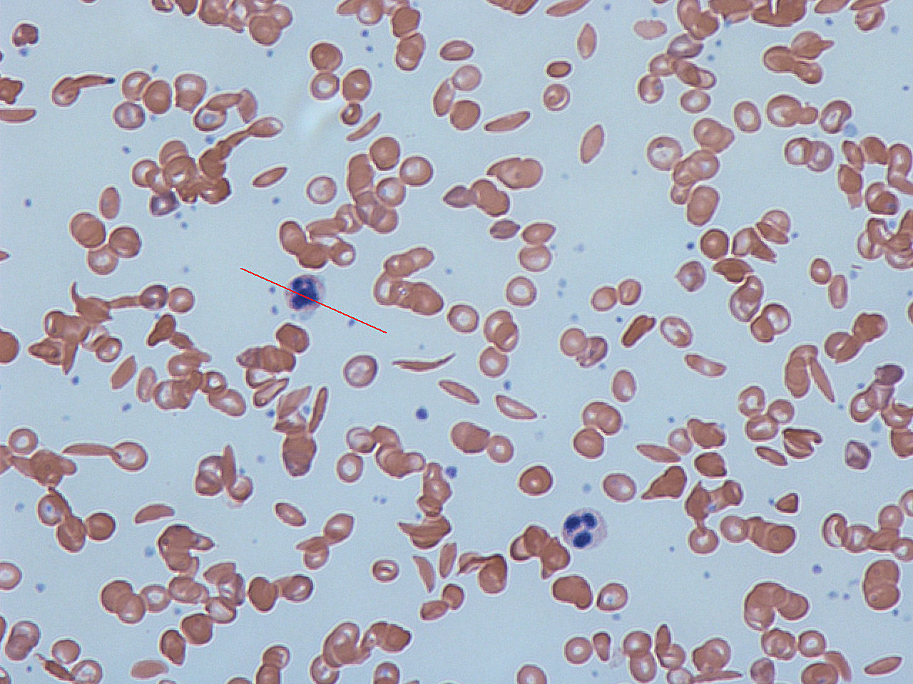
Image Source: Wikimedia Commons (2012). Sickle cells, Licensed under a creative commons licence. Available for free at: https://commons.wikimedia.org/wiki/File:Sickle_cells.jpg (Accessed on: 10 January 2023). |
Sickle cell diseases |
| Helmet cells (schistocytes) | Fragmented | 
Image Source: Wikimedia Commons (2013). Blood smear with typical schistocytes in TTP marked in blue, Licensed under a creative commons licence. Available for free at: https://commons.wikimedia.org/wiki/File:Blood_smear_with_typical_schistocytes_in_TTP_marked_in_blue_1.tif (Accessed on: 10 January 2023). |
Microangiopathic haemolytic anaemia (e.g. DIC, TTP, HUS)
Mechanical damage (e.g. metallic heart valves) |
| Pencil cells | Pencil shaped, hypochromic and microcytic. A variant of elliptocyte associated with iron deficiency. | 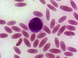
Image Source: Wikimedia Commons (2006). Elliptocytosis, Licensed under a creative commons licence. Available for free at: https://upload.wikimedia.org/wikipedia/commons/0/01/Elliptocytosis.jpg (Accessed on: 10 January 2023). |
Iron deficiency anaemia |
| Elliptocytes | Oval, elongated. | 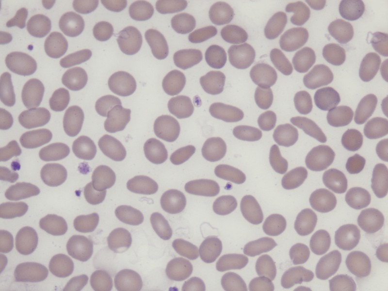
Image Source: Wikimedia Commons (2015). Hereditary Elliptocytosis in a 70-year-old man, Licensed under a creative commons licence. Available for free at: https://commons.wikimedia.org/wiki/File:Hereditary_Elliptocytosis_in_a_70-year-old_man.tif (Accessed on: 10 January 2023). |
Hereditary elliptocytosis Iron deficiency Thalassaemia Myelofibrosis |
| Spherocytes | Sphere shaped | 
Image Source: Wikimedia Commons (2010). Hereditary Spherocytosis smear, Licensed under a creative commons licence. Available for free at: https://commons.wikimedia.org/wiki/File:Hereditary_Spherocytosis_smear_2010-03-17.JPG (Accessed on: 10 January 2023). |
Hereditary spherocytosis Autoimmune haemolytic anaemia. |
| Target cells | Bullseye | 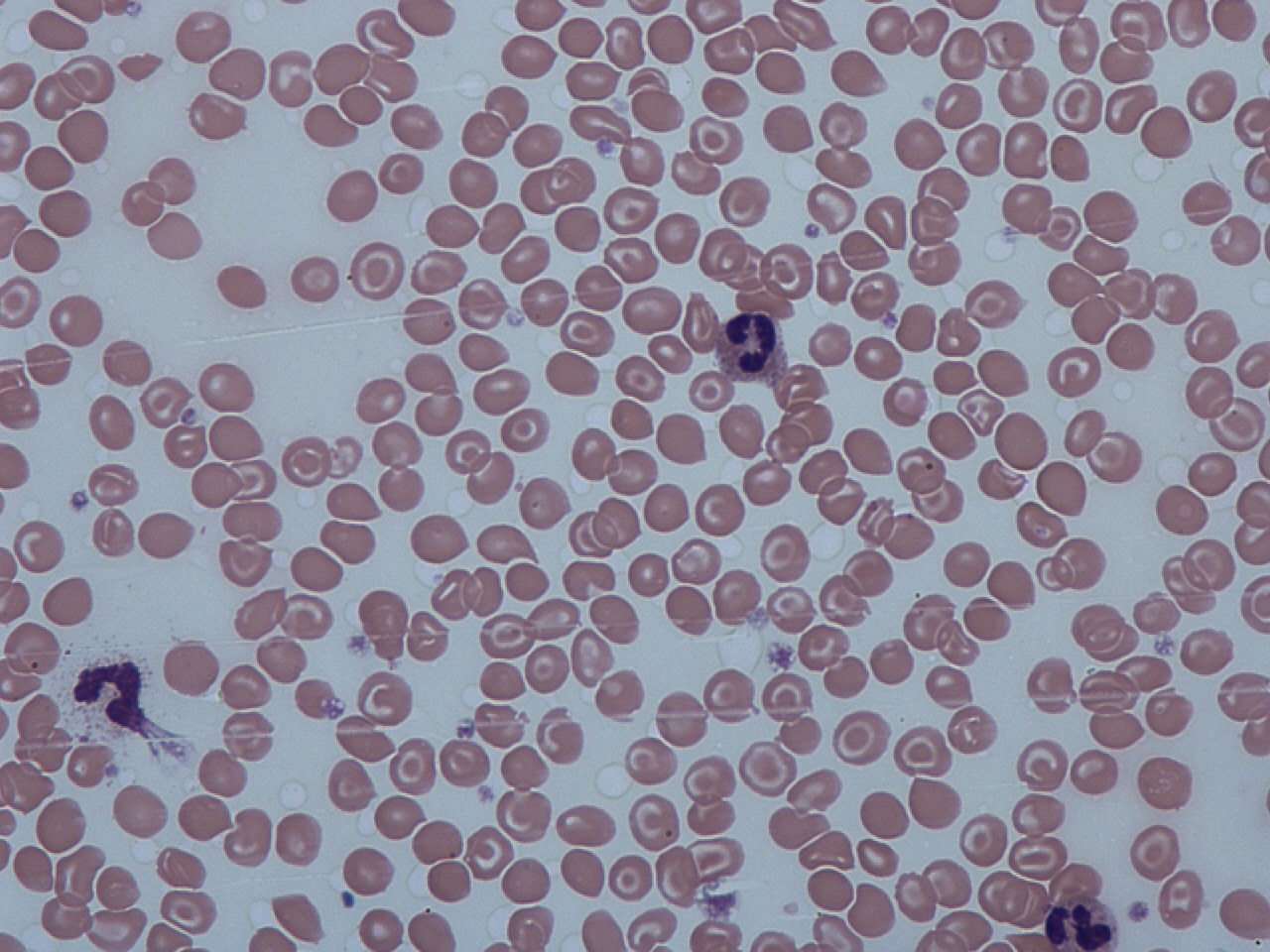
Image Source: Wikimedia Commons (2012). Target cells and spherocytes, Licensed under a creative commons licence. Available for free at: https://commons.wikimedia.org/wiki/File:Target_cells_and_spherocytes.jpg (Accessed on: 10 January 2023). |
Thalassaemia Haemoglobin C and S Liver disease Post-splenectomy Iron deficiency |
| Burr cells (echinocytes) | Smooth cytoplasmic projections | 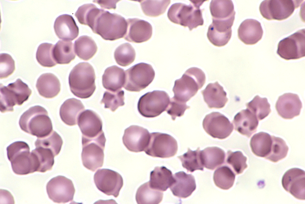
Image Source: Wikimedia Commons (2016). Echinocytes-12, Licensed under a creative commons licence. Available for free at: https://commons.wikimedia.org/wiki/File:Echinocytes-12.JPG (Accessed on: 10 January 2023). |
Liver disease Uraemia Pyruvate kinase deficiency |
| Bite cells | Bite marks | 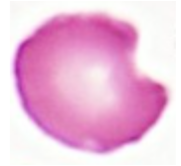
Image Source: Wikimedia Commons (2020). Bite Cells, Licensed under a creative commons licence. Available for free at: https://commons.wikimedia.org/wiki/File:BITE_CELLS.jpg (Accessed on: 10 January 2023). |
G6PD deficiency |
Some conditions result in erythrocytes with inclusion bodies.
| Inclusion bodies | Contents | Associations |
|---|---|---|
| Heinz bodies | Denatured haemoglobin | G6PD deficiency Thalassaemia |
| Howell-Jolly bodies | DNA | Hyposplenism |
| Basophilic stippling | RNA | Lead toxicity Sideroblastic anaemia Thalassaemia |
| Ringed sideroblasts | Iron | Sideroblastic anaemia |
| Pappenheimer bodies | Iron | Sideroblastic anaemia Haemolytic anaemia Hyposplenism |
Complete Guide to Clinical Genetics ST3 Application and Recruitment (2027)
Written by Medibuddy. Last updated: October 2026. Clinical Genetics is

Complete Guide to Clinical Radiology ST3 Application and Recruitment (2027)
Recruitment paused: NHS England is not running Clinical Radiology ST3+ recru

Complete Guide to Paediatric Surgery ST3 Application and Recruitment in United Kingdom (2026)
Written by Medibuddy. Last updated: October 2026. If you’re planning


Take your subscriptions with you
Our mobile app allows you to access your interview and exam question banks wherever you are.

