


 Close
Close
 26th January 2024
Autoimmune Connective Tissue Disorders
Systemic Lupus Erythematosus (SLE)
Background
There are four major types of lupus: systemic lupus erythematosus, lupus of the skin, drug-induced lupus erythematosus and neonatal lupus erythematosus.
Systemic lupus erythematosus (SLE) is the most common and is a chronic autoimmune disease that can affect multiple organs, including the skin, joints and kidneys.
Lupus of the skin can be subdivided into three main categories, each with their own distinctive lesions. These are 1) acute cutaneous lupus, 2) chronic cutaneous lupus erythematosus or discoid lupus erythematosus (DLE) and 3) subacute cutaneous lupus erythematosus.
Drug-induced lupus has a similar presentation to SLE and is caused by drugs such as hydralazine, procainamide, isoniazid and minocycline.
Neonatal lupus erythematosus is a rare condition that affects babies of women who have autoantibodies (Anti-Ro and Anti-La), which act on the neonates’ heart conduction system, leading to heat block.
Epidemiology
SLE is the most common form of Lupus – affecting approximately 70% of all patients diagnosed. There is a higher prevalence in females with the age of onset at diagnosis, most often between 15 and 45 years.
The prevalence of SLE is higher among Asian, African American, African Caribbean and Hispanic individuals compared to Caucasians.
Pathophysiology
The exact aetiology of SLE is unknown as it is thought to be multifactorial.
Genetic factors:
Generally, no single polymorphism is associated with a high risk for developing SLE. However, the combination of susceptibility genes and the absence of protective genes confer enough genetic susceptibility to cause disease development.
The following genetic factors are associated with SLE:
- The most common genetic predisposition is at the MHC locus – Class I HLA (HLA – A, B, C) and Class 11 HLA molecules (HLA – DR, DQ, DP)
- Loci of particular interest – Anti-La, Anti-Sm, Anti- dsDNA, Anti- Ro (associated with neonatal heart block)
- Deficiencies of complement components – C1q, C4A, B, C2, TREX1
- Gene Polymorphisms in DNA repair genes – ATG5, DNASE1
Hormonal and Immunological Factors
- An increase in CD4+ and CD8 + T cells due to excess or unopposed oestrogen release may lead to autoantibodies which eventually lead to the clinical findings of SLE. This may account for the higher prevalence in women.
- Elevated levels of circulating IFN-alpha correlate with active disease. The presence of genetic predisposing factors leads to increased expression of IFN-alpha-inducible RNA transcripts by mononuclear cells, which in turn causes these elevated levels.
- Dysregulation of apoptosis among different cell types – T lymphocytes and the Fas/Fas ligand pathway causes an abnormal programmed cell death and an aberrant lymphoid population, contributing to autoimmune activity.
- In lupus nephritis, there is deposition of immune complexes in the mesangium and activation of complement in the glomerular basement membrane, causing inflammation, which eventually leads to necrosis and scarring. There are also elevated circulating levels of TNF-alpha, which correlate with active disease.
Presentation
Articular Manifestations
Joint involvement is common and present in approximately 95% of patients diagnosed with SLE. In 75% of patients, it is the presenting complaint. The arthritis of SLE is typically non-erosive, symmetrical and polyarticular. Joints commonly affected include the proximal interphalangeal joints (PIPs), carpal joints and knees. On examination, joint deformities may resemble those seen in RA, such as ulnar deviation and thumb subluxation and are referred to as Jaccoud’s arthropathy. However, Jaccoud’s arthropathy results from soft tissue abnormalities and is a non-erosive, reversible joint disorder, unlike Rheumatoid Arthritis.

Image Source: Abushama S et al. (2021) A Sudanese male with Jaccoud’s arthropathy; case report. Biomed J Sci & Tech Res 33(2)-2021. Licensed under a commercial Creative Commons license. Available at: https://biomedres.us/pdfs/BJSTR.MS.ID.005387.pdf (Accessed: 21 December 2023).
Cutaneous Manifestations
Skin involvement is often the first sign in SLE, and about 80% of patients with SLE will have skin signs. Common skin signs include malar or “butterfly” rash, photosensitivity rashes, hair loss, discoid lupus – indurated, hyper-pigmented plaques, and muco-cutaneous involvement. On examination, active lesions of discoid lupus may present as erythematous, circular, scaly plaques, while healed lesions show a characteristic depigmented central area with surrounding hyper-pigmentation and an atrophic centre. The alopecia seen is scarring alopecia that often results in permanent hair loss.

Image Source: Wikimedia Commons (2022).Lupusfoto, Licensed under a creative commons licence. Available for free at: https://upload.wikimedia.org/wikipedia/commons/5/52/Lupusfoto.jpg (Accessed: 10 July 2022).

Image Source: Wikimedia Commons (2022). Discoid lupus erythematosus, Licensed under a creative commons licence. Available for free at: https://upload.wikimedia.org/wikipedia/commons/b/ba/Discoid_lupus_erythematosus.JPG (Accessed: 10 July 2022).

Image Source: Wikimedia Commons (2022). Discoid lupus etythermayouses new image, Licensed under a creative commons licence. Available for free at: https://upload.wikimedia.org/wikipedia/commons/9/91/Discoid_lupus_etythermayouses_new_image.jpg (Accessed: 10 July 2022).
Non-articular Systemic Manifestations of SLE
- General: fatigue (the most common complaint), intermittent fever, weight loss
- Cardiac: pericarditis with or without pericardial effusion (most common cardiac manifestation)
- Pulmonary: pleuritis, pneumonitis, interstitial lung disease, pulmonary hypertension
- Vascular: Raynaud’s phenomenon (occurs in up to 50% of patients with SLE), vasculitis – most commonly small vessel and can manifest as petechiae, palpable purpura, livedo reticularis, panniculitis and superficial ulcerations
- Renal: Renal involvement occurs in approximately 50% of SLE patients. Different forms of glomerulonephritis can occur and may present with proteinuria/nephrotic syndrome or asymptomatic haematuria.
- Neurologic and Neuropsychiatric: Symptoms and presentations can include cognitive dysfunction, delirium, psychosis, strokes, seizures, and peripheral neuropathies.
- Haematological: Anaemia of chronic disease is the most common presentation. Leukopenia is also common and occurs in approximately 50% of patients and generally correlates to disease activity.
- Ophthalmologic: Keratoconjunctivitis is the most common manifestation and is as a result of secondary Sjogren’s syndrome
Diagnosis
Laboratory Investigations
- Full Blood Count (FBC): may show anaemia, leukopenia, thrombocytopenia
- Renal function: elevated creatinine may be a sign of renal dysfunction
- ANA: positive in almost all patients with SLE
- Anti-dsDNA: highly specific and sensitive for SLE – seen in approximately 70% of patients
- Anti-Sm antibodies: highly specific but lacks sensitivity – seen in approximately 30% of patients
- Anti-Ro and Anti-La: more commonly associated with Sjogren’s syndrome but are present in approximately 20-30% of patients with SLE
- Anti-U1 RNP antibodies: are almost always found in high levels in patients with mixed connective tissue disease; however, it occurs in approximately 25% of patients with SLE.
Imaging
- X-rays of the hands may show evidence of Jaccoud’s arthropathy, which includes soft tissue deformities and the absence of erosions.
- MRI or Ultrasound: These may reveal synovial effusions which are usually small and can be used to guide aspiration of the joint that typically shows low protein and white cell counts.
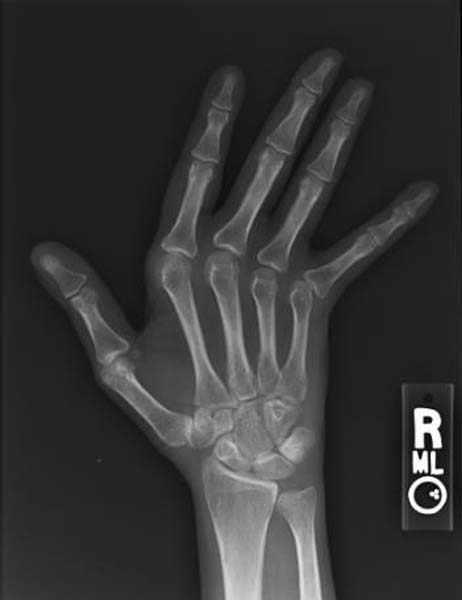
Image Source: Wikimedia Commons (2022). Jaccoud arthropathy hand x-ray front, Licensed under a creative commons licence. Available for free at: https://upload.wikimedia.org/wikipedia/commons/7/71/Jaccoud_arthropathy_hand_x-ray_front.jpg (Accessed: 10 July 2022).
Diagnostic Criteria
SLICC (Systemic Lupus International Collaborating Clinics) Criteria – 2012
Using the SLICC criteria, SLE is diagnosed if the patient has either of the following over time:
- Four criteria including ≥ one clinical criterion and ≥ one immunological criterion
- Biopsy-proven lupus nephritis and anti-nuclear antibodies or anti-double-stranded DNA antibodies
The clinical criteria include features such as cutaneous lupus, alopecia, oral or nasal ulcers, joint disease, serositis, neurological manifestations and laboratory findings such as anaemia, leukopenia, thrombocytopenia and proteinuria. The immunological criteria include positive ANA, Anti-ds DNA, Anti-Sm, Antiphospholipid and low complement (C3 and C4). A positive Direct Coombs Test can be considered in the absence of haemolytic anaemia.
| Clinical Criteria | Immunological criteria |
|---|---|
| Acute cutaneous lupus (lupus malar rash) | ANA |
| Chronic cutaneous lupus (discoid rash) | Anti-ds DNA |
| Alopecia | Anti-Sm |
| Oral or nasal ulcers | Antiphospholipid |
| Joint disease (synovitis or tenderness) | Low complement |
| Serositis | Direct Coombs Test |
| Renal (proteinuria, red cell casts) | |
| Neurological (seizures, psychosis) | |
| Haemolytic anaemia | |
| Leukopenia | |
| Thrombocytopenia |
Table 1: The SLICC classification criteria for systemic lupus erythematosus.
Table 1: UpToDate. Classification criteria for systemic lupus erythematosus [Internet]. 2022 [cited 2022 July 12]. Available from: https://www.uptodate.com/contents/image?imageKey=RHEUM%2F86633
EULAR/ACR (European Alliance of Associations for Rheumatology/American College of Rheumatology) Criteria – 2019
This criterion consists of 7 clinical and 3 immunological categories, each of which are weighted 2 – 10. Patients with a score of 10 points or more are classified as having SLE.
The clinical domains include constitutional, haematological, neuropsychiatric, mucocutaneous, serosal, musculoskeletal and renal. The immunological domains include antiphospholipid antibodies, complement proteins and SLE-specific antibodies.
| Clinical Criteria | Immunological criteria |
|---|---|
| Malar rash | ANA |
| Photosensitivity | Anti-ds DNA OR Anti-Sm OR Anti Phospholipid |
| Discoid rash | |
| Oral ulcers | |
| Arthritis | |
| Serositis | |
| Renal (proteinuria, cellular casts) | |
| Neurological (seizures or psychosis) | |
| Haematological (haemolytic anaemia, leukopenia, lymphopenia, thrombocytopenia) |
Table 2: The ACR classification criteria for systemic lupus erythematosus.
Table 2: UpToDate. Classification criteria for systemic lupus erythematosus [Internet]. 2022 [cited 2022 July 12]. Available from: https://www.uptodate.com/contents/image?imageKey=RHEUM%2F86633
Differential Diagnoses
SLE can be difficult to diagnose, particularly in the initial stages, and the symptoms can be vague and overlap with many other more common causes. The most common symptoms include arthralgia, myalgia, fatigue, weight loss and photosensitivity. A definitive diagnosis in many cases may depend on antibody testing.
Rheumatoid Arthritis
RA may also present with constitutional symptoms and arthralgia. However, characteristics such as morning joint stiffness and symmetrical, tender swollen joint would be more indicative of RA. In addition, joint deformities such as Boutonniere and Swan neck would be found in RA and not SLE.
Positive tests such as Rheumatoid factor may be positive in both SLE and RA; however, Anti-CCP positivity would be expected in RA, while Anti-ds DNA positivity would be expected in SLE.
Systemic Sclerosis
Systemic Sclerosis may also present with arthralgia, rashes, and Raynaud’s phenomenon. However, features such as decreased joint mobility and skin induration would be more common in systemic sclerosis.
Antibody testing is key here, where the presence of Anti-centromere and Anti-Scl-70 antibodies would be indicative of a diagnosis of sclerosis rather than SLE.
Sarcoidosis
Sarcoidosis may also present with features such as fatigue, fever, night sweats or a rash. However, symptoms such as cough and dyspnoea would be more common in sarcoidosis. In addition, an elevated angiotensin-converting enzyme and chest radiograph features such as bilateral hilar lymphadenopathy would be indicative of sarcoidosis rather than SLE.
Adult-Onset Still’s Disease
While similar constitutional features such as fever and arthralgia may be present, splenomegaly would be more characteristic of Still’s disease. In both cases, laboratory blood testing may show elevated ESR and anaemia; however, Anti-ds DNA would be expected to be positive in SLE and not Still’s disease.
Chronic Fatigue Syndrome
This is a diagnosis of exclusion. Patients may present with vague symptoms, and blood tests, including inflammatory markers, metabolic panel and antibody testing, will all be within the normal range.
Management
Treatment of SLE depends on disease activity and severity in addition to symptoms and organs affected. It requires management by a rheumatologist, and often, a multi-disciplinary approach is required.
Non-Pharmacological/Conservative
- UV protection: This is particularly significant in those affected with cutaneous SLE where photosensitivity may occur. Sunscreens of SPF (sun protection factor) of >55 and protection against UV-A and UV-B are recommended. Medications that induce photosensitivity should also be avoided.
Pharmacological
- Anti-malarials: Hydroxychloroquine and Chloroquine can help relieve constitutional symptoms in addition to musculoskeletal and mucocutaneous manifestations. Can be used in mild to moderate SLE where there are manifestations that are non-organ threatening (e.g. constitutional symptoms, skin, mucosal, joint or haematological involvement)
- Oral Corticosteroids: Prednisolone 5-15mg daily can be used as a short-term therapy. This can be tapered while the hydroxychloroquine or chloroquine takes effect.
- Intravenous Corticosteroids: Methylprednisolone IV is used in severe or life-threatening manifestations that occur secondary to major organ involvement. Patients are sometimes ‘pulsed’ on this high-dose IV corticosteroid for a short duration to rapidly reduce inflammation to achieve disease control.
- Azathioprine: This immunosuppressant is of value in treating the manifestations of SLE in patients of childbearing age and is one of the few medications that is generally safe to use in pregnancy.
- Adverse effects: Bone Marrow Suppression: there is an increased risk of this occurring in individuals with TPMT enzyme deficiency, which is why it is important to screen for TPMT prior to starting patients on azathioprine. Monitoring of full blood count, liver and renal function is required twice weekly for six weeks and, once the dose is stabilised, monthly for three months thereafter, which can be done at least every 12 weeks.
- Methotrexate: MTX competitively inhibits the enzyme dihydrofolate reductase. As such, daily folic acid replacement is required. It has been shown to reduce disease activity and can be effectively used as a steroid-sparing agent. Adverse effects: Methotrexate toxicity, hepatotoxicity, nephrotoxicity, myelosuppression, teratogenic, lung fibrosis, reactivation of latent TB. Monitoring guidelines suggest full blood count and liver and renal function every 1-2 weeks until therapy is stabilised, after which patients can be monitored every 2-3 months. In the case of methotrexate toxicity, the antidote is folinic acid (calcium folinate).
- Mycophenolate mofetil: MMF is an inhibitor of lymphocyte proliferation. It is used in combination with glucocorticoids in patients with focal or diffuse lupus nephritis. Adverse effects: Teratogenic, increased risk of infections, bone marrow suppression and rarely Progressive Multifocal Leukoencephalopathy (PML). As there is a risk of neutropenia, monitoring of full blood count is required every week for 4 weeks, then twice a month for 2 months, and then every month after that.
References
- Schur P, Hahn B. UpToDate. Epidemiology and pathogenesis of systemic lupus erythematosus [Internet]. 2021 [cited 2022 March 01]. Available from: https://www.uptodate.com/contents/epidemiology-and-pathogenesis-of-systemic-lupus-erythematosus?source=history
- Ho K, Reveille J. The clinical relevance of autoantibodies in scleroderma. Arthritis Res Ther. 2003; 5(2): 80-93. Available from https://www.ncbi.nlm.nih.gov/pmc/articles/PMC165038/
- Lam N, Ghetu M, Bieniek M. Systemic Lupus Erythematosus: Primary care approach to diagnosis and management. Am Fam Physician. 2016; 94(4): 284-294. Available from https://www.aafp.org/afp/2016/0815/p284.html
- Wallace D. Overview of the management and prognosis of systemic lupus erythromatosus in adults [Internet]. 2021 [cited 2022 March 01]. Available from: https://www.uptodate.com/contents/overview-of-the-management-and-prognosis-of-systemic-lupus-erythematosus-in-adults?source=history#H509844069
- Croyle L, Hoi A, Morand E, Characteristics of azathioprine use and cessation in a longitudinal lupus Cohort. Lupus Sci Med. 2015 Aug 20; 2(1): 105-110. Available from https://www.ncbi.nlm.nih.gov/pmc/articles/PMC4548066/
- Gansauge S, Breitbart A, Rinaldi N, et al. Methotrexate in patients with moderate systemic lupus erythematosus (exclusion of renal and central nervous system disease) Annals of the Rheumatic Diseases 1997;56:382-385.
- Falk R, Dall’Era M, Appel G. UpToDate. Lupus Nephritis: Initial and subsequent therapy for focal or diffuse lupus nephritis [Internet]. 2022. [cited 2022 March 18]. Available from: https://www.uptodate.com/contents/lupus-nephritis-initial-and-subsequent-therapy-for-focal-or-diffuse-lupus-nephritis?topicRef=4675&source=see_link#H3594109834
Systemic Sclerosis
Background
Systemic sclerosis is an immune-mediated, multi-system disease that is characterised by fibrosis of the skin and internal organs and widespread vascular dysfunction. The diagnosis and related disorders are based upon clinical findings and supported by specific serological abnormalities.
Epidemiology
The majority of patients with systemic sclerosis are female and tend to have a younger age of onset, more limited disease and increased risk of peripheral vascular disease and pulmonary hypertension.
Men have an increased risk of diffuse cutaneous disease, cardiac involvement, renal crisis and frequent interstitial lung disease.
African Americans tend to have an earlier age at onset, more severe phenotypes and increased risk of pulmonary fibrosis and renal crisis.
Pathophysiology
The exact pathogenesis of systemic sclerosis is complex and not fully understood, but immunological and vascular processes are known to play a key role. There is increased production and deposition of structurally normal collagen as a result of extracellular matrix interactions.
Vascular and endothelial changes cause vasoconstriction, which is mediated by endothelins, nitric oxide, hypoxia, physical stress, and humoral and inflammatory mediators.
Autoantibodies are present in approximately 90% of patients with systemic sclerosis. The most common include topoisomerase I (previously Scl-70), centromere antigens and RNA-polymerase.
Anti-topoisomerase-I (Scl-70) antibodies are not sensitive but highly specific for systemic sclerosis and are associated with a higher risk of interstitial lung disease, more extensive skin involvement and higher disease activity.
Anti-centromere antibodies are associated with limited cutaneous involvement.
Presentation
Cutaneous Manifestations
Skin involvement is a universal feature which commonly presents early in the face and hands. Features include:
- Tight/taut skin
- Shiny skin
- Pigmentation changes – hyperpigmentation and depigmentation
- Telangiectasia
- Lipoatrophy

Image Source: Wikimedia Commons (2022). Systemic sclerosis 2 fingers, Licensed under a creative commons licence. Available for free at: https://upload.wikimedia.org/wikipedia/commons/1/15/Systemic_sclerosis%2C_fingers.jpg (Accessed: 10 July 2022).
Musculoskeletal manifestations
- Swelling and stiffness of the fingers
- Large and small joint contractures
- Joint pain and immobility
- Arthritis
- Tendonitis
- Tendon friction rubs
- Calcinosis cutis (soft tissue calcifications and resorption of the distal phalangeal tufts can be seen on radiographs)

Image Source: Wikimedia Commons (2022). Calcinosis of CREST syndrome, Licensed under a creative commons licence. Available for free at: https://upload.wikimedia.org/wikipedia/commons/c/c8/Calcinosis_of_CREST_syndrome.jpg (Accessed:10 July 2022).
Vascular manifestations
Digital ischemic ulcers can develop in up to 50% of patients with systemic sclerosis. Progressive changes within the small blood vessels occur and can cause blood flow to be impaired permanently. It is more commonly seen in patients with diffuse disease and those with positive anti-topoisomerase I (Scl-70) antibodies. This can result in gangrene, infection and, in extreme cases, amputation.
Gastrointestinal manifestations:
Almost 90% of patients with systemic sclerosis have evidence of gastrointestinal involvement, and the oesophagus is the most frequently affected area. Symptoms include dysphagia, early satiety, bloating, constipation, small bowel bacterial overgrowth, pseudo-obstruction, faecal incontinence and gastro-oesophageal reflux.
Gastrointestinal bleeding and subsequent anaemia may occur due to angiodysplasia in the antrum of the stomach, which is sometimes referred to as “watermelon stomach.”
Pulmonary manifestations
Pulmonary involvement may be seen in more than 80% of patients and mainly manifests as interstitial lung disease or pulmonary fibrosis. This can eventually lead to pulmonary arterial hypertension.
Renal manifestations
Renal involvement may occur in 50-80% of patients and can present with microalbuminuria, mild elevation in plasma creatinine or hypertension. Rarely (in 10-15% of patients) scleroderma renal crisis can occur and is characterised by malignant hypertension, acute oliguria, microangiopathic haemolysis, thrombocytopenia and mild proteinuria.
Diagnosis
Limited or diffuse systemic sclerosis can be diagnosed in patients with skin thickening of the fingers and hands, which extend from the proximal interphalangeal joints to the metacarpophalangeal joints. In many cases, there would be positive anti-topoisomerase (anti-Scl-70) antibodies, anti-centromere or anti-RNA polymerase III antibody or a positive ANA (anti-nuclear antibody).
In differentiating between both limited and cutaneous systemic sclerosis, it is important to note that patients with diffuse cutaneous systemic sclerosis will have widespread skin involvement, which includes proximal limbs and significant internal organ involvement. In addition, diffuse cutaneous systemic sclerosis would be more associated with Anti-Scl-70 (Anti-topoisomerase) or Anti-RNA polymerase III positivity.
Investigations such as pulmonary function tests may show restrictive changes, while CT may show evidence of interstitial pulmonary disease. Echocardiography may reveal pulmonary arterial hypertension. Dermoscopy may show abnormal capillaries.
Sine sclerosis is a rare autoimmune disease diagnosed in patients with no evidence of skin manifestations of systemic sclerosis but have positive clinical features such as Raynaud’s phenomenon, digital ischemia, abnormal capillaries, and pulmonary and renal involvement. It can be very difficult to diagnose due to the lack of external cutaneous manifestations; however, in these cases, antibodies such as ANA would be positive, but antibodies such as Anti-Scl-70 and Anti-centromere antibodies would be expected to be negative.
Scleroderma overlap syndrome occurs in approximately 20% of patients where there is cutaneous sclerosis and features of another connective tissue disease (most often an inflammatory myopathy such as polymyositis and dermatomyositis). However, these patients can also have symptoms of other autoimmune illnesses such as rheumatoid arthritis, Sjogren’s syndrome and SLE. In these cases, it is more common to find other antibodies such as Anti-U1RNP, Anti-Ro, Anti-La and Anti-Jo rather than Anti-Scl-70 and Anti-centromere.
Table 3: The American College of Rheumatology/European League Against Rheumatism criteria for the classification of systemic sclerosis (2013). It incorporates the clinical features of systemic sclerosis and corresponding scores for each aspect. A score of 9 or more is required to confirm the diagnosis.
| Clinical Features | Score |
|---|---|
| Skin thickening of the fingers of both hands extending proximal to metacarpal phalangeal joint | 9 |
| Skin thickening of the fingers only or puffy fingers | 2 |
| Sclerodactyly | 4 |
| Fingertip lesions (digital tip ulcers) | 2 |
| Fingertip pitted scars | 3 |
| Telangiectasias | 2 |
| Abnormal nail fold capillaries | 2 |
| Pulmonary disease (pulmonary arterial hypertension OR interstitial lung disease) | 2 |
| Raynaud phenomenon | 3 |
| Systemic Sclerosis antibodies (Anti-Scl-70 OR Anti-centromere OR Anti-RNA polymerase III) | 3 |
Table 3: Hoogen et al. 2013 Classification Criteria for Systemic Sclerosis: An American College of Rheumatology/European League Against Rheumatism Collaborative Initiative. Arthritis & Rheumatism. 2013 Nov; 65 (11):2737-2747. Available from: https://www.rheumatology.org/Portals/0/Files/2013%20ACR%20EULAR%20SSc%20Classification%20Criteria_Complete%20article.pdf
Differential Diagnosis
Although the skin changes in scleroderma are characteristic, other conditions may have similar skin changes. For example, indurated skin (scleroderma) may be seen in conditions such as hypothyroidism, diabetes mellitus, amyloidosis, rheumatoid arthritis, Sjogren’s syndrome and multiple myeloma. Scleroderma presents with hardening of the skin and pigment changes; however, there are plaques with a red/brown type of appearance as opposed to the pigmentary changes seen in cutaneous sclerosis, which tend to be hyperpigmented and/or depigmented. The fingers are usually spared in scleroderma, while in cutaneous sclerosis, skin induration and thickening are commonly found on the palms and fingers.
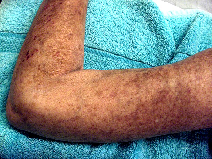
Image Source: Wikimedia Commons (2022). Left Arm Scleroderma Patient, Licensed under a creative commons licence. Available for free at: https://upload.wikimedia.org/wikipedia/commons/6/60/Left_Arm_Scleroderma_Patient.jpg (Accessed: 10 July 2022).
Raynaud’s phenomenon occurs in more than 90% of patients with systemic sclerosis; however, Raynaud’s phenomenon may occur in isolation and, as such, is referred to as primary Raynaud’s phenomenon. However, there are no digital ulcerations or ischemia associated with primary RP.

Image Source: Wikimedia Commons (2022). Raynaud syndrome on female airman’s hand, Licensed under a creative commons licence. Available for free at: https://upload.wikimedia.org/wikipedia/commons/1/1f/Raynaud_syndrome_on_female_airman%27s_hand.jpg (Accessed: 10 July 2022).
Management
The management of systemic sclerosis depends on the disease subset and the presence or absence of internal organ involvement.
Patients with diffuse skin involvement or inflammatory organ involvement are treated with systemic immunosuppressive therapy. These patients may have skin involvement that is severe or diffuse, interstitial lung disease, myocarditis, or arthritis.
Methotrexate and mycophenolate mofetil are both used in patients who exhibit diffuse and progressive skin involvement but no visceral involvement. Generally, methotrexate is used in patients with arthritis or myositis, while mycophenolate mofetil is used in those who have significant interstitial lung disease. For patients who are refractory to the use of these, cyclophosphamide is usually the next step.
In cases of renal sclerosis, the mainstay of treatment is angiotensin-converting enzyme inhibitors (ACE-i). Initially, Captopril may be stated acutely for example, in renal scleroderma crisis within the first 72 hours, after which ACE-i such as Enalapril or Ramipril are used for maintenance. It is essential to closely monitor the serum creatinine (small, transient increases are expected and not usually an indication to stop therapy). There is no evidence that supports the use of ARBs or non-ACE-i therapy.
Gastrointestinal manifestations are treated symptomatically. For instance, proton pump inhibitors are used for oesophageal reflux and gastritis and can be titrated to up to twice daily. Oesophageal dysmotility, which may present with dysphagia, can be treated using prokinetic drugs such as Domperidone and Metoclopramide. Small intestinal bacterial overgrowth is another sequela and is usually treated with antibiotics, namely Metronidazole.
Raynaud’s phenomenon, either because of systemic sclerosis or primary RP, is treated with long-acting dihydropyridine calcium channel blockers such as Amlodipine or Nifedipine. If calcium channel blockers are not tolerated or ineffective, phosphodiesterase type 5 inhibitors such as Sildenafil may be used. Topical nitrates can be considered for patients with ischemic digits as a result of RP. However, it is not recommended to use topical nitrates with PDE 5 inhibitors due to the risk of hypotension.
Arthralgia and arthritis are treated initially with NSAIDs or Paracetamol for symptomatic relief. However, if these are contraindicated or ineffective, alternatives such as low-dose prednisone or hydroxychloroquine can be used. Methotrexate is another option, but only in those patients who are not at risk of pulmonary fibrosis.
References
- Denton C. UpToDate. Pathogenesis of systemic sclerosis (scleroderma) [Internet]. 2021 [cited 2022 March 23]. Available from https://www.uptodate.com/contents/pathogenesis-of-systemic-sclerosis-scleroderma?search=systemic%20sclerosis%20diagnosis&topicRef=7539&source=see_link#H25
- Pearson D, Werth V, Taffer L. Systemic sclerosis: Current concepts of skin and systemic manifestations. Clinics in Dermatology. 2018; 36(4): 459-474. Available from https://www.sciencedirect.com/science/article/abs/pii/S0738081X18300634
- Hughes M , Allanore Y, Denton C, Matucci-Cerinic M. Systemic Sclerosis.
- EMJ Rheumatol. 2020;7[1]:100-109. Available from https://www.emjreviews.com/rheumatology/article/systemic-sclerosis/
- Saracino A, Denton C, Oakley A. DermNetNZ. Systemic Sclerosis [Internet]. 2020 [cited March 30]. Available from: https://dermnetnz.org/topics/systemic-sclerosis
- Varga J. Clinical manifestations and diagnosis of systemic sclerosis (scleroderma) in adults [Internet]. 2021 [cited March 30]. Available from: https://www.uptodate.com/contents/clinical-manifestations-and-diagnosis-of-systemic-sclerosis-scleroderma-in-adults?search=systemic%20sclerosis%20diagnosis&source=search_result&selectedTitle=1~150&usage_type=default&display_rank=1#H6
- Denton C. UpToDate. Overview of the treatment and prognosis of systemic sclerosis (scleroderma) in adults[Internet]. 2021 [Cited 2022 April 10]. Available from: https://www.uptodate.com/contents/overview-of-the-treatment-and-prognosis-of-systemic-sclerosis-scleroderma-in-adults?search=management%20of%20systemic%20sclerosis&source=search_result&selectedTitle=1~150&usage_type=default&display_rank=1
- Varga J, Fenves A. UpToDate. Kidney disease in systemic sclerosis (scleroderma), including scleroderma renal crisis [Internet]. 2021 [Cited 2022 April 5]. Available from: https://www.uptodate.com/contents/kidney-disease-in-systemic-sclerosis-scleroderma-including-scleroderma-renal-crisis?sectionName=Treatment&search=management%20of%20systemic%20sclerosis&topicRef=7542&anchor=H18&source=see_link#H19
- Kaye-Barrett S, Denton C. UpToDate. Treatment of gastrointestinal disease in systemic sclerosis (scleroderma) [Internet]. 2021 [cited 2022 April 4]. Available from: https://www.uptodate.com/contents/treatment-of-gastrointestinal-disease-in-systemic-sclerosis-scleroderma?search=management%20of%20systemic%20sclerosis&topicRef=7542&source=see_link
Inflammatory Myopathy – Myositis
Background
Inflammatory myopathies can take many forms. Those relevant to the MRCP examination include mainly polymyositis, dermatomyositis and inclusion body myositis. It is chronic progressive inflammation of muscles, and each form of myositis presents differently. Thus, the management is tailored accordingly.
Epidemiology
Inflammatory myopathies are rare- estimated to occur in 1 in 100,000 persons. While dermatomyositis and polymyositis occur more commonly in females, inclusion body myositis is more prevalent in males. The average age of those affected ranges from 40-60 years, with inclusion body myositis mainly affecting those above 50 years of age.
Pathophysiology
In polymyositis, lymphocytes such as CD 8+ cytotoxic T cells invade myofibers in the muscle. Triggers for this include viruses such as HIV and parvovirus and drugs such as Phenytoin and Penicillamine. A small percentage (8%) may occur due to malignancies.
Dermatomyositis results from an exposure to a type 1 interferon, causing capillary, myofiber and keratinocyte injury. It is a humorally mediated process driven by CD4 + T cells, causing capillary damage and ischaemic muscle injury.
Inclusion body myositis is primarily a degenerative disorder with a likely secondary cytotoxic inflammatory component. The muscles are infiltrated by T cells, myeloid dendritic cells, macrophages and plasma cells, which aggregate in clusters in the perimysial spaces and cause myofiber necrosis.
Presentation
Dermatomyositis presents with proximal, symmetrical muscle weakness, elevated muscle enzymes and distinctive cutaneous findings. Myalgia is common during the active phase and respiratory muscle, and there may sometimes be myocardial and respiratory muscle involvement and signs of dysphasia.
Characteristic cutaneous findings include a heliotrope rash, a violaceous, oedematous rash affecting the eyelids and Gottron’s papules over the MCP and PIP joints and extensor surfaces. Other less common skin presentations include calcinosis cutis, poikiloderma, periungual telangiectasias and cuticular hypertrophy.
Polymyositis presents with similar muscle weakness as dermatomyositis. However, there are no characteristic skin changes, making it more difficult to diagnose clinically. The hip extensors are often a severely affected muscle group, causing difficulty in climbing stairs and standing from a seated position. It is defined by the presence of anti-synthetase autoantibodies and features such as interstitial lung disease, inflammatory myopathy and small joint symmetrical polyarthritis.
Inclusion body myositis tends to have a more insidious onset, with the average onset of symptoms 5 years prior to diagnosis. There is asymmetric, distal muscle involvement, such as finger flexors and forearm flexors and as such, these patients may complain of weakness in grip strength and difficulty in doing tasks such as opening jars. Myalgia, muscle atrophy and dysphagia can occur but not as commonly.

Image Source: Wikimedia Commons (2022). Dermatomyositis10, Licensed under a creative commons licence. Available for free at: https://upload.wikimedia.org/wikipedia/commons/a/a1/Dermatomyositis10.jpg (Accessed: 10 July 2022).

Image Source: Wikimedia Commons (2022). Dermatomyositis2, Licensed under a creative commons licence. Available for free at: https://upload.wikimedia.org/wikipedia/commons/7/7b/Dermatomyositis2.jpg (Accessed: 10 July 2022).

Diagnosis
Dermatomyositis
Most patients with myositis will have one or more muscle enzymes elevated, such as creatinine kinase, lactate dehydrogenase, aldolase, aspartate aminotransferase or alanine aminotransferase.
Blood Tests: In dermatomyositis, there are specific autoantibodies that may be found, such as Anti Mi-2, which is associated with cutaneous disease but seems to have a lower risk of associated malignancy. Anti-melanocyte differentiation-associated protein 5 (anti-MDA5) is associated with rapidly progressive interstitial lung disease and characteristic skin changes, including ulcerations, Gottron’s papules, painful palmar and macular papules and non-scarring alopecia.
The diagnostic criterion generally used is the 2017 EULAR/ACR classification criteria. To make a diagnosis of dermatomyositis, there needs to be features such as the previously mentioned skin changes (heliotrope rash and Gottron’s papules or Gottron’s sign) AND symmetrical, progressive muscle weakness (usually the proximal upper extensors and/or lower extensors or neck flexors relatively weaker than extensors.
Muscle Biopsy: If this is required, the histopathological changes would show inflammation in the perimysial region (around the muscle bundles) and include CD4+ cells.
Polymyositis
Polymyositis is seen in patients with muscle weakness without characteristic skin changes.
Laboratory Tests: These may reveal similar results to that of other inflammatory myopathies, and as such, a muscle biopsy is frequently required for diagnosis.
Muscle Biopsy: Histopathology, in this case, would show predominantly cellular infiltrates in the endomysial and inflammatory cells invading individual muscle fibres. Myocyte injury is mediated by CD8+ cytotoxic T lymphocytes that surround and invade the myofibers.
Inclusion Body Myositis
Inclusion body myositis also presents without any skin changes, and thus, diagnosis is dependent upon laboratory investigations, electromyography and muscle biopsy.
Blood Tests: Autoantibodies against cytoplasmic 5′-nucleotidase 1A (cN1A) may help distinguish IBM from other forms of myositis.
Neurophysiology studies: Changes in electromyography include fibrillation potentials, bizarre high-frequency repetitive discharges and short, small, low-amplitude polyphasic motor unit potentials.
Muscle biopsy: This is likely to be diagnostic in many cases and histologically has characteristic features of sarcoplasmic ‘rimmed’ vacuoles that are red-rimmed on modified trichrome stain and blue-rimmed on H&E stain.
Differential Diagnoses
- Muscular dystrophies may present similarly to inflammatory myopathy. However, features such as calf hypertrophy, winging of the scapular would point more to a diagnosis of muscular dystrophy, which can be confirmed by muscle biopsy.
- Myasthenia gravis may also have similar features, such as muscle weakness and dysphagia. However, symptoms in MG tend to be intermittent and physical examination findings would show muscle fatigability, whereas those of myositis would have a more progressive or insidious onset. Laboratory testing for autoantibodies and electrophysiology (EMG) studies may help distinguish between both.
- Drug-induced myopathy can mimic inflammatory myopathies, and as such, a thorough history needs to be taken to determine whether the patient is on any drugs that may possibly trigger this, including glucocorticoids, statins, antimalarials, Penicillamine, anti-retroviral therapies, Colchicine, alcohol or cocaine. If the diagnosis remains unclear, then an EMG and/or muscle biopsy may aid in making a definitive diagnosis.
- Inherited metabolic myopathies (disorders of carbohydrate and lipid metabolism) – patients tend to develop chronic muscle weakness. However, this would be preceded by years of acute episodes of intermittent muscle pain and tenderness induced by exertion followed by myoglobinuria, which is not a finding in inflammatory myopathies.
- Motor Neuron disease (Amyotrophic Lateral Sclerosis) can present with muscle weakness but will also have features of hyperreflexia and muscle spasticity (long-tract signs), unlike myositis. Investigations such as EMG would not show myopathic changes, and muscle enzymes such as creatinine kinase will be normal or only mildly elevated.
Management
Dermatomyositis and Polymyositis
Systemic glucocorticoids are the mainstay of acute treatment. Patients are started on a tapering dose usually for one year, which improves the strength and preservation of muscle function.
A glucocorticoid-sparing agent is started simultaneously as the Prednisolone is weaned off. Azathioprine is frequently used and is usually started at a dose of 50mg/day. Prior to initiating Azathioprine, it is important to check for thiopurine methyltransferase deficiency (TPMT) deficiency, as these patients would be at significant risk of having myelosuppression and neutropenic sepsis on Azathioprine. Regular monitoring of liver function tests and complete blood count is done every three months.
Methotrexate is also used as a steroid-sparing agent and is generally started at 15mg once weekly dosing and is titrated up. Folic acid is administered with Methotrexate to prevent folate deficiency, which can have symptoms such as mouth ulcers, paresthesia, fatigue, and muscle weakness. Methotrexate can have other adverse effects such as methotrexate pneumonitis and methotrexate toxicity.
Antimalarials such as Hydroxychloroquine can be used to help to control skin disease. However, it is not beneficial for muscle disease.
Inclusion body myositis
The management of inclusion body myositis primarily aims to optimise muscle strength and function. Non-pharmacological interventions which have proven to be helpful include physiotherapy and speech therapy. Physiotherapy can provide a tailored exercise program, and with the assistance of occupational therapy, appropriate assistive devices can be sourced.
It is not generally recommended to use immunosuppressive therapy in inclusion body myositis. However, in some cases, particularly where there is suspicion of overlap with another autoimmune disease, a trial of Prednisolone can be done, and if there is a response, consideration may be given to tapering prednisolone and commencing a steroid-sparing agent such as azathioprine or methotrexate.
References
- Greenberg S. UpToDate. Pathogenesis of inflammatory myopathies [Internet]. 2022 [cited 2022 April 20]. Available from https://www.uptodate.com/contents/pathogenesis-of-inflammatory-myopathies?search=myositis%20adult&source=search_result&selectedTitle=3~150&usage_type=default&display_rank=3
- Christopher-Stine L, Amato A, Vleugels R. UpToDate. Clinical manifestations of dermatomyositis and polymyositis in adults [Internet]. 2022 [cited 2022 April 20]. Available from https://www.uptodate.com/contents/clinical-manifestations-of-dermatomyositis-and-polymyositis-in-adults?source=history_widget
- Lloyd T, Christopher-Stine L. UpToDate. Clinical manifestations and diagnosis of inclusion body myositis [Internet]. 2022 [cited 2022 April 20]. Available from https://www.uptodate.com/contents/clinical-manifestations-and-diagnosis-of-inclusion-body-myositis?source=history_widget
- Leclair V, Lundberg I. New Myositis classification criteria – What we have learned since Bohan and Peter. Curr Rheumatol Rep. 2018; 20(4): 18. Available from: https://www.ncbi.nlm.nih.gov/pmc/articles/PMC5857275/
- Christopher-Stine L, Amato A, Vleugels R. UpToDate. Diagnosis and differential diagnosis of dermatomyositis and polymyositis in adults [Internet]. 2022 [cited 2022 April 23]. Available from: https://www.uptodate.com/contents/diagnosis-and-differential-diagnosis-of-dermatomyositis-and-polymyositis-in-adults?source=history_widget#H69088617
- Lloyd T, Christopher-Stine L. UpToDate. Clinical manifestations and diagnosis of inclusion body myositis [Internet]. 2022 [cited 2022 April 23]. Available from: https://www.uptodate.com/contents/clinical-manifestations-and-diagnosis-of-inclusion-body-myositis?source=history_widget#H10
- The Myositis Association. Types of Myositis[Internet]. 2022. [cited 2022 April 23]. Available from: https://www.myositis.org/about-myositis/types-of-myositis/
- Lloyd T. UpToDate. Management of inclusion body myositis. [Internet]. 2022 [cited 2022 May 7]. Available from: https://www.uptodate.com/contents/management-of-inclusion-body-myositis?source=history_widget
- Targoff I. UpToDate. Initial treatment of dermatomyositis and polymyositis in adults [Internet]. 2021 [cited 2022 May 7]. Available from: https://www.uptodate.com/contents/initial-treatment-of-dermatomyositis-and-polymyositis-in-adults?source=history_widget#H8
Sjogren’s Syndrome
Background
Sjogren’s syndrome is a chronic autoimmune inflammatory disorder characterised by dryness of the eyes and mouth, fatigue and musculoskeletal pain that is the result of diminished lacrimal and salivary gland function.
Epidemiology
The overall incidence is 7 per 100,000 people per year. The prevalence is highest in the Asian population and lowest in the African population. It is ten times more common in women and is usually diagnosed during middle age (40s) or after menopause. Older adults tend to have more severe symptoms of dry eyes and dry mouth; however, this is due to confounding factors such as adverse effects of medications, age-related fibrosis, and atrophy of secreting tissues.
Pathophysiology
The pathophysiology of SS is thought to be a multifactorial process. Autoantibodies in genetically predisposed individuals are triggered by environmental factors such as viral infection, which result in B-cell stimulation and chronic inflammation of the salivary and lacrimal glands.
Multiple polymorphisms are associated with SS, the most common haplotype being HLA-DR3, but there are also associations with HLA-B8 and HLA-DQ2. The most common viral association is Epstein-Barr virus (EBV). However, HTLV-1, HIV and Hepatitis C have also been noted to have associations with SS. The presence of autoantibodies such as Anti-Ro/SSA and Anti-La/SSB are found in 60-80% of individuals affected with SS.
Presentation
- Systemic Autoimmunity: Thyroid dysfunction and/or other co-existing autoimmune conditions are found in approximately 45% of patients with primary SS.
- Fatigue: Extreme fatigue despite getting more rest, occurs in approximately 50% of patients and is sometimes caused or made worse due to underlying hypothyroidism.
- Cutaneous manifestations: Approximately 55% of patients complain of skin dryness, which is a main characteristic feature of SS. Some patients complain of a rash that is vasculitic in nature and sometimes may be due to underlying vasculitis. Raynaud phenomenon may be observed in about 30% of cases. However, it is usually very mild.
- Pulmonary manifestations: Up to 30% of patients have subclinical pulmonary disease, and lung function tests may show small airway obstruction. Other pulmonary complications include lymphocytic alveolitis, lymphocytic interstitial pneumonitis and fibrosis, and pseudolymphoma.
- Gastrointestinal manifestations: Oesophageal dysmotility is the most common GI presentation and is treated symptomatically with prokinetic drugs. Rarely, mild pancreatitis or autoimmune hepatitis may occur.
- Renal manifestations: Some patients with SS may have tubulointerstitial involvement and glomerular lesions leading to symptoms such as haematuria, proteinuria and renal insufficiency.
- Neurological involvement: The most common significant manifestation is sensory peripheral neuropathy, which is found in approximately 22% of patients.
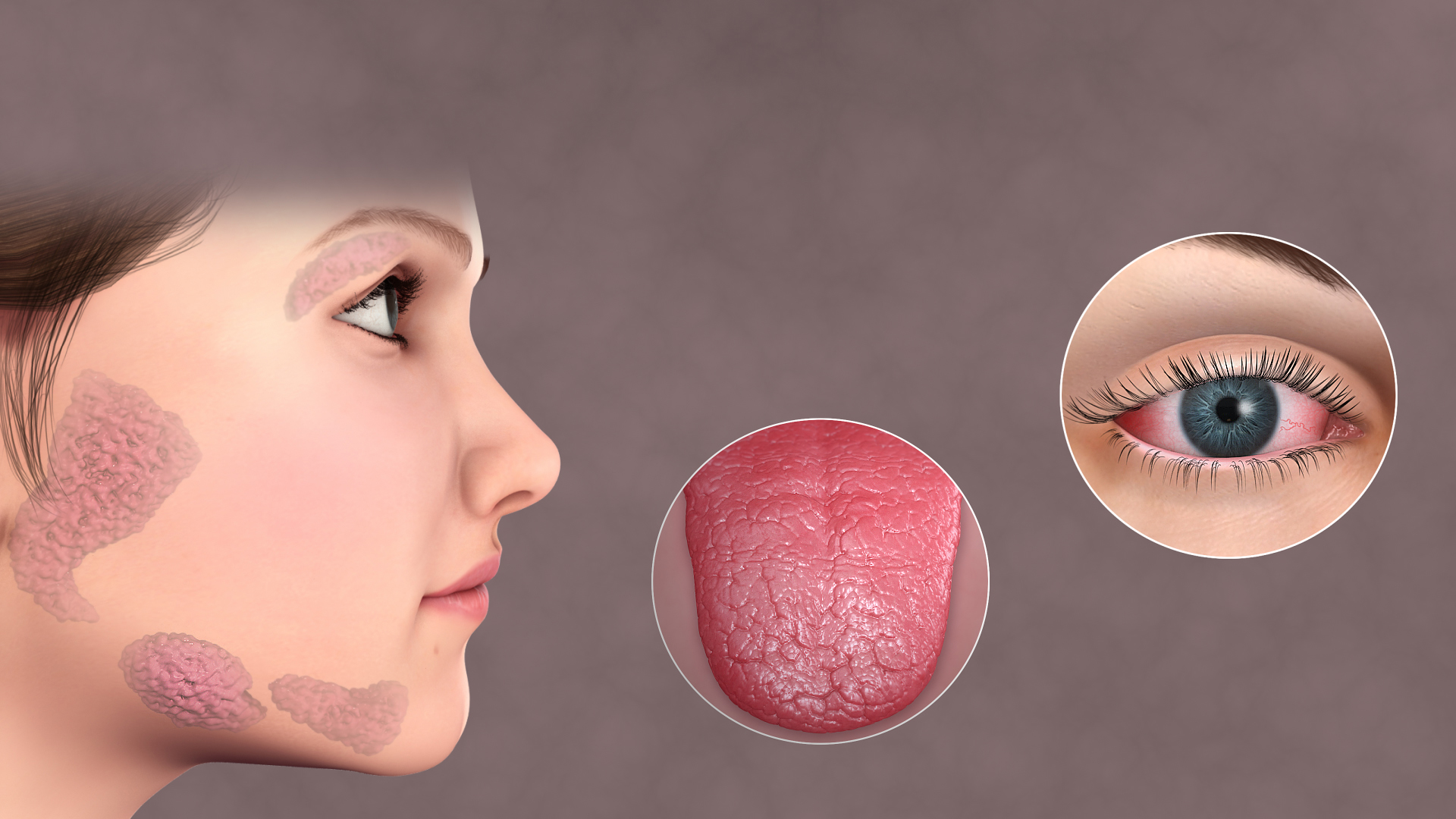
Image Source: Wikimedia Commons (2022). Sjogrens Syndrome, Licensed under a creative commons licence. Available for free at: https://upload.wikimedia.org/wikipedia/commons/5/54/Sjogrens_Syndrome.jpg (Accessed: 10 July 2022).
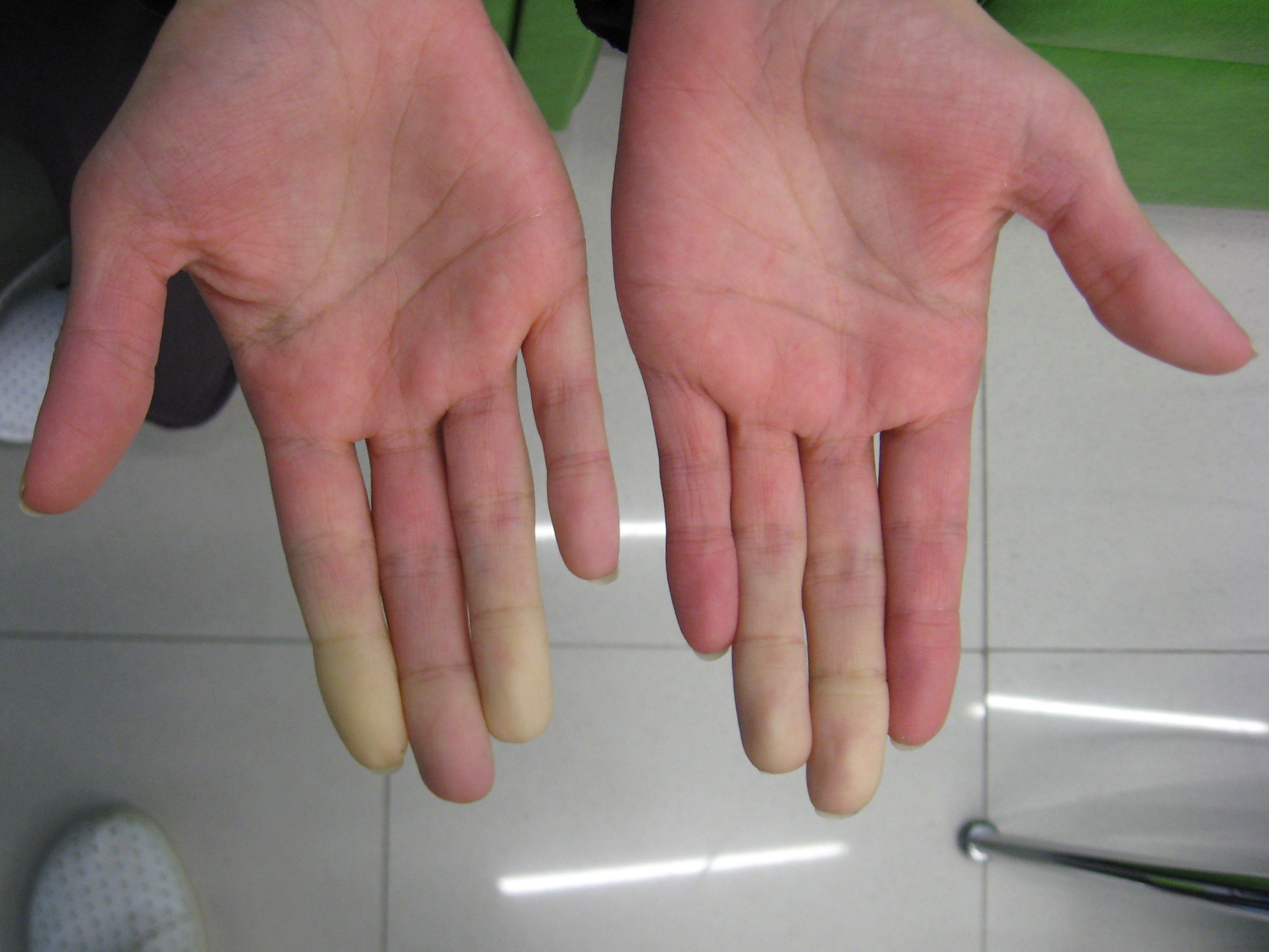
Image Source: Wikimedia Commons (2022). Secondary Raynaud’s in Sjögren’s syndrome 1, Licensed under a creative commons licence. Available for free at: https://upload.wikimedia.org/wikipedia/commons/8/8b/Secondary_Raynaud%27s_in_Sj%C3%B6gren%27s_syndrome_1.JPG (Accessed: 10 July 2022).
Diagnosis
The diagnosis of primary SS can be made in patients with objective findings of ocular and/or oral dryness and evidence of autoimmune exocrine glandular dysfunction. These clinical features, in combination with serological and in some cases, histopathological or radiological findings, assist in making a diagnosis of SS.
- Ophthalmological tests used to investigate for dry eyes include the Schirmer test, tear break-up time and ocular surface staining.
- Evaluation of salivary hypofunction: On physical examination, lack of salivary pooling under the tongue may be found, and testing such as whole sialometry (also called Saxon test) may be done. Imaging such as US may show characteristic salivary gland parenchymal changes, including a hypoechoic linear band, cysts and calcifications. MRI may show a nodular pattern of parenchymal changes such as fatty infiltration, fibrosis, and ductal dilatation.
- Serological testing: Positive testing for Anti-Ro/SSA antibodies with or without Anti/La/SSB antibodies. Positive ANA and Rheumatoid factor, in combination with other clinical features, may also support a diagnosis of SS.
- Salivary gland biopsy: A labial salivary gland biopsy is taken from the lower lip, and the key histological feature is focal collections of tightly aggregated lymphocytes (lymphocytic foci).
Diagnostic criteria
The 2016 ACR/EULAR classification criteria for diagnosis of primary SS are based on a weighted scoring system. They can only be applied to an individual with at least one symptom of ocular or oral dryness or suspicion of SS based on glandular enlargement or the presence of characteristic extra-glandular involvement. An individual can be classified as having primary SS if the total score is four or more. The domains include labial salivary gland with focal lymphocytic sialadenitis, Anti-SSA/Ro positivity (each one 3 points), ocular staining score >5 in at least one eye, Schirmer’s test <5mm/5minutes in at least one eye and an unstimulated whole saliva flow rate <0.1ml/minute (each one is 1 point).
Differential Diagnosis
Most differential diagnoses of SS can be excluded by taking an in-depth history, physical examination and serological testing.
- Age-related sicca syndrome: Age-related ocular and oral dryness may mimic features of SS. However, patients with sicca syndrome would not have positive autoantibodies (Anti-Ro/SSA and Anti-la/SSB), which are present in 60-80% of patients with SS.
- Sarcoidosis: Sarcoidosis may share some extra-glandular features similar to SS, including arthritis and interstitial nephritis. However, biopsy findings and serology for autoantibodies would help in differentiating both conditions.
- Rheumatoid Arthritis: RA may have similar features to SS, such as arthritis and serological testing in both cases may show positive ANA autoantibodies and Rheumatoid factor positive antibodies. However, symptoms such as oral and ocular dryness would make a diagnosis of SS more likely.
Management
The goals of management necessitate a multi-disciplinary team approach through pre-treatment evaluation, which allows tailored treatment to individual patients and their particular disease manifestations.
Non-pharmacologic interventions
- Self-management of sicca symptoms using artificial tears and artificial saliva.
- Immunisations: Appropriate immunisations should be received prior to the start of immunosuppressive therapy, for example, COVID-19, herpes zoster and pneumococcal vaccinations.
- Smoking cessation: This is of particular importance as smoking cigarettes or cannabis can cause worsening symptoms of dry eyes and mouth, increase the risk of dental complications and reduce the efficacy of medications such as hydroxychloroquine.
Pharmacological Interventions:
- Lubricating ocular ointments such as topical Cyclosporine or Lifitegrast and oral secretagogues such as Pilocarpine or cevimeline may be used to treat moderate to severe sicca symptoms.
- Glucocorticoids can be used to treat systemic manifestations of SS in a similar way to other autoimmune disease. However, weaning of steroids is necessary as long-term corticosteroid use is associated with many adverse effects.
- Immunosuppressive and DMARDs:
- Hydroxychloroquine can be used to improve symptoms such as arthralgias and myalgias.
- Methotrexate is effective in controlling dry mouth and eyes, arthralgias, arthritis and musculoskeletal pain.
- Azathioprine at high dose can be used to manage extra-glandular manifestations such as interstitial pneumonitis, myelopathy and autoimmune hepatitis.
- Mycophenolate mofetil has been shown to lead to a reduction in hypergammaglobulinemia and rheumatoid factor and an increase in complement and white blood cell count, making it effective in treating agranulocytosis as a result of primary SS.
References
- Baer A, Deshmukh U. UpToDate. Pathogenesis of Sjogren’s Syndrome [Internet] 2021. [cited 2022 May 12]. Available from: https://www.uptodate.com/contents/pathogenesis-of-sjogrens-syndrome
- Baer A. UpToDate. Diagnosis and Classification of Sjogren’s Syndrome [Internet] 2021. [cited 2022 May 12]. Available from: https://www.uptodate.com/contents/diagnosis-and-classification-of-sjogrens-syndrome?source=history_widget
- Kassan SS, Moutsopoulos HM. Clinical Manifestations and Early Diagnosis of Sjögren Syndrome. Arch Intern Med. 2004;164(12):1275–1284.
- Baer A, Vivino F. UpToDate. Treatment of Sjögren’s syndrome: Constitutional and non-sicca organ-based manifestations [Internet] 2022. [cited 2022 May 21]. Available from: https://www.uptodate.com/contents/treatment-of-sjogrens-syndrome-constitutional-and-non-sicca-organ-based-manifestations?source=history#H2606837497
Mixed Connective Tissue Disease (MCTD)
Background
Epidemiology
Mixed Connective Tissue Disease is an autoimmune overlap syndrome that presents with clinical features of SLE, systemic sclerosis and polymyositis. It is characterised by presence of high titers of a distinctive autoantibody, now called anti-U1 ribonucleoprotein (RNP) (previously termed antibody to extractable nuclear antigens, anti-ENA).
Worldwide, the prevalence is estimated to be between 2-3.8 cases per 100,000 population. It is much more common in females than males, with an estimated ratio of 3:1 and the typical age of onset is between 15-25 years; however, it can occur at any age. MCTD occurs in patients of all ethnicities, but the clinical manifestations, particularly the frequency of end-organ involvement can vary.
Pathophysiology
Whilst the exact genetic cause is unknown, DNA methylation levels may be relevant as patients with MCTD have been found to have decreased levels of DNA methylation. There has also been a genetic association with major histocompatibility genes such as HLA-B*08 and HLA-B1*04.
These genetic abnormalities lead to B-lymphocyte hyperactivity, resulting in high levels of auto-antibodies (namely anti–U1-RNP and anti–U1-70 kd autoantibodies). These auto-antibodies then cause T-lymphocyte activation, which leads to lymphocytic and plasmacytic infiltration of tissues, causing increased vascular endothelial apoptosis.
Presentation
- The initial clinical symptoms of MCTD tend to be non-specific features such as malaise, arthralgia, fever and Raynaud’s phenomenon.
- The most common presenting symptoms are arthralgia with swollen hands, Raynaud’s and gastro-oesophageal reflux.
- RP almost always presents initially and occurs in about 96% of patients with MCTD.
- Arthritis: Swollen fingers and diffuse oedema, sometimes with arthritis and bone deformities in the hands, are present in 96% of patients.
- Oesophageal hypomotility: symptoms of GORD are present in 66% of patients
- Myalgias: Muscle aches are common. However, elevation of inflammatory muscle enzymes and changes on EMG are not usually seen.
- Rash: A skin rash may be observed in 53% of patients and is usually vasculitic in nature.
- Pulmonary involvement: This occurs in approximately 75% of patients and may present as pleural effusion, interstitial lung disease, thromboembolic events or rarely, pulmonary hypertension.
- Haematological manifestations: Leukopenia is present in 53% of patients, and findings of anaemia or hypergammaglobulinemia are also common.

Image Source: Rahmouni S et al. (2020) Mixed connective tissue disease: Not always an obvious diagnosis. Clinical case reports. 2020 Oct; 8(10): 1979-1983. Licensed under a commercial Creative Commons license Available at: https://onlinelibrary.wiley.com/doi/full/10.1002/ccr3.3045 (Accessed: 21 Decemvber 2023).

Image Source: Rahmouni S et al. (2020) Mixed connective tissue disease: Not always an obvious diagnosis. Clinical case reports. 2020 Oct; 8(10): 1979-1983. Licensed under a commercial Creative Commons license. Available at: https://onlinelibrary.wiley.com/doi/full/10.1002/ccr3.3045 (Accessed: 21 December 2023).
Diagnosis
In the early stages, differentiating MCTD from other autoimmune diseases, such as SLE and polymyositis, can be difficult. However, the most prominent initial features, such as puffy fingers, arthralgia and myalgia, should be further investigated by specific antibody testing, including ANA, ENA and Anti-U1 RNP.
One of the most widely used diagnostic criteria is that of the Alarcon-Segovia’s criteria, which has a sensitivity of 63% and a specificity of 86%. To fulfil the criteria, patients must have Anti-RNP antibodies with a hemagglutination titer of ≥1:1600 and three or more of the following clinical features (one of which must include myositis or synovitis): swollen hands, Raynaud’s phenomenon, acrosclerosis, synovitis and myositis.
The laboratory testing for MCTD hinges on the presence of the specific auto-antibodies, as mentioned previously, in particular ENA and Anti-U1 RNP. However, antibodies Sm, Ro, and La are occasionally, transiently seen in patients with MCTD.
Differential Diagnosis
MCTD was initially thought to be a type of overlap syndrome, and thus, other autoimmune illnesses can present similarly.
Systemic Lupus Erythematosus (SLE)
Both MCTD and SLE patients have similar symptoms of arthritis and synovitis. However, symptoms such as photosensitivity are prominent with SLE but not MCTD and antibodies such Anti-ds DNA would be expected to be positive in SLE while Anti- U1 RNP would be positive in MCTD.
Rheumatoid Arthritis (RA)
Both RA and MCTD may present with symptoms such as fatigue, arthralgia and some degree of swelling. However, in RA, the swelling would be more around the joints, whereas in MCTD, the swelling would be more in the subcutaneous connective tissue of the hands. This swelling can be determined both on physical examination as well as on radiographs of the hands. In addition, the auto-antibodies again will differ where Anti-CCP would be more specific with RA and Anti-U1 RNP more specific for MCTD.
Inflammatory Myopathy – Polymyositis
In both polymyositis and MCTD, there are features of myositis and end-organ involvement, such as interstitial lung disease and oesophageal dysmotility. However, there are significantly elevated inflammatory markers such as CK and CRP in polymyositis, but in MCTD, these markers would be expected to be within the normal range or mildly elevated.
Systemic Sclerosis
Raynaud’s phenomenon can occur both in systemic sclerosis and MCTD. However, it tends to be more severe in systemic sclerosis, and the characteristic skin changes seen in systemic sclerosis, such as skin thickening and telangiectasia, are not seen in MCTD. In addition, renal involvement is rarely seen in MCTD and is a more common feature of systemic sclerosis. Laboratory testing again would help differentiate by testing for autoantibodies where Anti-centromere or Anti-Scl-70 would be expected to be positive in systemic sclerosis while Anti-U1 RNP is positive in MCTD.
Management
Although there have been no controlled trials to guide the management of MCTD, general principles are guided by effective therapies for SLE, systemic sclerosis and polymyositis.
- Glucocorticoids: Oral prednisolone is the steroid of choice initially and is effective in treating pleurisy, pericarditis, myocarditis and oesophageal dysmotility. Corticosteroids need to be weaned due to the adverse effects of long-term steroid therapy.
- Immunosuppressants/Steroid-sparing agents: Methotrexate or Hydroxychloroquine may be started while weaning off steroids; they are effective in preventing flares and symptom control.
- Anti-hypertensives: Pulmonary arterial hypertension is a major cause of death in patients with MCTD. As such, routine echocardiography is recommended. Treatment for PAH includes anti-hypertensives such as calcium channel blockers and ACE-i. Other treatments include intravenous prostacyclins and anti-coagulation, endothelin -1 antagonist (e.g. Bosentan) and phosphodiesterase inhibitors (e.g. Sildenafil).
- Rituximab: This is used in patients who have steroid-resistant thrombocytopenia, autoimmune haemolytic anaemia, myositis and severe eruptive skin disease.
References:
- Greidinger, Eric L.; Hoffman, Robert W. Autoantibodies in the pathogenesis of mixed connective tissue disease. Rheumatic Disease Clinics of North America. 31 (3): 437–450.
- Axford J. UpToDate. Definition and diagnosis of mixed connective tissue disease [Internet]. 2022 [cited 2022 May 22]. Available from: https://www.uptodate.com/contents/definition-and-diagnosis-of-mixed-connective-tissue-disease?search=mixed%20connective%20tissue%20disease%20adult&source=search_result&selectedTitle=1~121&usage_type=default&display_rank=1#H4
- Axford J. UpToDate. Prognosis and treatment of mixed connective tissue disease [Internet]. 2022 [cited 2022 May 22]. Available from: https://www.uptodate.com/contents/prognosis-and-treatment-of-mixed-connective-tissue-disease?search=management%20of%20mixed%20connective%20tissue%20disease&source=search_result&selectedTitle=1~121&usage_type=default&display_rank=1#H5
Complete Guide to Clinical Genetics ST3 Application and Recruitment (2027)
Written by Medibuddy. Last updated: October 2026. Clinical Genetics is

Complete Guide to Clinical Radiology ST3 Application and Recruitment (2027)
Recruitment paused: NHS England is not running Clinical Radiology ST3+ recru

Complete Guide to Paediatric Surgery ST3 Application and Recruitment in United Kingdom (2026)
Written by Medibuddy. Last updated: October 2026. If you’re planning


Take your subscriptions with you
Our mobile app allows you to access your interview and exam question banks wherever you are.

