


 Close
Close
 26th January 2024
Systemic Lupus Erythematosus (SLE)
Background
There are four major types of lupus: systemic lupus erythematosus, lupus of the skin, drug-induced lupus erythematosus and neonatal lupus erythematosus.
Systemic lupus erythematosus (SLE) is the most common and is a chronic autoimmune disease that can affect multiple organs, including the skin, joints and kidneys.
Lupus of the skin can be subdivided into three main categories, each with their own distinctive lesions. These are 1) acute cutaneous lupus, 2) chronic cutaneous lupus erythematosus or discoid lupus erythematosus (DLE) and 3) subacute cutaneous lupus erythematosus.
Drug-induced lupus has a similar presentation to SLE and is caused by drugs such as hydralazine, procainamide, isoniazid and minocycline.
Neonatal lupus erythematosus is a rare condition that affects babies of women who have autoantibodies (Anti-Ro and Anti-La), which act on the neonates’ heart conduction system, leading to heat block.
Epidemiology
SLE is the most common form of Lupus – affecting approximately 70% of all patients diagnosed. There is a higher prevalence in females with the age of onset at diagnosis, most often between 15 and 45 years.
The prevalence of SLE is higher among Asian, African American, African Caribbean and Hispanic individuals compared to Caucasians.
Pathophysiology
The exact aetiology of SLE is unknown as it is thought to be multifactorial.
Genetic factors:
Generally, no single polymorphism is associated with a high risk for developing SLE. However, the combination of susceptibility genes and the absence of protective genes confer enough genetic susceptibility to cause disease development.
The following genetic factors are associated with SLE:
- The most common genetic predisposition is at the MHC locus – Class I HLA (HLA – A, B, C) and Class 11 HLA molecules (HLA – DR, DQ, DP)
- Loci of particular interest – Anti-La, Anti-Sm, Anti- dsDNA, Anti- Ro (associated with neonatal heart block)
- Deficiencies of complement components – C1q, C4A, B, C2, TREX1
- Gene Polymorphisms in DNA repair genes – ATG5, DNASE1
Hormonal and Immunological Factors
- An increase in CD4+ and CD8 + T cells due to excess or unopposed oestrogen release may lead to autoantibodies which eventually lead to the clinical findings of SLE. This may account for the higher prevalence in women.
- Elevated levels of circulating IFN-alpha correlate with active disease. The presence of genetic predisposing factors leads to increased expression of IFN-alpha-inducible RNA transcripts by mononuclear cells, which in turn causes these elevated levels.
- Dysregulation of apoptosis among different cell types – T lymphocytes and the Fas/Fas ligand pathway causes an abnormal programmed cell death and an aberrant lymphoid population, contributing to autoimmune activity.
- In lupus nephritis, there is deposition of immune complexes in the mesangium and activation of complement in the glomerular basement membrane, causing inflammation, which eventually leads to necrosis and scarring. There are also elevated circulating levels of TNF-alpha, which correlate with active disease.
Presentation
Articular Manifestations
Joint involvement is common and present in approximately 95% of patients diagnosed with SLE. In 75% of patients, it is the presenting complaint. The arthritis of SLE is typically non-erosive, symmetrical and polyarticular. Joints commonly affected include the proximal interphalangeal joints (PIPs), carpal joints and knees. On examination, joint deformities may resemble those seen in RA, such as ulnar deviation and thumb subluxation and are referred to as Jaccoud’s arthropathy. However, Jaccoud’s arthropathy results from soft tissue abnormalities and is a non-erosive, reversible joint disorder, unlike Rheumatoid Arthritis.

Image Source: Abushama S et al. (2021) A Sudanese male with Jaccoud’s arthropathy; case report. Biomed J Sci & Tech Res 33(2)-2021. Licensed under a commercial Creative Commons license. Available at: https://biomedres.us/pdfs/BJSTR.MS.ID.005387.pdf (Accessed: 21 December 2023).
Cutaneous Manifestations
Skin involvement is often the first sign in SLE, and about 80% of patients with SLE will have skin signs. Common skin signs include malar or “butterfly” rash, photosensitivity rashes, hair loss, discoid lupus – indurated, hyper-pigmented plaques, and muco-cutaneous involvement. On examination, active lesions of discoid lupus may present as erythematous, circular, scaly plaques, while healed lesions show a characteristic depigmented central area with surrounding hyper-pigmentation and an atrophic centre. The alopecia seen is scarring alopecia that often results in permanent hair loss.

Image Source: Wikimedia Commons (2022).Lupusfoto, Licensed under a creative commons licence. Available for free at: https://upload.wikimedia.org/wikipedia/commons/5/52/Lupusfoto.jpg (Accessed: 10 July 2022).

Image Source: Wikimedia Commons (2022). Discoid lupus erythematosus, Licensed under a creative commons licence. Available for free at: https://upload.wikimedia.org/wikipedia/commons/b/ba/Discoid_lupus_erythematosus.JPG (Accessed: 10 July 2022).

Image Source: Wikimedia Commons (2022). Discoid lupus etythermayouses new image, Licensed under a creative commons licence. Available for free at: https://upload.wikimedia.org/wikipedia/commons/9/91/Discoid_lupus_etythermayouses_new_image.jpg (Accessed: 10 July 2022).
Non-articular Systemic Manifestations of SLE
- General: fatigue (the most common complaint), intermittent fever, weight loss
- Cardiac: pericarditis with or without pericardial effusion (most common cardiac manifestation)
- Pulmonary: pleuritis, pneumonitis, interstitial lung disease, pulmonary hypertension
- Vascular: Raynaud’s phenomenon (occurs in up to 50% of patients with SLE), vasculitis – most commonly small vessel and can manifest as petechiae, palpable purpura, livedo reticularis, panniculitis and superficial ulcerations
- Renal: Renal involvement occurs in approximately 50% of SLE patients. Different forms of glomerulonephritis can occur and may present with proteinuria/nephrotic syndrome or asymptomatic haematuria.
- Neurologic and Neuropsychiatric: Symptoms and presentations can include cognitive dysfunction, delirium, psychosis, strokes, seizures, and peripheral neuropathies.
- Haematological: Anaemia of chronic disease is the most common presentation. Leukopenia is also common and occurs in approximately 50% of patients and generally correlates to disease activity.
- Ophthalmologic: Keratoconjunctivitis is the most common manifestation and is as a result of secondary Sjogren’s syndrome
Diagnosis
Laboratory Investigations
- Full Blood Count (FBC): may show anaemia, leukopenia, thrombocytopenia
- Renal function: elevated creatinine may be a sign of renal dysfunction
- ANA: positive in almost all patients with SLE
- Anti-dsDNA: highly specific and sensitive for SLE – seen in approximately 70% of patients
- Anti-Sm antibodies: highly specific but lacks sensitivity – seen in approximately 30% of patients
- Anti-Ro and Anti-La: more commonly associated with Sjogren’s syndrome but are present in approximately 20-30% of patients with SLE
- Anti-U1 RNP antibodies: are almost always found in high levels in patients with mixed connective tissue disease; however, it occurs in approximately 25% of patients with SLE.
Imaging
- X-rays of the hands may show evidence of Jaccoud’s arthropathy, which includes soft tissue deformities and the absence of erosions.
- MRI or Ultrasound: These may reveal synovial effusions which are usually small and can be used to guide aspiration of the joint that typically shows low protein and white cell counts.
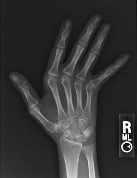
Image Source: Wikimedia Commons (2022). Jaccoud arthropathy hand x-ray front, Licensed under a creative commons licence. Available for free at: https://upload.wikimedia.org/wikipedia/commons/7/71/Jaccoud_arthropathy_hand_x-ray_front.jpg (Accessed: 10 July 2022).
Diagnostic Criteria
SLICC (Systemic Lupus International Collaborating Clinics) Criteria – 2012
Using the SLICC criteria, SLE is diagnosed if the patient has either of the following over time:
- Four criteria including ≥ one clinical criterion and ≥ one immunological criterion
- Biopsy-proven lupus nephritis and anti-nuclear antibodies or anti-double-stranded DNA antibodies
The clinical criteria include features such as cutaneous lupus, alopecia, oral or nasal ulcers, joint disease, serositis, neurological manifestations and laboratory findings such as anaemia, leukopenia, thrombocytopenia and proteinuria. The immunological criteria include positive ANA, Anti-ds DNA, Anti-Sm, Antiphospholipid and low complement (C3 and C4). A positive Direct Coombs Test can be considered in the absence of haemolytic anaemia.
| Clinical Criteria | Immunological criteria |
|---|---|
| Acute cutaneous lupus (lupus malar rash) | ANA |
| Chronic cutaneous lupus (discoid rash) | Anti-ds DNA |
| Alopecia | Anti-Sm |
| Oral or nasal ulcers | Antiphospholipid |
| Joint disease (synovitis or tenderness) | Low complement |
| Serositis | Direct Coombs Test |
| Renal (proteinuria, red cell casts) | |
| Neurological (seizures, psychosis) | |
| Haemolytic anaemia | |
| Leukopenia | |
| Thrombocytopenia |
Table 1: The SLICC classification criteria for systemic lupus erythematosus.
Table 1: UpToDate. Classification criteria for systemic lupus erythematosus [Internet]. 2022 [cited 2022 July 12]. Available from: https://www.uptodate.com/contents/image?imageKey=RHEUM%2F86633
EULAR/ACR (European Alliance of Associations for Rheumatology/American College of Rheumatology) Criteria – 2019
This criterion consists of 7 clinical and 3 immunological categories, each of which are weighted 2 – 10. Patients with a score of 10 points or more are classified as having SLE.
The clinical domains include constitutional, haematological, neuropsychiatric, mucocutaneous, serosal, musculoskeletal and renal. The immunological domains include antiphospholipid antibodies, complement proteins and SLE-specific antibodies.
| Clinical Criteria | Immunological criteria |
|---|---|
| Malar rash | ANA |
| Photosensitivity | Anti-ds DNA OR Anti-Sm OR Anti Phospholipid |
| Discoid rash | |
| Oral ulcers | |
| Arthritis | |
| Serositis | |
| Renal (proteinuria, cellular casts) | |
| Neurological (seizures or psychosis) | |
| Haematological (haemolytic anaemia, leukopenia, lymphopenia, thrombocytopenia) |
Table 2: The ACR classification criteria for systemic lupus erythematosus.
Table 2: UpToDate. Classification criteria for systemic lupus erythematosus [Internet]. 2022 [cited 2022 July 12]. Available from: https://www.uptodate.com/contents/image?imageKey=RHEUM%2F86633
Differential Diagnoses
SLE can be difficult to diagnose, particularly in the initial stages, and the symptoms can be vague and overlap with many other more common causes. The most common symptoms include arthralgia, myalgia, fatigue, weight loss and photosensitivity. A definitive diagnosis in many cases may depend on antibody testing.
Rheumatoid Arthritis
RA may also present with constitutional symptoms and arthralgia. However, characteristics such as morning joint stiffness and symmetrical, tender swollen joint would be more indicative of RA. In addition, joint deformities such as Boutonniere and Swan neck would be found in RA and not SLE.
Positive tests such as Rheumatoid factor may be positive in both SLE and RA; however, Anti-CCP positivity would be expected in RA, while Anti-ds DNA positivity would be expected in SLE.
Systemic Sclerosis
Systemic Sclerosis may also present with arthralgia, rashes, and Raynaud’s phenomenon. However, features such as decreased joint mobility and skin induration would be more common in systemic sclerosis.
Antibody testing is key here, where the presence of Anti-centromere and Anti-Scl-70 antibodies would be indicative of a diagnosis of sclerosis rather than SLE.
Sarcoidosis
Sarcoidosis may also present with features such as fatigue, fever, night sweats or a rash. However, symptoms such as cough and dyspnoea would be more common in sarcoidosis. In addition, an elevated angiotensin-converting enzyme and chest radiograph features such as bilateral hilar lymphadenopathy would be indicative of sarcoidosis rather than SLE.
Adult-Onset Still’s Disease
While similar constitutional features such as fever and arthralgia may be present, splenomegaly would be more characteristic of Still’s disease. In both cases, laboratory blood testing may show elevated ESR and anaemia; however, Anti-ds DNA would be expected to be positive in SLE and not Still’s disease.
Chronic Fatigue Syndrome
This is a diagnosis of exclusion. Patients may present with vague symptoms, and blood tests, including inflammatory markers, metabolic panel and antibody testing, will all be within the normal range.
Management
Treatment of SLE depends on disease activity and severity in addition to symptoms and organs affected. It requires management by a rheumatologist, and often, a multi-disciplinary approach is required.
Non-Pharmacological/Conservative
- UV protection: This is particularly significant in those affected with cutaneous SLE where photosensitivity may occur. Sunscreens of SPF (sun protection factor) of >55 and protection against UV-A and UV-B are recommended. Medications that induce photosensitivity should also be avoided.
Pharmacological
- Anti-malarials: Hydroxychloroquine and Chloroquine can help relieve constitutional symptoms in addition to musculoskeletal and mucocutaneous manifestations. Can be used in mild to moderate SLE where there are manifestations that are non-organ threatening (e.g. constitutional symptoms, skin, mucosal, joint or haematological involvement)
- Oral Corticosteroids: Prednisolone 5-15mg daily can be used as a short-term therapy. This can be tapered while the hydroxychloroquine or chloroquine takes effect.
- Intravenous Corticosteroids: Methylprednisolone IV is used in severe or life-threatening manifestations that occur secondary to major organ involvement. Patients are sometimes ‘pulsed’ on this high-dose IV corticosteroid for a short duration to rapidly reduce inflammation to achieve disease control.
- Azathioprine: This immunosuppressant is of value in treating the manifestations of SLE in patients of childbearing age and is one of the few medications that is generally safe to use in pregnancy.
- Adverse effects: Bone Marrow Suppression: there is an increased risk of this occurring in individuals with TPMT enzyme deficiency, which is why it is important to screen for TPMT prior to starting patients on azathioprine. Monitoring of full blood count, liver and renal function is required twice weekly for six weeks and, once the dose is stabilised, monthly for three months thereafter, which can be done at least every 12 weeks.
- Methotrexate: MTX competitively inhibits the enzyme dihydrofolate reductase. As such, daily folic acid replacement is required. It has been shown to reduce disease activity and can be effectively used as a steroid-sparing agent. Adverse effects: Methotrexate toxicity, hepatotoxicity, nephrotoxicity, myelosuppression, teratogenic, lung fibrosis, reactivation of latent TB. Monitoring guidelines suggest full blood count and liver and renal function every 1-2 weeks until therapy is stabilised, after which patients can be monitored every 2-3 months. In the case of methotrexate toxicity, the antidote is folinic acid (calcium folinate).
- Mycophenolate mofetil: MMF is an inhibitor of lymphocyte proliferation. It is used in combination with glucocorticoids in patients with focal or diffuse lupus nephritis. Adverse effects: Teratogenic, increased risk of infections, bone marrow suppression and rarely Progressive Multifocal Leukoencephalopathy (PML). As there is a risk of neutropenia, monitoring of full blood count is required every week for 4 weeks, then twice a month for 2 months, and then every month after that.
References
- Schur P, Hahn B. UpToDate. Epidemiology and pathogenesis of systemic lupus erythematosus [Internet]. 2021 [cited 2022 March 01]. Available from: https://www.uptodate.com/contents/epidemiology-and-pathogenesis-of-systemic-lupus-erythematosus?source=history
- Ho K, Reveille J. The clinical relevance of autoantibodies in scleroderma. Arthritis Res Ther. 2003; 5(2): 80-93. Available from https://www.ncbi.nlm.nih.gov/pmc/articles/PMC165038/
- Lam N, Ghetu M, Bieniek M. Systemic Lupus Erythematosus: Primary care approach to diagnosis and management. Am Fam Physician. 2016; 94(4): 284-294. Available from https://www.aafp.org/afp/2016/0815/p284.html
- Wallace D. Overview of the management and prognosis of systemic lupus erythromatosus in adults [Internet]. 2021 [cited 2022 March 01]. Available from: https://www.uptodate.com/contents/overview-of-the-management-and-prognosis-of-systemic-lupus-erythematosus-in-adults?source=history#H509844069
- Croyle L, Hoi A, Morand E, Characteristics of azathioprine use and cessation in a longitudinal lupus Cohort. Lupus Sci Med. 2015 Aug 20; 2(1): 105-110. Available from https://www.ncbi.nlm.nih.gov/pmc/articles/PMC4548066/
- Gansauge S, Breitbart A, Rinaldi N, et al. Methotrexate in patients with moderate systemic lupus erythematosus (exclusion of renal and central nervous system disease) Annals of the Rheumatic Diseases 1997;56:382-385.
- Falk R, Dall’Era M, Appel G. UpToDate. Lupus Nephritis: Initial and subsequent therapy for focal or diffuse lupus nephritis [Internet]. 2022. [cited 2022 March 18]. Available from: https://www.uptodate.com/contents/lupus-nephritis-initial-and-subsequent-therapy-for-focal-or-diffuse-lupus-nephritis?topicRef=4675&source=see_link#H3594109834
Complete Guide to Clinical Genetics ST3 Application and Recruitment (2027)
Written by Medibuddy. Last updated: October 2026. Clinical Genetics is

Complete Guide to Clinical Radiology ST3 Application and Recruitment (2027)
Recruitment paused: NHS England is not running Clinical Radiology ST3+ recru

Complete Guide to Paediatric Surgery ST3 Application and Recruitment in United Kingdom (2026)
Written by Medibuddy. Last updated: October 2026. If you’re planning


Take your subscriptions with you
Our mobile app allows you to access your interview and exam question banks wherever you are.

