


 Close
Close
 26th January 2024
Systemic Sclerosis
Background
Systemic sclerosis is an immune-mediated, multi-system disease that is characterised by fibrosis of the skin and internal organs and widespread vascular dysfunction. The diagnosis and related disorders are based upon clinical findings and supported by specific serological abnormalities.
Epidemiology
The majority of patients with systemic sclerosis are female and tend to have a younger age of onset, more limited disease and increased risk of peripheral vascular disease and pulmonary hypertension.
Men have an increased risk of diffuse cutaneous disease, cardiac involvement, renal crisis and frequent interstitial lung disease.
African Americans tend to have an earlier age at onset, more severe phenotypes and increased risk of pulmonary fibrosis and renal crisis.
Pathophysiology
The exact pathogenesis of systemic sclerosis is complex and not fully understood, but immunological and vascular processes are known to play a key role. There is increased production and deposition of structurally normal collagen as a result of extracellular matrix interactions.
Vascular and endothelial changes cause vasoconstriction, which is mediated by endothelins, nitric oxide, hypoxia, physical stress, and humoral and inflammatory mediators.
Autoantibodies are present in approximately 90% of patients with systemic sclerosis. The most common include topoisomerase I (previously Scl-70), centromere antigens and RNA-polymerase.
Anti-topoisomerase-I (Scl-70) antibodies are not sensitive but highly specific for systemic sclerosis and are associated with a higher risk of interstitial lung disease, more extensive skin involvement and higher disease activity.
Anti-centromere antibodies are associated with limited cutaneous involvement.
Presentation
Cutaneous Manifestations
Skin involvement is a universal feature which commonly presents early in the face and hands. Features include:
- Tight/taut skin
- Shiny skin
- Pigmentation changes – hyperpigmentation and depigmentation
- Telangiectasia
- Lipoatrophy

Image Source: Wikimedia Commons (2022). Systemic sclerosis 2 fingers, Licensed under a creative commons licence. Available for free at: https://upload.wikimedia.org/wikipedia/commons/1/15/Systemic_sclerosis%2C_fingers.jpg (Accessed: 10 July 2022).
Musculoskeletal manifestations
- Swelling and stiffness of the fingers
- Large and small joint contractures
- Joint pain and immobility
- Arthritis
- Tendonitis
- Tendon friction rubs
- Calcinosis cutis (soft tissue calcifications and resorption of the distal phalangeal tufts can be seen on radiographs)

Image Source: Wikimedia Commons (2022). Calcinosis of CREST syndrome, Licensed under a creative commons licence. Available for free at: https://upload.wikimedia.org/wikipedia/commons/c/c8/Calcinosis_of_CREST_syndrome.jpg (Accessed:10 July 2022).
Vascular manifestations
Digital ischemic ulcers can develop in up to 50% of patients with systemic sclerosis. Progressive changes within the small blood vessels occur and can cause blood flow to be impaired permanently. It is more commonly seen in patients with diffuse disease and those with positive anti-topoisomerase I (Scl-70) antibodies. This can result in gangrene, infection and, in extreme cases, amputation.
Gastrointestinal manifestations:
Almost 90% of patients with systemic sclerosis have evidence of gastrointestinal involvement, and the oesophagus is the most frequently affected area. Symptoms include dysphagia, early satiety, bloating, constipation, small bowel bacterial overgrowth, pseudo-obstruction, faecal incontinence and gastro-oesophageal reflux.
Gastrointestinal bleeding and subsequent anaemia may occur due to angiodysplasia in the antrum of the stomach, which is sometimes referred to as “watermelon stomach.”
Pulmonary manifestations
Pulmonary involvement may be seen in more than 80% of patients and mainly manifests as interstitial lung disease or pulmonary fibrosis. This can eventually lead to pulmonary arterial hypertension.
Renal manifestations
Renal involvement may occur in 50-80% of patients and can present with microalbuminuria, mild elevation in plasma creatinine or hypertension. Rarely (in 10-15% of patients) scleroderma renal crisis can occur and is characterised by malignant hypertension, acute oliguria, microangiopathic haemolysis, thrombocytopenia and mild proteinuria.
Diagnosis
Limited or diffuse systemic sclerosis can be diagnosed in patients with skin thickening of the fingers and hands, which extend from the proximal interphalangeal joints to the metacarpophalangeal joints. In many cases, there would be positive anti-topoisomerase (anti-Scl-70) antibodies, anti-centromere or anti-RNA polymerase III antibody or a positive ANA (anti-nuclear antibody).
In differentiating between both limited and cutaneous systemic sclerosis, it is important to note that patients with diffuse cutaneous systemic sclerosis will have widespread skin involvement, which includes proximal limbs and significant internal organ involvement. In addition, diffuse cutaneous systemic sclerosis would be more associated with Anti-Scl-70 (Anti-topoisomerase) or Anti-RNA polymerase III positivity.
Investigations such as pulmonary function tests may show restrictive changes, while CT may show evidence of interstitial pulmonary disease. Echocardiography may reveal pulmonary arterial hypertension. Dermoscopy may show abnormal capillaries.
Sine sclerosis is a rare autoimmune disease diagnosed in patients with no evidence of skin manifestations of systemic sclerosis but have positive clinical features such as Raynaud’s phenomenon, digital ischemia, abnormal capillaries, and pulmonary and renal involvement. It can be very difficult to diagnose due to the lack of external cutaneous manifestations; however, in these cases, antibodies such as ANA would be positive, but antibodies such as Anti-Scl-70 and Anti-centromere antibodies would be expected to be negative.
Scleroderma overlap syndrome occurs in approximately 20% of patients where there is cutaneous sclerosis and features of another connective tissue disease (most often an inflammatory myopathy such as polymyositis and dermatomyositis). However, these patients can also have symptoms of other autoimmune illnesses such as rheumatoid arthritis, Sjogren’s syndrome and SLE. In these cases, it is more common to find other antibodies such as Anti-U1RNP, Anti-Ro, Anti-La and Anti-Jo rather than Anti-Scl-70 and Anti-centromere.
Table 3: The American College of Rheumatology/European League Against Rheumatism criteria for the classification of systemic sclerosis (2013). It incorporates the clinical features of systemic sclerosis and corresponding scores for each aspect. A score of 9 or more is required to confirm the diagnosis.
| Clinical Features | Score |
|---|---|
| Skin thickening of the fingers of both hands extending proximal to metacarpal phalangeal joint | 9 |
| Skin thickening of the fingers only or puffy fingers | 2 |
| Sclerodactyly | 4 |
| Fingertip lesions (digital tip ulcers) | 2 |
| Fingertip pitted scars | 3 |
| Telangiectasias | 2 |
| Abnormal nail fold capillaries | 2 |
| Pulmonary disease (pulmonary arterial hypertension OR interstitial lung disease) | 3 |
| Raynaud phenomenon | 3 |
| Systemic Sclerosis antibodies (Anti-Scl070 OR Anti-centromere OR Anti-RNA polymerase III) | 3 |
Table 3: Hoogen et al. 2013 Classification Criteria for Systemic Sclerosis: An American College of Rheumatology/European League Against Rheumatism Collaborative Initiative. Arthritis & Rheumatism. 2013 Nov; 65 (11):2737-2747. Available from: https://www.rheumatology.org/Portals/0/Files/2013%20ACR%20EULAR%20SSc%20Classification%20Criteria_Complete%20article.pdf
Differential Diagnosis
Although the skin changes in scleroderma are characteristic, other conditions may have similar skin changes. For example, indurated skin (scleroderma) may be seen in conditions such as hypothyroidism, diabetes mellitus, amyloidosis, rheumatoid arthritis, Sjogren’s syndrome and multiple myeloma. Scleroderma presents with hardening of the skin and pigment changes; however, there are plaques with a red/brown type of appearance as opposed to the pigmentary changes seen in cutaneous sclerosis, which tend to be hyperpigmented and/or depigmented. The fingers are usually spared in scleroderma, while in cutaneous sclerosis, skin induration and thickening are commonly found on the palms and fingers.
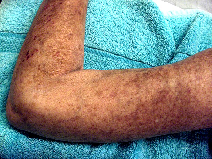
Image Source: Wikimedia Commons (2022). Left Arm Scleroderma Patient, Licensed under a creative commons licence. Available for free at: https://upload.wikimedia.org/wikipedia/commons/6/60/Left_Arm_Scleroderma_Patient.jpg (Accessed: 10 July 2022).
Raynaud’s phenomenon occurs in more than 90% of patients with systemic sclerosis; however, Raynaud’s phenomenon may occur in isolation and, as such, is referred to as primary Raynaud’s phenomenon. However, there are no digital ulcerations or ischemia associated with primary RP.

Image Source: Wikimedia Commons (2022). Raynaud syndrome on female airman’s hand, Licensed under a creative commons licence. Available for free at: https://upload.wikimedia.org/wikipedia/commons/1/1f/Raynaud_syndrome_on_female_airman%27s_hand.jpg (Accessed: 10 July 2022).
Management
The management of systemic sclerosis depends on the disease subset and the presence or absence of internal organ involvement.
Patients with diffuse skin involvement or inflammatory organ involvement are treated with systemic immunosuppressive therapy. These patients may have skin involvement that is severe or diffuse, interstitial lung disease, myocarditis, or arthritis.
Methotrexate and mycophenolate mofetil are both used in patients who exhibit diffuse and progressive skin involvement but no visceral involvement. Generally, methotrexate is used in patients with arthritis or myositis, while mycophenolate mofetil is used in those who have significant interstitial lung disease. For patients who are refractory to the use of these, cyclophosphamide is usually the next step.
In cases of renal sclerosis, the mainstay of treatment is angiotensin-converting enzyme inhibitors (ACE-i). Initially, Captopril may be stated acutely for example, in renal scleroderma crisis within the first 72 hours, after which ACE-i such as Enalapril or Ramipril are used for maintenance. It is essential to closely monitor the serum creatinine (small, transient increases are expected and not usually an indication to stop therapy). There is no evidence that supports the use of ARBs or non-ACE-i therapy.
Gastrointestinal manifestations are treated symptomatically. For instance, proton pump inhibitors are used for oesophageal reflux and gastritis and can be titrated to up to twice daily. Oesophageal dysmotility, which may present with dysphagia, can be treated using prokinetic drugs such as Domperidone and Metoclopramide. Small intestinal bacterial overgrowth is another sequela and is usually treated with antibiotics, namely Metronidazole.
Raynaud’s phenomenon, either because of systemic sclerosis or primary RP, is treated with long-acting dihydropyridine calcium channel blockers such as Amlodipine or Nifedipine. If calcium channel blockers are not tolerated or ineffective, phosphodiesterase type 5 inhibitors such as Sildenafil may be used. Topical nitrates can be considered for patients with ischemic digits as a result of RP. However, it is not recommended to use topical nitrates with PDE 5 inhibitors due to the risk of hypotension.
Arthralgia and arthritis are treated initially with NSAIDs or Paracetamol for symptomatic relief. However, if these are contraindicated or ineffective, alternatives such as low-dose prednisone or hydroxychloroquine can be used. Methotrexate is another option, but only in those patients who are not at risk of pulmonary fibrosis.
References
- Denton C. UpToDate. Pathogenesis of systemic sclerosis (scleroderma) [Internet]. 2021 [cited 2022 March 23]. Available from https://www.uptodate.com/contents/pathogenesis-of-systemic-sclerosis-scleroderma?search=systemic%20sclerosis%20diagnosis&topicRef=7539&source=see_link#H25
- Pearson D, Werth V, Taffer L. Systemic sclerosis: Current concepts of skin and systemic manifestations. Clinics in Dermatology. 2018; 36(4): 459-474. Available from https://www.sciencedirect.com/science/article/abs/pii/S0738081X18300634
- Hughes M , Allanore Y, Denton C, Matucci-Cerinic M. Systemic Sclerosis.
- EMJ Rheumatol. 2020;7[1]:100-109. Available from https://www.emjreviews.com/rheumatology/article/systemic-sclerosis/
- Saracino A, Denton C, Oakley A. DermNetNZ. Systemic Sclerosis [Internet]. 2020 [cited March 30]. Available from: https://dermnetnz.org/topics/systemic-sclerosis
- Varga J. Clinical manifestations and diagnosis of systemic sclerosis (scleroderma) in adults [Internet]. 2021 [cited March 30]. Available from: https://www.uptodate.com/contents/clinical-manifestations-and-diagnosis-of-systemic-sclerosis-scleroderma-in-adults?search=systemic%20sclerosis%20diagnosis&source=search_result&selectedTitle=1~150&usage_type=default&display_rank=1#H6
- Denton C. UpToDate. Overview of the treatment and prognosis of systemic sclerosis (scleroderma) in adults[Internet]. 2021 [Cited 2022 April 10]. Available from: https://www.uptodate.com/contents/overview-of-the-treatment-and-prognosis-of-systemic-sclerosis-scleroderma-in-adults?search=management%20of%20systemic%20sclerosis&source=search_result&selectedTitle=1~150&usage_type=default&display_rank=1
- Varga J, Fenves A. UpToDate. Kidney disease in systemic sclerosis (scleroderma), including scleroderma renal crisis [Internet]. 2021 [Cited 2022 April 5]. Available from: https://www.uptodate.com/contents/kidney-disease-in-systemic-sclerosis-scleroderma-including-scleroderma-renal-crisis?sectionName=Treatment&search=management%20of%20systemic%20sclerosis&topicRef=7542&anchor=H18&source=see_link#H19
- Kaye-Barrett S, Denton C. UpToDate. Treatment of gastrointestinal disease in systemic sclerosis (scleroderma) [Internet]. 2021 [cited 2022 April 4]. Available from: https://www.uptodate.com/contents/treatment-of-gastrointestinal-disease-in-systemic-sclerosis-scleroderma?search=management%20of%20systemic%20sclerosis&topicRef=7542&source=see_link
Complete Guide to Clinical Genetics ST3 Application and Recruitment (2027)
Written by Medibuddy. Last updated: October 2026. Clinical Genetics is

Complete Guide to Clinical Radiology ST3 Application and Recruitment (2027)
Recruitment paused: NHS England is not running Clinical Radiology ST3+ recru

Complete Guide to Paediatric Surgery ST3 Application and Recruitment in United Kingdom (2026)
Written by Medibuddy. Last updated: October 2026. If you’re planning


Take your subscriptions with you
Our mobile app allows you to access your interview and exam question banks wherever you are.

