


 Close
Close
 21st February 2024
Extraocular Muscles
The extraocular muscles are a group of six muscles responsible for the precise movement and control of the eyeball within the orbit. They are attached to the wall of the orbit and work in conjunction to allow coordinated eye movements, maintaining binocular vision.
The recti muscles (superior, inferior, lateral and medial) all originate from the common tendinous ring, a ring of fibrous tissue found at the back of the orbit which surrounds the optic canal. The oblique muscles (superior and inferior) take an angled approach into the eyeball and attach to the posterior surface of the sclera.
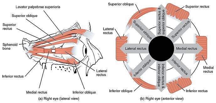
Image Source: Wikimedia Commons (2015). 1107 The Extrinsic Eye Muscles right eye lat, Licensed under a creative commons licence. Available for free at: https://commons.wikimedia.org/wiki/File:1107_The_Extrinsic_Eye_Muscles_right_eye_lat.png (Accessed: 28 September 2023).
Superior Rectus
Originates from the superior part of the common tendinous ring and attaches to the superior and anterior sclera. Its main function is to elevate, and it also contributes to adduction and medial rotation. Its supply is the oculomotor nerve.
Inferior Rectus
Originates from the inferior part of the common tendinous ring and attaches to the inferior and anterior sclera. Its main function is to depress the eyeball, but it also contributes to adduction and lateral rotation. It is supplied by the oculomotor nerve.
Medial Rectus
Originates from the medial part of the common tendinous ring and attaches to the medial and anterior sclera. Its main function is to adduct the eyeball. It is supplied by the oculomotor nerve.
Lateral Rectus
Originates from the lateral part of the common tendinous ring and attaches to the lateral and anterior sclera. Its main function is to abduct the eyeball. It is supplied by the abducens nerve.
Superior Oblique
Originates from the sphenoid bone; its tendon passes through a trochlea and attaches just behind the superior rectus. Its main function is to depress, abduct and medially rotate the eyeball.
Inferior Oblique
Originates from the inferomedial aspect of the orbital floor, passes above the inferior rectus and attaches posteriorly to the lateral rectus.
Complete Guide to Clinical Genetics ST3 Application and Recruitment (2027)
Written by Medibuddy. Last updated: October 2026. Clinical Genetics is

Complete Guide to Clinical Radiology ST3 Application and Recruitment (2027)
Recruitment paused: NHS England is not running Clinical Radiology ST3+ recru

Complete Guide to Paediatric Surgery ST3 Application and Recruitment in United Kingdom (2026)
Written by Medibuddy. Last updated: October 2026. If you’re planning


Take your subscriptions with you
Our mobile app allows you to access your interview and exam question banks wherever you are.

