


 Close
Close
 4th June 2023
Anatomy of the cerebral cortex
The brain, along with the spinal cord, is the main organ in the central nervous system. The brain is made up of the cerebrum, cerebellum, and brainstem. The cerebrum is made up of two hemispheres, left and right. Each hemisphere is further divided into the cerebral cortex and structures beneath it known as subcortical structures. The cerebral cortex is divided in to six lobes. These are the frontal, temporal, parietal, occipital, insular and limbic lobes (see image below). Each of these lobes is associated with specific functions. As a result, lesions in a particular part of the brain will have specific symptoms and signs.

Lobes of the brain.
Image Source: Wikimedia Commons (2014). Blausen 0101 Brain Lateral View, Licensed under a creative commons licence. Available for free at: https://commons.wikimedia.org/wiki/File:Blausen_0101_Brain_LateralView.png (Accessed on: 11 January 2023).

| Lobe | Function |
|---|---|
| Frontal | Control of voluntary movement, involved in attention, short term memory tasks, motivation, planning, speech |
| Parietal | Integrates proprioceptive and mechanoreceptive stimuli, involved in language processing |
| Temporal | Processing visual and auditory sensory input for visual memory and language comprehension |
| Occipital | Visual processing |
| Insular | Processing and integration of taste sensation, visceral and pain sensation, and vestibular functions |
| Limbic | Modulation of emotions, visceral and autonomic functions, learning, memory |
Table 1: Lobes of the brain and their function
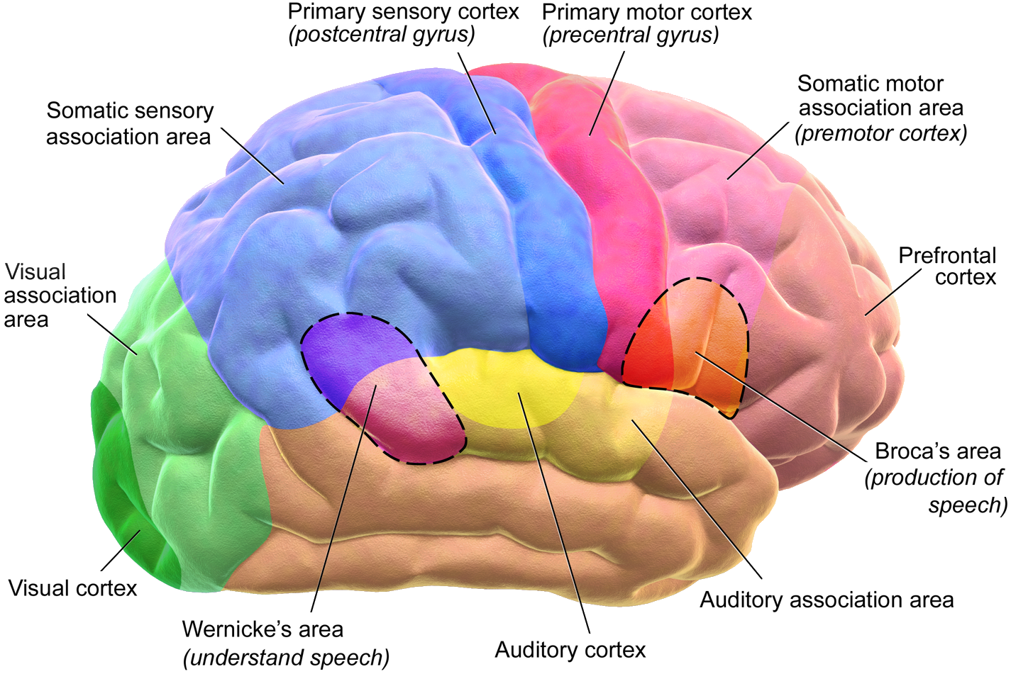
Functional areas of the brain. Dashed areas shown are commonly left hemisphere dominant.
Image Source: Wikimedia Commons (2014). Blausen 0102 Brain Motor & Sensory (flipped), Licensed under a creative commons licence. Available for free at: https://commons.wikimedia.org/wiki/File:Blausen_0102_Brain_Motor%26Sensory_%28flipped%29.png (Accessed on: 11 January 2023).

Knowing the function of the different lobes of the brain helps to localise the site of damage in the CNS based on the patient’s signs and symptoms. It is important to note Wernicke’s and Broca’s areas (see image above) as these are important when thinking about the localisation of lesions in patients presenting with aphasia.
This may be receptive aphasia, affecting reading, listening (seen in damage to Wernicke’s area) or expressive aphasia, affecting writing and talking (seen in damage to Broca’s area).
For aphasia to occur, the lesion must be in the dominant hemisphere; right-handed individuals will invariably be left-hemisphere dominant; the majority of left-handed individuals will also be left-hemisphere dominant, however, there is an increased proportion than right-handers of having a dominant right hemisphere. Lesions to the corresponding area of the non-dominant hemisphere will result in visual or auditory inattention to the opposite side.
Complete Guide to Clinical Genetics ST3 Application and Recruitment (2027)
Written by Medibuddy. Last updated: October 2026. Clinical Genetics is

Complete Guide to Clinical Radiology ST3 Application and Recruitment (2027)
Recruitment paused: NHS England is not running Clinical Radiology ST3+ recru

Complete Guide to Paediatric Surgery ST3 Application and Recruitment in United Kingdom (2026)
Written by Medibuddy. Last updated: October 2026. If you’re planning


Take your subscriptions with you
Our mobile app allows you to access your interview and exam question banks wherever you are.

