


 Close
Close
 20th February 2024
Methods of gene analysis
Background
DNA analysis is critical to aid with the diagnosis of genetic disorders. Recent developments in scientific methods have altered how genetic mutations are investigated, and this remains an evolving field. Candidates are not expected to know specific details but to understand the basic techniques of detecting genetic mutations.
Samples of a patient’s tissue are required for testing, and this can be obtained from blood, buccal mucosa or within affected tissues. Prenatal testing for foetal genetic abnormalities can be performed on amniotic fluid or chorionic villus samples. Genetic testing can be carried out in a variety of methods:
Karyotyping
Karyotyping involves whole chromosomal analysis using microscopy. Cells are cultured within a medium, and colchicine arrests cell division during metaphase. The chromosomes are made visible using various stains and arranged based on their appearance to form the karyotype.
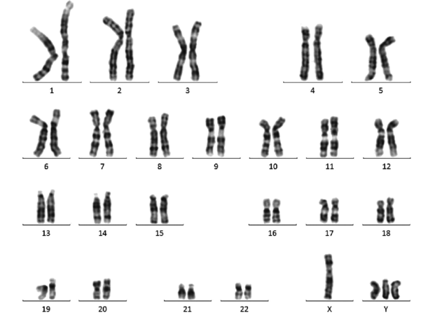
Image Source: Wikimedia Commons (2017). Peripheral blood karyotype result belonging to the subject compatible with 48-XYYY, Licensed under a creative commons licence. Available for free at: https://commons.wikimedia.org/wiki/File:Peripheral-blood-karyotype-result-belonging-to-the-subject-compatible-with-48-XYYY.png (Accessed: 4 January 2024).
Karyotypes have a role in identifying large genetic abnormalities, including aneuploidy, reciprocal and Robertsonian translocation, and large microdeletions or duplications. Its use has largely been superseded by microarray analysis but still has a role in identifying balanced translocations.
Nucleic acid hybridisation
Hybridisation detects mutations using single-stranded DNA probes to detect specific DNA/RNA sequences within the genetic material.
Several methods utilise hybridisation to detect mutations:
- Southern blotting – DNA samples are broken down into smaller fragments, and electrophoresis is employed to separate DNA fragments based on size. DNA probes with a marker are applied to allow hybridisation, and chemiluminescence or X-rays are used to detect the probes. This only detects significant mutations or DNA sequences that PCR cannot amplify.
- Fluorescence in-situ hybridisation (FISH) – Single-stranded DNA probes are exposed to denatured chromosomes within the interphase, prophase or metaphase stage of cell division, and fluorescence is used to detect its bonding to sample DNA. FISH occasionally detect aneuploidies, translocations, and microdeletions/duplications.
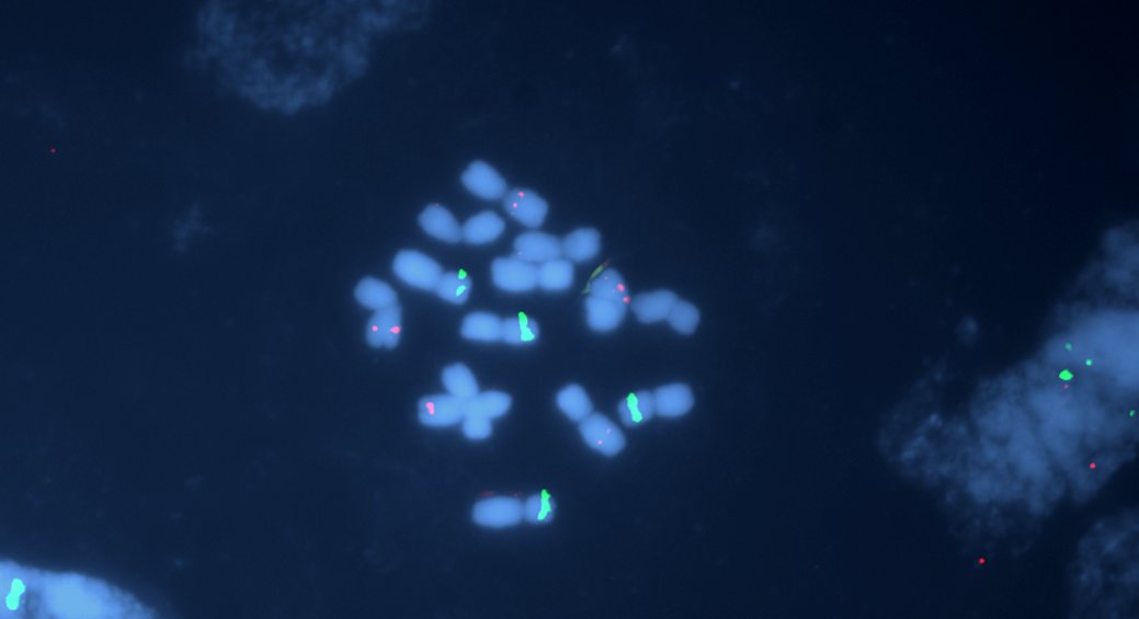
Image Source: Wikimedia Commons (2009). Hordeum vulgare stained by fluorescent in situ hybridization, Licensed under a creative commons licence. Available for free at: https://commons.wikimedia.org/wiki/File:Hordeum_vulgare_stained_by_fluorescent_in_situ_hybridization.jpg (Accessed: 4 January 2024).
- Array comparative genomic hybridisation (aCGH) – A microarray containing thousands to millions of oligonucleotide probes is exposed to a sample and a control DNA sample. Both DNA samples are marked with a fluorescent dye and are compared under a fluorescence microscope. This method detects aneuploidy, deletions, duplications (both large and small) and unbalanced translocations. It is possible to detect single nucleotide polymorphism with different probes. aCGH rarely detects balanced translocation as no excess genetic material exists within the sample DNA.

Image Source: Wikimedia Commons (2012). Array-CGH protocol, Licensed under a creative commons licence. Available for free at: https://commons.wikimedia.org/wiki/File:Array-CGH_protocol.svg (Accessed: 4 January 2024).
Polymerase chain reaction (PCR)
PCR is performed by denaturing the sample DNA and annealing a DNA primer to the denatured DNA to allow expression of the sequence of interest. DNA polymerase is used to amplify the sequence, exponentially increasing the amount of the DNA sequence of interest available. PCR is often only successful in sequences within the hundreds to thousands of base pairs, with sequences over 20kbp almost impossible to amplify.
Gene analysis uses various methods, including electrophoresis, restriction enzyme analysis, allele-specific oligonucleotide hybridisation and multiplex ligation-dependant probe amplification. PCR is versatile, detecting insertions, deletions, point mutations and trinucleotide expansion mutations.
DNA sequencing
DNA sequencing determines the precise sequence of A, C, T and G nucleotides within the gene of interest. It is helpful in detecting insertion, deletion, point mutations and nucleotide polymorphisms. At its core, sequencing is performed using either the Sanger method or next-generation sequencing (NGS).
The Sanger method analyses a DNA sequence by starting with multiple DNA strands and a primer to target a sequence of interest. Dideoxynucleoside molecules terminate the nucleotide chain during the reaction, creating multiple chains of DNA with differing lengths. Each dideoxynucleoside is attached to its fluorescent dye, and a computer program compares each fragment’s size and the fluorescent dye present to determine the precise DNA sequence. This process is labour-intensive but allows a specific gene to be sequenced accurately.
NGS allows rapid sequencing of an entire genome. The genome is divided into millions of fragments, each up to several hundred base pairs in size. Fragments are amplified using PCR, complementary base pairs are attached, and laser scanning helps to identify the fluorescent dye. It is useful to detect rare genetic diseases caused by mutations of various genes.
References
- Ratan, Z. A., Zaman, S. B., Mehta, V., Haidere, M. F., Runa, N. J., & Akter, N. (2017). Application of Fluorescence in Situ Hybridization (FISH) Technique for the Detection of Genetic Aberration in Medical Science. Cureus, 9(6), e1325. https://doi.org/10.7759/cureus.1325
- Wallace SE, Bean LJH. Educational Materials — Genetic Testing: Current Approaches. 2017 Mar 14 [Updated 2020 Jun 18]. In: Adam MP, Ardinger HH, Pagon RA, et al., editors. GeneReviews® [Internet]. Seattle (WA): University of Washington, Seattle; 1993-2021. Available from: https://www.ncbi.nlm.nih.gov/books/NBK279899/
- Gomes, A., & Korf, B. (2018). Chapter 5 – Genetic Testing Techniques. In N. H. Robin & M. B. Farmer (Eds.), Pediatric Cancer Genetics (pp. 47-64): Elsevier.
- Turnpenny, P. D., & Ellard, S. (2017). Laboratory Techniques for Diagnosis of Monogenic Disorders. In Emery’s Elements of Medical Genetics. China: Elsevier.
- Kingston, H. M. (2002). Techniques of DNA analysis. In ABC of Clinical Genetics. Malaysia: BMJ Books.
- Jorde, L. B., Carey, J. C., & Bamshad, M. J. (2016). Genetic Variation: Its Origin and Detection. In Medical Genetics (5ᵗʰ ed.). Canada: Elsevier.
- Read, A., & Donnai, D. (2021). Chapter 4: How can a patient’s DNA be studied? In New Clinical Genetics: A guide to genomic medicine (4ᵗʰ ed.). United Kingdom: Scoin Publishing Limited.
Complete Guide to Clinical Genetics ST3 Application and Recruitment (2027)
Written by Medibuddy. Last updated: October 2026. Clinical Genetics is

Complete Guide to Clinical Radiology ST3 Application and Recruitment (2027)
Recruitment paused: NHS England is not running Clinical Radiology ST3+ recru

Complete Guide to Paediatric Surgery ST3 Application and Recruitment in United Kingdom (2026)
Written by Medibuddy. Last updated: October 2026. If you’re planning


Take your subscriptions with you
Our mobile app allows you to access your interview and exam question banks wherever you are.

