


 Close
Close
 21st July 2023
Osteoporosis and Fragility Fracture
Introduction
Osteoporosis is a disease of significantly reduced bone mineral density (BMD) that increases the risk of fractures, primarily due to old age. Secondary causes include high dose steroid use, hyperthyroidism, hyperparathyroidism, increased alcohol intake, smoking, BMI <18.5 kg/m², decreased testosterone, early menopause, kidney failure and liver failure. It is asymptomatic, with many people being diagnosed following a fragility fracture.
Fragility fractures occur secondary to low trauma events such as a fall from a standing height. Examples of these fractures include pubic rami or neck of femur fractures, vertebral fractures (see image below) (often appearing as vertebral collapse on imaging) and distal radial fractures (e.g., Smith or Colles fracture). It is important to investigate secondary causes, which include osteoporotic and non-osteoporotic causes, such as multiple myeloma, Paget’s disease, and metastatic bone cancer.

T12 osteoporotic compression fracture
Image Source: Wikimedia Commons (2016). T12compressionfracMark, Licensed under a creative commons licence. Available for free at: https://commons.wikimedia.org/wiki/File:T12compressionfracMark.png (Accessed: 2 March 2022).
Diagnosis
The World Health Organisation (WHO) defines osteoporosis as a Dual Energy X-ray Absorptiometry (DEXA) scan T-score of less than -2.5 or a geriatric patient presenting with a low trauma fracture. DEXA scans are the only diagnostic investigation. This uses low dose x-rays to measure BMD, typically of the vertebra, pelvis, and femur. T-scores on a DEXA scan are a standard deviation of the BMD compared to that of an average healthy 30-year-old adult. The Z-score on a DEXA scan is a standard deviation of the BMD adjusted to the patient’s age and gender.
Tools can be used to predict the risk of a patient suffering from an osteoporotic fracture, such as the Fracture Risk Assessment Tool (FRAX®) or Q Risk (fracture risk) calculator. These tools consider a person’s risk factors to calculate their 10-year probability of having a major osteoporotic or hip fracture.
NICE recommends DEXA scans for those over 50 years with a history of a fragility fracture or those <40 years with a high-risk factor for osteoporosis (as the FRAX® tool is not used in those <40 years). Those >40 years with risk factors (without a fragility fracture) require a FRAX® score, for which a fracture risk of greater than 10% would indicate the need for a DEXA scan.
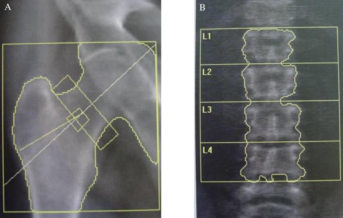
DEXA scan images of pelvic and L1-L4 vertebrae
Image Source: Wikimedia Commons (2011). Morbus Fabry DXA 01, Licensed under a creative commons licence. Available for free at: https://commons.wikimedia.org/wiki/File:Morbus_Fabry_DXA_01.jpg (Accessed: 17 February 2022).
Due to the potential differential diagnoses and secondary causes of osteoporosis, it is important to conduct a full history, physical examination, and blood tests in addition to the above.
Blood tests would be unremarkable in osteoporosis in primary osteoporosis.
Management
Initial non-pharmacological treatment:
- Treating any underlying or secondary causes
- Calcium and vitamin D supplements
- Reduce modifiable risk factors with lifestyle choices
Pharmacological treatment:
This usually commenced in men over 50 or post-menopausal women with a DEXA scan T-score result in keeping with osteoporosis. It is also given to people with a high risk of fracture according to the FRAX® score or to those with intermediate risk if awaiting a DEXA scan will significantly delay treatment as recommended by the National Osteoporosis Guideline Group (NOGG). In other specific circumstances, treatment is usually started by specialists, such as when patients are commenced on high dose prednisolone. Treatment options are listed below, with each medication tried individually and not in combination.
- Bisphosphonates – synthetic analogues of bone pyrophosphate. First line treatment is initially given orally (alendronate, risedronate or ibandronate). It can also be given as a one-yearly intravenous infusion of zoledronic acid. Due to the risk of atypical fractures with long term bisphosphonates, they are usually reviewed after taking the medication for 3-5 years.
- Denosumab – 6 monthly subcutaneous injections, monoclonal antibody, RANK Ligand inhibitor.
- Teriparatide – daily subcutaneous synthetic parathyroid hormone (PTH)
- Raloxifene – a selective oestrogen receptor modulator. Used in post-menopausal women.
- Strontium Ranelate – stimulates bone formation and reduces bone resorption
Complete Guide to Clinical Genetics ST3 Application and Recruitment (2027)
Written by Medibuddy. Last updated: October 2026. Clinical Genetics is

Complete Guide to Clinical Radiology ST3 Application and Recruitment (2027)
Recruitment paused: NHS England is not running Clinical Radiology ST3+ recru

Complete Guide to Paediatric Surgery ST3 Application and Recruitment in United Kingdom (2026)
Written by Medibuddy. Last updated: October 2026. If you’re planning


Take your subscriptions with you
Our mobile app allows you to access your interview and exam question banks wherever you are.

