


 Close
Close
 26th June 2023
Types of brain tumours
Glioma
Gliomas originate from glial cells. Glial cells are non-neuronal cells that have many functions involving neuronal pruning, producing myelin and modulating neuronal growth. Glial cells include astrocytes, oligodendrocytes, and ependymal cells. Tumours of these cells can give rise to astrocytomas or glioblastomas, oligodendrogliomas, or ependymomas, respectively. Astrocytomas are by far the most common type of glioma in both adults and children; they can be low grade (Grade 1 and 2 – slow-growing) or high grade (Grade 3 and 4 – fast-growing). Grade 4 astrocytomas are more commonly called glioblastoma multiforme (GBM). The imaging shows a ring enhancing lesion with central necrosis, typical of GBM. Prognosis for GBM is poor; the average survival time is just 12-18 months.

Glioma on MRI brain.
Image Source: Wikimedia Commons (2009). Glioma, Licensed under a creative commons licence. Available for free at: https://commons.wikimedia.org/wiki/File:Glioma.gif (Accessed on: 30 January 2023).

Meningioma
Meningiomas are slow-growing tumours arising from the lining (meninges) that encapsulates the brain and spinal cord. This is a benign tumour that rarely metastasises and is usually monitored with surgical resection if impeding on neurological structures. Meningiomas account for 25% of all brain tumours.

Meningioma on CT.
Image Source: Wikimedia Commons (2007). Contrast enhanced meningioma, Licensed under a creative commons licence. Available for free at: https://commons.wikimedia.org/wiki/File:Contrast_enhanced_meningioma.jpg (Accessed on: 11 January 2023).

Vestibular Schwannoma
These are slow-growing tumours that form along the vestibulocochlear nerve (CNVIII). These are benign tumours of vestibulocochlear Schwann cells. Patients may present with tinnitus, unilateral sensorineural hearing loss, vestibular dysfunction causing dizziness and rarely ipsilateral disturbances of facial pain (CNV) and facial weakness (CNVII). These tumours can be managed conservatively by MRI monitoring; however, if compressing adjacent structures, surgical resection and stereotactic radiosurgery are the main management options.

Vestibular Schwannoma on MRI brain
Image Source: Wikimedia Commons (2008). Vestibular-schwannoma-003, Licensed under a creative commons licence. Available for free at: https://commons.wikimedia.org/wiki/File:Vestibular-schwannoma-003.jpg (Accessed on: 30 January 2023).

Medulloblastoma
Medulloblastomas are high-grade solid tumours located in the cerebellum, usually in children. Metastases can occur within the CSF to other parts of the brain and spinal cord; therefore, a full spine MRI is important as metastases are present in 1/3rd of children at presentation. Treatment is tumour-specific, often involving radiation, chemotherapy, and surgery.

Medulloblastoma on MRI brain
Image Source: Wikimedia Commons (2005). CT brain scan of child with medulloblastoma and resulting hydrocephalus, Licensed under a creative commons licence. Available for free at: https://commons.wikimedia.org/wiki/File:CT_brain_scan_of_child_with_medulloblastoma_and_resulting_hydrocephalus.jpg (Accessed on: 30 January 2023).

Craniopharyngioma
Arising from the pituitary stalk, these benign tumours can affect the production of hormones from the hypothalamus or pituitary glands and may even present as pituitary insufficiency. As the tumour site is situated above the optic chiasm, patients may develop a bitemporal inferior quadrantinopia, which may progress to a bitemporal hemianopia as the tumour grows. Clinical features may include headache, visual changes, excessive urination and dehydration (due to diabetes insipidus), delayed puberty in children or sexual dysfunction in adults, symptoms of hypothyroidism such as weight gain, or symptoms of adrenal insufficiency.

Craniopharyngioma on MRI brain.
Image Source: Wikimedia Commons (2011). Craniopharyngioma-t1sagkm-006, Licensed under a creative commons licence. Available for free at: https://commons.wikimedia.org/wiki/File:Craniopharyngioma-t1sagkm-006.jpg (Accessed on: 11 January 2023).

Pituitary Adenoma
Pituitary adenomas are benign tumours arising from the pituitary gland (not to be confused with craniopharyngiomas which arise from the pituitary stalk, above the pituitary gland). They mostly affect the anterior lobe of the pituitary and are the most common cause of hypopituitarism. About half of pituitary adenomas do not secrete hormones and are called ‘non-functioning’ adenomas; however, they can still produce symptoms resulting from compression of surrounding structures, e.g. bitemporal hemianopia due to compression of the optic chiasm. Functioning adenomas secrete hormones and can result in symptoms of the following depending on the hormone secreted:
- Cushingoid symptoms
- Acromegaly
- Diabetes insipidus
- Hyperpituitarism
- Amenorrhoea/galactorrhoea/impotence
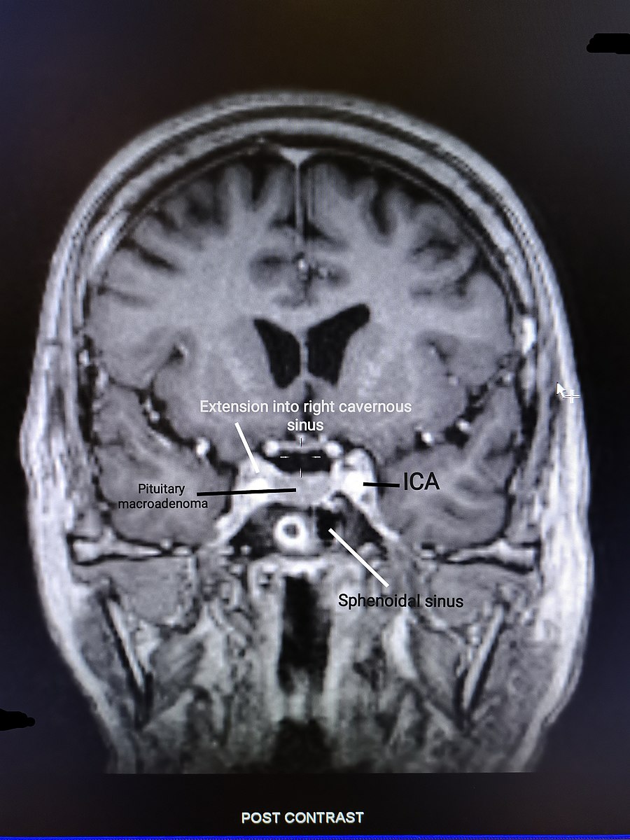
Pituitary adenoma on MRI brain
Image Source: Wikimedia Commons (2022). Coronal section of non-functioning pituitary adenoma, Licensed under a creative commons licence. Available for free at: https://commons.wikimedia.org/wiki/File:Coronal_section_of_non-functioning_pituitary_adenoma.jpg (Accessed on: 30 January 2023).

Haemangioblastoma
Haemangioblastomas are slow-growing benign tumours of the vascular system; they predominantly affect the cerebellum but can occasionally affect other sites, including the spinal cord and cerebrum. Von Hippel-Lindau syndrome is a rare inherited condition which is characterised by the formation of tumours and visceral cysts; haemangioblastomas are characteristic of Von Hippel-Lindau syndrome.
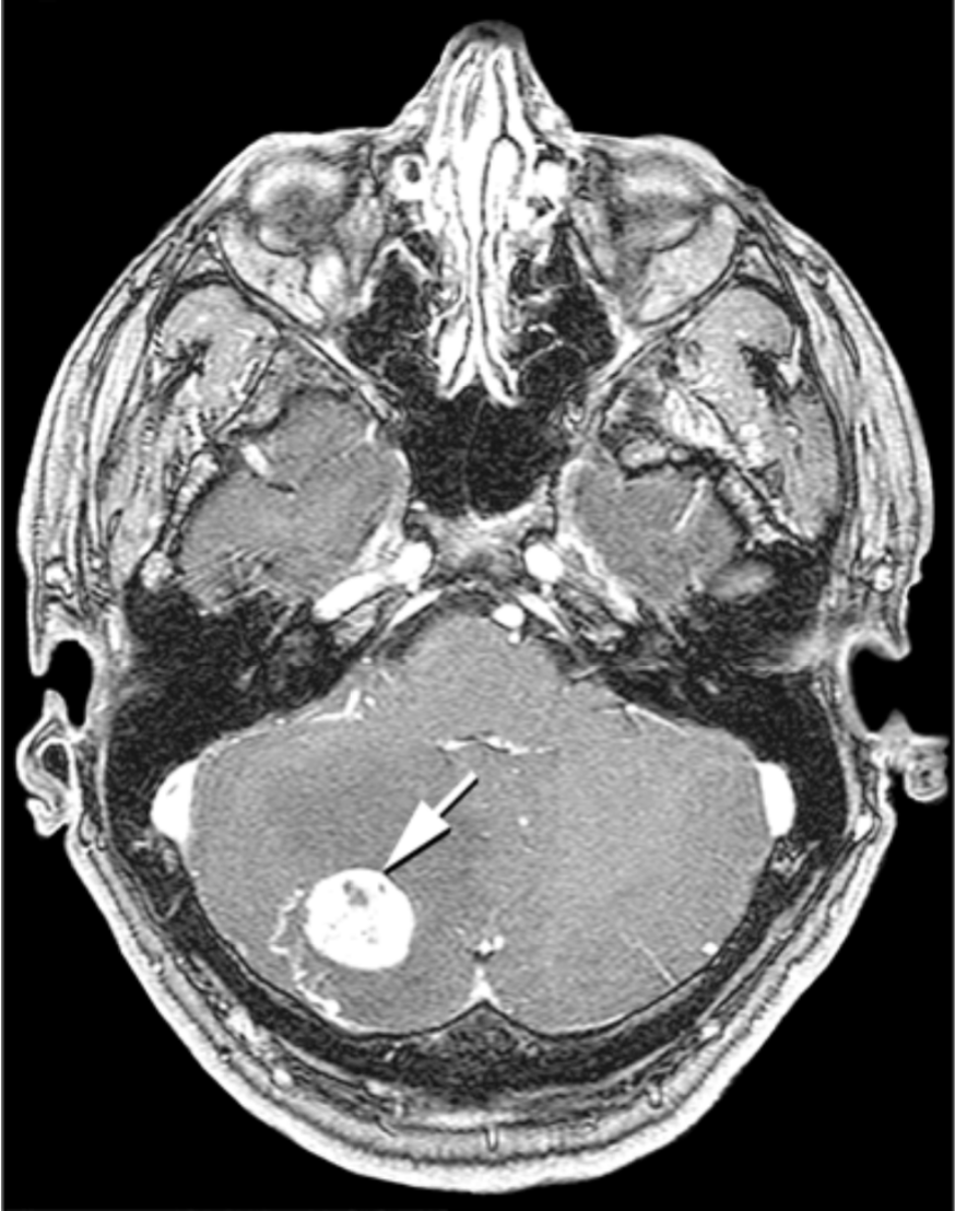
Cerebellar haemangioma on MRI.
Image Source: Wikimedia Commons (2007). Hippel Lindau, Licensed under a creative commons licence. Available for free at: https://commons.wikimedia.org/wiki/File:Hippel_Lindau.gif (Accessed on: 30 January 2023).

Complete Guide to Clinical Genetics ST3 Application and Recruitment (2027)
Written by Medibuddy. Last updated: October 2026. Clinical Genetics is

Complete Guide to Clinical Radiology ST3 Application and Recruitment (2027)
Recruitment paused: NHS England is not running Clinical Radiology ST3+ recru

Complete Guide to Paediatric Surgery ST3 Application and Recruitment in United Kingdom (2026)
Written by Medibuddy. Last updated: October 2026. If you’re planning


Take your subscriptions with you
Our mobile app allows you to access your interview and exam question banks wherever you are.

