


 Close
Close
 16th February 2024
Cardiac system
Pericardium
There are two layers of the pericardium: the outer tough fibrous (parietal) pericardium and the inner membranous serous pericardium, with a trace of pericardial fluid between the layers. This acts as a physical barrier between the heart and the other organs. It also prevents overfilling and fixes the heart within the mediastinum.
Cardiac anatomy
The heart consists of four chambers: two atria and two ventricles. The right atrium receives deoxygenated blood from the body via the superior and inferior vena cava and pumps into the right ventricle through the tricuspid valve. The right ventricle then pumps this blood through the pulmonary trunk into the pulmonary circulation for oxygenation, and the oxygenated blood is returned via the pulmonary veins into the left atrium.
Blood flows from the left atrium through the mitral valve into the left ventricle. Then, this oxygenated blood is supplied to the body, starting from the aorta to arteries, arterioles and the capillaries. The atria contract, and the AV valves (mitral and tricuspid) open in diastole. The ventricles contract, and outlet valves (Pulmonic and aortic) open in systole.
The heart’s conduction system begins at the sinoatrial node within the right atrial wall. The pacemaker cells spontaneously generate an electrical impulse, and these travel across the atrial walls. The impulses arrive at the atrioventricular (AV) node within the atrioventricular wall septum close to the coronary sinus. The impulses will travel from the AV node into the atrioventricular bundle (Bundle of His). The AV bundle will divide into the left and right bundle branches to supply the left and right ventricles. These bundles terminate as the Purkinje fibres as the electrical impulses are transmitted across the ventricular walls.

Image Source: Wikimedia Commons (2015). Cardiac conduction system, Licensed under a creative commons licence. Available for free at: https://upload.wikimedia.org/wikipedia/commons/3/3d/Cardiac_conduction_system.jpg (Accessed: 9 January 2024).
Cardiac blood supply
The blood supply to the heart is by the left and right coronary arteries. The left coronary artery comes off the left coronary cusp, while the right coronary artery comes off the right coronary cusp above the aortic valve. The left coronary artery starts as the left main stem and then divides into the left anterior descending (LAD) and left circumflex artery. The LAD lies within the anterior interventricular groove and travels towards the apex of the heart. The left marginal artery travels along the coronary sulcus towards the posterior heart surface.
The right coronary artery travels along the coronary sulcus, with the right marginal artery branching off the artery. The right coronary artery continues as the posterior interventricular artery within the posterior interventricular groove.
The distribution of the arteries’ blood supply in a typical right-dominant system is as follows:
- Right coronary artery – Right atrium, SA node, AV node
- Right marginal artery – Lateral right ventricular wall.
- Right interventricular artery – Inferior aspect of the heart
- LAD – Anterior interventricular septum and anterior left ventricular wall
- Left circumflex artery – Lateral left ventricular wall
Venous return from the heart is through the coronary sinus into the right atrium. The main tributaries for the coronary sinus include the great cardiac, small, middle, and posterior cardiac veins.
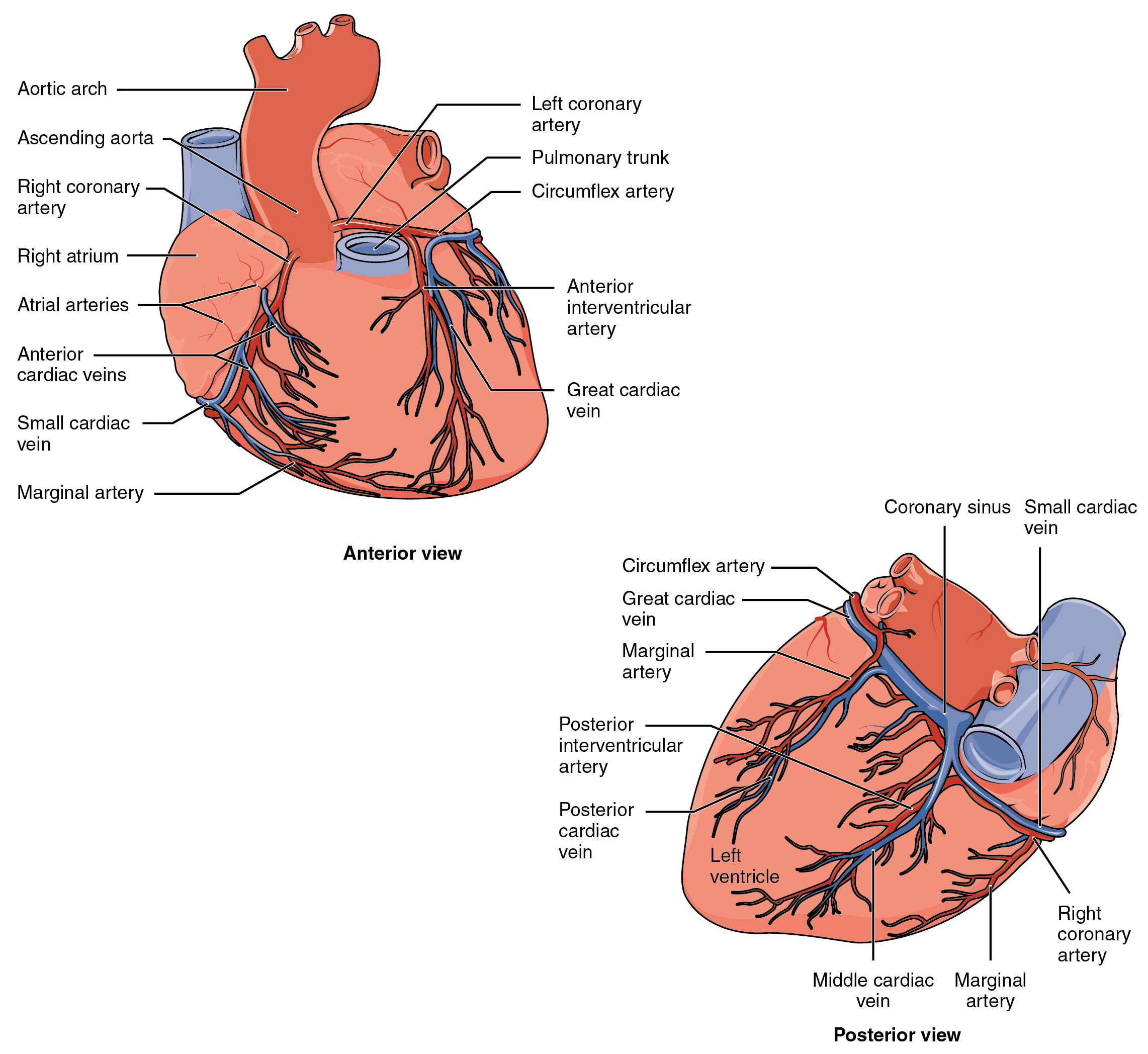 Image Source: Wikimedia Commons (2013). 2014ab Coronary Blood Vessels, Licensed under a creative commons licence. Available for free at: https://upload.wikimedia.org/wikipedia/commons/7/7c/2014ab_Coronary_Blood_Vessels.jpg (Accessed: 9 January 2024).
References
Wineski, L. E., & Snell, R. S. (2019). Snell’s clinical anatomy by regions.
Chaurasia, B. D. (2019). Human anatomy: Regional and applied, dissection and clinical. New Delhi, India: CBS Publishers & Distributors.
Moore, K. L., Dalley, A. F., & Agur, A. M. R. (2014). Moore’s Clinically Orientated Anatomy (7th ed.). China: Lippincott.
Complete Guide to Clinical Genetics ST3 Application and Recruitment (2027)
Written by Medibuddy. Last updated: October 2026. Clinical Genetics is

Complete Guide to Clinical Radiology ST3 Application and Recruitment (2027)
Recruitment paused: NHS England is not running Clinical Radiology ST3+ recru

Complete Guide to Paediatric Surgery ST3 Application and Recruitment in United Kingdom (2026)
Written by Medibuddy. Last updated: October 2026. If you’re planning


Take your subscriptions with you
Our mobile app allows you to access your interview and exam question banks wherever you are.

