


 Close
Close
 6th March 2024
Arrhythmias
Background
Any aberration of the normal cardiac rhythm is considered a cardiac arrhythmia. This may be because of a change in rate (normal heart rate 60-100 bpm) or rhythm, in terms of regularity or frequency of either atrial and or ventricular impulses. Cardiac arrhythmia can be benign or life-threatening through haemodynamic instability.
Normal cardiac rhythm refers to electrical activity originating from the sino-atrial (SA) node within the posterior wall of the right atrium. This spreads across the atria via internodal pathways and Bachmann’s bundle, allowing for atrial contraction. The conduction arrives at the atrio-ventricular (AV) node where it is delayed allowing for sufficient ventricular filling. It continues along the bundle of His, the left and right bundle branches; the left divides into two fascicles (anterior and posterior) and the right into one fascicle, to reach the Purkinje fibres resulting in ventricular contraction.
Any deviation from this, such as disease of SA or AV node, accessory pathways, or ectopic pacemaker activity, will result in a cardiac arrhythmia that may manifest with symptoms of palpitations, breathlessness, syncope/presyncope or cardiac arrest. Common arrhythmias include sinus bradycardia or tachycardia, atrial fibrillation or flutter, SA or AV node disease, re-entry tachycardias, and ventricular tachycardia or fibrillation. Common causes for arrhythmia include:
- Myocardial ischaemia – hypoxia causes inappropriate depolarisation of cardiac cells affecting impulse conduction and/or formation.
- Electrolyte abnormalities – K+, Mg2+and/or Ca2+ disturbance.
- Drug-induced arrhythmias including antiarrhythmic medication – this may be through influence on autonomic activity, electrolyte balance or QT interval.
- Structural causes including accessory pathways and changes secondary to heart failure.
We can assess arrhythmias by detecting the electrical impulses which translate to waveforms on an ECG. Persistent arrhythmias can be detected on a standard ECG, however intermittent arrhythmias require monitoring over a period using a Holter monitor, typically monitoring between 24 hours to a few days. Longer monitoring is possible by means of an implantable loop recorder. More complex arrhythmias can be investigated using electrophysiological studies.
Electrical conduction of the heart
The action potentials at each level of the conduction pathway have unique properties complementary to their function. Cardiac action potentials are differentiated by their ability for spontaneous depolarisation or pacemaker potential and the speed at which they depolarise. Pacemaker cells found at the SA and AV nodes spontaneously generate slow response action potentials. The AV node conducts at a rate of around 0.05m/s.
In contrast, cardiac myocytes lack the same degree of automaticity; however, they depolarise at a much faster rate. Note that the His-Purkinje system has both features of automaticity and fast conduction, with a conduction velocity of around 2-4m/s, respectively. The whole conduction occurs very quickly and any delays are translated to changes on the ECG such as prolonged PR (>200ms), QT interval or QRS duration (>120ms). We will consider the cardiac action potentials in more detail as it bears relevance to the actions of antiarrhythmic medication.
Nodal action potential
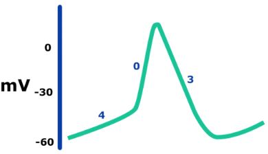
The pacemaker or nodal action potential consists of 3 phases: 4, 0 and 3. Phase 4 starts at a membrane potential of around -60mV with the opening of funny (If) channels. This allows an influx of Na⁺ ions into nodal cells and spontaneous depolarisation. This is propagated by the opening of Ca2+ channels (T-type at -50mV and L-type at -40mV) to allow an influx of Ca²⁺ ions. At the same time, there is reduced efflux of K+ due to closure of K+ ion channels.
Once a threshold (pacemaker potential) is reached, which is around -40 to -30 mV, there is an influx of Ca2+ ions through L-type channels leading to depolarisation, which constitutes phase 0.
The nodal cell then repolarises in phase 3 through the opening of K+ channels to allow K+ efflux and the closing of L-type Ca2+ channels, decreasing Ca²⁺ influx. This achieves a membrane potential of around -60mV and the cycle restarts again with no resting potential.
Non-nodal cardiac potential
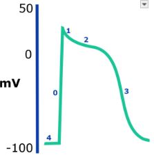
The non-nodal cardiac myocyte potential consists of 5 phases: 4, 0, 1, 2 and 3. Phase 4 is the resting membrane potential which sits at around -90mV. In a single action potential, there is rapid depolarisation in phase 0 due to the opening of fast sodium channels allowing Na + influx and closing of potassium channels to reduce K+ efflux. Once depolarised, the myocyte begins to repolarise with the opening of K+ ion channels, allowing for K+ efflux in phase 1. This is delayed at the plateau phase in 2 due to influx of Ca2+ via the L-type Ca2+ channels. Phase 3 follows with repolarisation due to an increase in K+ influx and a decrease in Ca2+ efflux through the closing of L-type Ca2+ channels, and the membrane potential returns to the threshold level (phase 4).
Antiarrhythmic Medication
The mainstay in arrhythmia management is antiarrhythmic medication. Electrical cardioversion is used where there is haemodynamic compromise or electively to return to sinus rhythm, for instance, in AF. Alternatively, electrophysiological studies may be performed to identify arrhythmias and ablate areas along the conductive pathway to eliminate the arrhythmia.
Antiarrhythmic drugs target ion channels in cardiac myocytes and/or nodal cells to manipulate the cardiac action potential and negate arrhythmias. Since parasympathetic and sympathetic activity also influence cardiac conductivity, drugs that affect their activity can also be used. There are numerous ways to categorise antiarrhythmics but the Vaughan-Williams classification system remains the most prevalent:
- Class I: Na⁺ Channel Blockers. They work by reducing conduction velocity at phase 0 but can also affect phase 3 of non-nodal repolarisation and thereby change the corrected QT interval. These drugs can be further subclassified by the degree to which they block Na+ channels as well as their effect on the QTc interval. All class I drugs can be used in VT, however only Ia and Ic can be used in AF/flutter and SVT, this is because Ib drugs have little influence on atrial myocytes.
- Ia: Moderate Na blockade, QTc prolongation – e.g., Quinidine, procainamide, disopyramide. These drugs are limited by their pro-arrhythmic effects.
- Ib: Weak Na blockade, QTc shortening – e.g., Lidocaine and mexiletine – only used for ventricular arrhythmias. They have a greater influence on myocytes following ischaemia, so they are useful in post-MI arrhythmias.
- Ic: Strong Na blockage, no effect on QTc – Flecainide and propafenone. They should be avoided in patients with ischaemic heart disease or structural heart disease as they are found to cause increased mortality.
- Class II: Beta blockers – e.g., bisoprolol, metoprolol and atenolol. They reduce sympathetic influence by blocking beta-adrenoreceptors in the heart. There is reduced SA node firing and AV node conduction, which reduces rate and conduction velocity, respectively. Side effects include bradycardia, AV block, bronchospasm, fatigue, and reduced exercise tolerance due to negative chronotropy.
- Class III – K+ channel blockers – e.g., amiodarone, dronedarone, sotalol, ibutilide, dofetilide. They affect potassium efflux, thus prolonging the QT interval. Amiodarone is commonly used in clinical practice for VT, VF, AF and flutter and has additional class I, II, and IV action. Side effects include thyroid dysfunction, deranged LFTs and pulmonary fibrosis.
- Class IV – Ca²⁺ channel blocker (non-dihydropyridines) – diltiazem and verapamil. They block L-type calcium channels which reduces both rate and velocity of conduction, most effective at nodal cells. Side effects include AV block, bradycardia and reduced contractility, therefore contraindicated in heart failure.
| Class | Mechanism | Useful in |
|---|---|---|
| I | Na+ blockade | AF/Flutter, SVT, VT. |
| I | Beta-blockade | Sinus tachycardia, AF/Flutter, SVT, PVC, VT |
| III | K+ blockade | AF/Flutter, SVT, VT |
| IV | Ca2+ blockade | Sinus tachycardia, AF/Flutter, SVT, PVC |
Table 1: Showing mode of action of common conditions in which these medications are used.
References
- Klabunde RE. Arrhythmias. Cardiovascular Physiology Concepts. Last updated October 11th 2012. Available at https://www.cvphysiology.com/Arrhythmias/A008/
- Wei X, Yohannan S, Richards JR. Physiology, Cardiac Repolarization Dispersion and Reserve. StatPearls. Last updated April 21st 2022. Available at https://www.ncbi.nlm.nih.gov/books/NBK537194/
- Klabunde RE. Vaughan-Williams Classification of Antiarrhythmic Drugs. Cardiovascular Physiology Concepts. Last updated March 19th 2010. Available at https://www.cvpharmacology.com/antiarrhy/Vaughan-Williams/
- King GS, Goyal A, Grigorova Y, et al. Antiarrhythmic Medications. StatPearls. Last updated February 16th 2022. Available at https://www.ncbi.nlm.nih.gov/books/NBK482322/
Complete Guide to Clinical Genetics ST3 Application and Recruitment (2027)
Written by Medibuddy. Last updated: October 2026. Clinical Genetics is

Complete Guide to Clinical Radiology ST3 Application and Recruitment (2027)
Recruitment paused: NHS England is not running Clinical Radiology ST3+ recru

Complete Guide to Paediatric Surgery ST3 Application and Recruitment in United Kingdom (2026)
Written by Medibuddy. Last updated: October 2026. If you’re planning


Take your subscriptions with you
Our mobile app allows you to access your interview and exam question banks wherever you are.

