


 Close
Close
 6th March 2024
Electrical conduction of the heart
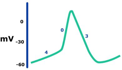
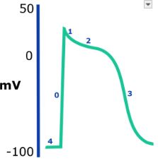
The action potentials at each level of the conduction pathway have unique properties complementary to their function. Cardiac action potentials are differentiated by their ability for spontaneous depolarisation or pacemaker potential and the speed at which they depolarise. Pacemaker cells found at the SA and AV nodes spontaneously generate slow response action potentials. The AV node conducts at a rate of around 0.05m/s.
In contrast, cardiac myocytes lack the same degree of automaticity; however, they depolarise at a much faster rate. Note that the His-Purkinje system has both features of automaticity and fast conduction, with a conduction velocity of around 2-4m/s, respectively. The whole conduction occurs very quickly and any delays are translated to changes on the ECG such as prolonged PR (>200ms), QT interval or QRS duration (>120ms). We will consider the cardiac action potentials in more detail as it bears relevance to the actions of antiarrhythmic medication.
Complete Guide to Clinical Genetics ST3 Application and Recruitment (2027)
Written by Medibuddy. Last updated: October 2026. Clinical Genetics is

Complete Guide to Clinical Radiology ST3 Application and Recruitment (2027)
Recruitment paused: NHS England is not running Clinical Radiology ST3+ recru

Complete Guide to Paediatric Surgery ST3 Application and Recruitment in United Kingdom (2026)
Written by Medibuddy. Last updated: October 2026. If you’re planning


Take your subscriptions with you
Our mobile app allows you to access your interview and exam question banks wherever you are.

