


 Close
Close
 23rd June 2023
Chronic Kidney Disease
Overview of chronic kidney disease
Classification of chronic kidney disease
Chronic kidney disease (CKD) is defined as a persistent reduction in renal function (for greater than three months); this may be progressive in nature and have consequences for patient health. Classification of CKD is based on glomerular filtration rate (GFR), amount of albuminuria and underlying cause.
GFR categories
| Stage | GFR (ml/min/1.73m²) | Terminology and interventions |
|---|---|---|
| G1 | ≥90 – considered CKD if accompanied by urinary or imaging markers suggestive of kidney damage | Normal or high |
| G2 | 60-89 – considered CKD if accompanied by urinary or imaging markers suggestive of kidney damage | Mildly reduced |
| G3a | 45-59 | Mildly to moderately reduced |
| G3b | 30-44 | Moderately to severely reduced |
| G4 | 15-29 | Severely reduced – likely to need treatment of CKD complications and preparation for renal replacement therapy (RRT) if appropriate |
| G5 | ≤14 | Renal failure – consider commencing RRT if appropriate |
Table 1: Staging of CKD based on GFR, adapted from the KDIGO 2012 Clinical Practice Guideline for the ‘Evaluation and Management of Chronic Kidney Disease’ and the NICE guideline on ‘Chronic Kidney Disease: Assessment and Management’.
Albuminuria categories
| Stage | Albumin-to-creatinine ratio (mg/mmol) | Terminology |
|---|---|---|
| A1 | <3 | Normal to mildly increased |
| A2 | 3-30 | Moderately increased |
| A3 | >30 | Severely increased – note this category includes the nephrotic range (usually >200mg/mmol) |
Table 2: Staging of CKD based on albuminuria, adapted from the KDIGO 2012 Clinical Practice Guideline for the ‘Evaluation and Management of Chronic Kidney Disease’ and the NICE guideline on ‘Chronic Kidney Disease: Assessment and Management’
It is important to note factors that may influence the interpretation of the measurements above, for example, those of particularly high muscle mass or lower than expected muscle mass (e.g. amputees, muscle wasting disorders). It is normal for GFR to gradually decline with increasing age without a patient necessarily having CKD. There are also a number of drugs that cause a rise in plasma creatinine levels due to reduced tubular secretion (e.g. cimetidine, trimethoprim); this will result in a false reduction in calculated GFR.
There are a number of formulae available to calculate estimated GFR (eGFR); the MDRD (Modification of Diet in Renal Disease) was developed in 1999 and previously widely used, however more recent guidance suggests the use of the CKD-EPI equation (Chronic Kidney Disease Epidemiology Collaboration), the first version of which was developed in 2009. The main four variables used in both original equations are serum creatinine, age, sex, and race. The CKD-EPI equation is less biased, especially at higher GFRs, and its latest iteration (2021) does not include race.
Causes of chronic kidney disease
The frequency of different causes of CKD will vary depending on the population concerned. The most common causes in the UK are:
- Diabetes mellitus
- Hypertension
- Atherosclerotic disease
- Glomerulonephritis, e.g. IgA nephropathy
- Congenital causes, e.g. autosomal dominant polycystic kidney disease, congenital renal dysplasia
- Chronic pyelonephritis/reflux nephropathy
- Obstructive uropathy
- Other, e.g. analgesic nephropathy, TB, HIV, interstitial nephritis
Management of chronic kidney disease
of chronic kidney disease has four key components:
- Treatment of the underlying cause, if possible, e.g. specific treatment for a vasculitis or myeloma
- Prevent the progression of CKD:
- Controlling blood pressure – NICE recommends a target of <140/90 for patients with CKD and a target of <130/80 if significant proteinuria (ACR of >70mg/mmol)
- Reducing proteinuria – this has traditionally been addressed with an angiotensin receptor blocker (ARB) or an angiotensin-converting enzyme (ACE) inhibitor titrated up to the highest dose tolerated. More recent evidence has supported the use of sodium glucose co-transporter-2 (SGLT2) inhibitors in addition to the above.
- Improving glycaemic control in diabetics.
- Dietary modifications – low phosphate, low potassium, salt-restriction, and fluid-restriction if needed
- Identify and treat any complications of CKD – e.g. anaemia, renal bone disease, hyperkalaemia, metabolic acidosis, fluid overload, cardiovascular disease.
- Prepare for renal replacement therapy if appropriate, or consider discussions about conservative/palliative care.
Complications of chronic kidney disease
Anaemia
Mechanism
The healthy kidneys normally produce erythropoietin (EPO) in response to cell hypoxia, this stimulates bone marrow red blood cell production. Reduced production in chronic kidney disease results in less erythropoiesis and a normochromic normocytic anaemia.
However, renal patients are also at risk of other causes of anaemia, with iron deficiency commonly seen. Iron deficiency can be absolute (reduced stores) or functional (normal stores but inability to use).
- Absolute – due to gastrointestinal losses (uraemic platelet dysfunction increases the risk of bleeding), serial phlebotomy, circuit clotting during haemodialysis, or reduced absorption due to medication such as acid inhibitors and phosphate binders.
- Functional – due to underlying inflammation preventing the body from using stored iron (haemodialysis itself is a pro-inflammatory state due to interaction with the dialysis membrane), high hepcidin levels (increased in inflammatory states, decreased renal clearance) also reduces the body’s ability to absorb and use iron.
Investigation
- Full blood count – target haemoglobin (Hb) in CKD is 100-120g/L (higher levels are linked with an increased risk of cardiovascular disease). Review the mean corpuscular volume and the mean cell haemoglobin concentration; anaemia related to reduced EPO alone should be normochromic and normocytic.
- Serum ferritin level (target of 200-500 μg/l in haemodialysis patients and 100-500μg/l in CKD patients not on dialysis) and transferrin saturations (target of >20%) – targets from the UK Renal Association
- Serum B12 and folate levels
Management
- Iron replacement – it should be ensured that a patient is iron-replete before considering erythropoiesis-stimulating agents. The intravenous route is preferable to oral, especially in more advanced CKD/haemodialysis patients, though this does carry a small but significant risk of drug reaction.
- Erythropoiesis-stimulating agents – recombinant erythropoietin addresses the lack of endogenous production; it can be given subcutaneously or intravenously. Notable cautions/contraindications include pure red cell aplasia, uncontrolled hypertension, intercurrent infection, malignancy, or other inflammatory disease.
- Red cell transfusion – generally avoided. The risks and benefits of transfusion need to be considered carefully in patients who may be eligible for a renal transplant, as antibody formation may limit future organ matching options.
Renal bone disease
Mechanism
The overall term for this condition and its complications is CKD mineral and bone disorder (CKD-MBD). The pathophysiology centres around levels of calcium and phosphate – this is affected by factors including parathyroid hormone (PTH), 1,25(OH)₂D (calcitriol) and FGF-23 (fibroblast growth factor-23).

Clinical consequences
- Bone – renal osteodystrophy encompasses a range of skeletal abnormalities, including osteomalacia, osteoporosis, osteosclerosis, adynamic bone disease (low turnover state) and osteitis fibrosis cystica (high turnover state).
- Skin/soft tissue – calcinosis cutis, calcium phosphate deposition
- Cardiovascular – vascular calcification/stiffness and left ventricular hypertrophy
Management
The aim of management strategies in CKD-MBD is to maintain normal serum levels of calcium and phosphate and prevent very raised or low PTH levels (aim 2-9 times the upper limit of normal) to maintain normal bone metabolism and prevent parathyroid gland hyperplasia.
- Dietary phosphate restriction – foods high in phosphate include dairy products and eggs.
- Phosphate binders – must be taken with meals to allow the binding of phosphate in the gut. Examples include calcium carbonate (note concerns about the calcium load in patients at risk of vascular calcification) and non-calcium agents such as sevelamer or lanthanum carbonate.
- Sodium bicarbonate – acidosis exacerbates renal bone disease; it can be improved with oral replacement.
- Renal replacement therapy – treatments such as regular dialysis can reduce phosphate levels
- Vitamin D analogues – 1α-hydroxylated analogues such as alfacalcidol are often used. It is important that phosphate levels are corrected first and to monitor the patient for side effects of hypercalcaemia or hyperphosphataemia.
- Cinacalcet – used in tertiary hyperparathyroidism, it is a calcimimetic that stimulates the calcium-sensing receptors of the parathyroid gland, reducing PTH secretion.
- Parathyroidectomy – surgical intervention is reserved for severe cases where medical management has failed.
Cardiovascular disease
Multiple factors in CKD interact to significantly increase the risk of cardiovascular disease. Minimising this includes factors such as:
- Controlling hypertension
- Lipid-lowering therapy, e.g. statins
- Addressing renal bone disease to reduce vascular calcification
- Consideration of antiplatelet therapy as secondary prevention, e.g. aspirin
- Management of other risk factors, e.g. smoking cessation, diabetic control
Renal replacement therapy
Haemodialysis
Mechanism
- During haemodialysis, blood is removed from the body via a form of vascular access and is exposed to dialysate fluid across a semipermeable membrane.
- Small molecules that are at a high concentration in blood will diffuse down their concentration gradient and into the dialysate fluid via the membrane pores. Blood and dialysate fluid flow in opposite directions to maintain these concentration gradients.
- Fluid removal is primarily via ultrafiltration, i.e. moves out of the blood due to a generated pressure gradient or the ‘transmembrane pressure (TMP)’. Some solutes are removed in addition to the fluid via convection, this process is known as ‘solute drag’.
- Haemodialysis clears small molecules well, however, is less effective for larger ‘middle’ molecules such as beta-2 microglobulin.
- Different related processes can be used in different clinical contexts:
- Haemofiltration – more frequently seen in critical care. Process relies on clearance via convection due to a generated TMP and movement down a pressure gradient. Favoured in haemodynamically unstable patients as it can be performed with slow pump speeds continuously over 24 hours. Fluid replacement is required to achieve solute clearance; a large amount of fluid must be filtered from the blood. Good at clearing larger molecules.
- Haemodiafiltration – this process combines clearance via diffusion and convection, therefore, requires both dialysate and replacement fluid. Benefits over haemodialysis alone include improved haemodynamic stability and better clearance of middle molecules.
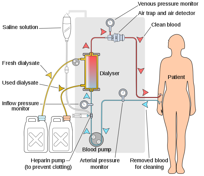
The process of haemodialysis.
Image Source: Wikimedia Commons (2008). Hemodialysis-en, Licensed under a creative commons licence. Available for free at: https://commons.wikimedia.org/wiki/File:Hemodialysis-en.svg (Accessed on: 13 February 2023).
Requirements and process
- Vascular access – dialysis requires the fast removal and return of large volumes of blood.
- Arteriovenous fistula (AVF) – this is the preferred mode of vascular access, generally formed by the connection of the brachial or radial artery with the cephalic or basilic vein. Requires suitable anatomy and advance planning (an AVF will take at least 6-8 weeks to ‘mature’ and become usable).
- Polytetrafluoroethylene (PTFE) graft – used to create an anastomosis between an artery and vein if native anatomy is unfavourable.
- Tunnelled cuffed dialysis catheter – similar to temporary dialysis catheters but are tunnelled through subcutaneous tissue to a separate exit site and have a cuff that is located in the tunnel to reduce ascending infection.
- Temporary dialysis catheter – these are used for emergency renal replacement therapy and are for short-term use only. Commonly inserted into the internal jugular or femoral veins.
- Dialysis prescription – a minimum of twelve hours per week is recommended for most patients, this is generally split into three sessions. Patients will have a set ‘dry weight’ that acts as a target for fluid removal each session.
- Anticoagulation – required to prevent clotting in the dialysis circuit external to the body, usually heparin or citrate.
Complications
- Related to dialysis access:
- Bacteraemia – tunnelled and temporary dialysis lines are indwelling devices that carry a high risk of infection.
- Central venous stenosis or occlusion
- Fistula – thrombosis (risk if volume deplete, requires urgent declotting), infection (unusual), aneurysm, ‘steal’ (blood flow redirected through the fistula may reduce distant arterial supply and compromise fingers etc.).
- Related to the process:
- Intradialytic hypotension – can occur when fluid from the extracellular space does not replace that taken from the intravascular space during dialysis at a fast enough rate.
- Dialysis disequilibrium syndrome – a risk when a patient has high urea levels that are lowered too quickly.
- Clotting of the extracorporeal circuit.
- Accidental disconnection – risk of serious blood loss.
- Air embolism.
- Longer term risks:
- Vascular calcification and cardiovascular disease.
- Dialysis-related amyloidosis – beta-2 microglobulin is poorly cleared via haemodialysis, it can accumulate and deposit in various tissues. Risk is reduced by using haemodiafiltration or a high-flux membrane (larger pores).
Peritoneal dialysis
Mechanism
- Peritoneal dialysis (PD) utilises the patient’s peritoneum as the semi-permeable membrane. Dialysate fluid is introduced into the peritoneal cavity via a catheter where it can then be in contact with the membrane and its blood supply.
- Fluid moves from the blood through the peritoneum to the higher osmolality dialysate fluid via osmosis, other molecules either diffuse down their concentration gradients or move via convection alongside fluid.
- The qualities of each patient’s membrane varies:
- ‘High transporters’ will remove solutes quickly and will also absorb glucose from fluid quickly.
- ‘Low transporters’ have a slower rate of solute removal.
- Membrane qualities will change over time and will generally move towards high transporter status.
Requirements and process
- Access – peritoneal dialysis requires the presence of an indwelling peritoneal catheter.
- Types of fluid – dextrose is commonly used to create high osmolality dialysate fluid, the concentration can be varied to affect the degree of fluid removal. However, glucose from the fluid can be absorbed and metabolised, abolishing the gradient – this is not a risk with alternatives such as glucose polymers (e.g. icodextrin).
- Regimes:
- Continuous Ambulatory Peritoneal Dialysis – usually four exchanges of dialysate fluid per 24-hour period, performed manually by the patient. Relatively long dwell periods are, therefore, generally more suitable for low transporters.
- Automated Peritoneal Dialysis (APD) – multiple exchanges of dialysate fluid performed by a machine overnight with shorter dwell periods. Patients may have fluid left in the abdomen during the day or perform an additional manual exchange during the day. Generally more suitable for high transporters.
- Tidal APD – the machine is set to leave a small amount of fluid in the abdomen when draining. This avoids discomfort associated with the removal of the full volume, however, it means dialysis is less efficacious.
Contraindications
- Anatomical – hernias (inguinal, umbilical, diaphragmatic), abdominal wall defect (e.g. a stoma).
- Infection – intra-abdominal or of the abdominal wall.
Technical/practical – patient/carer needs adequate dexterity and skill to perform safe exchanges, and the home environment needs to be suitable for the required equipment. - Cautions:
- Multiple previous abdominal surgeries causing adhesions
- Severe obesity
- Severe respiratory disease (increased intra-abdominal pressure can cause diaphragmatic splinting)
- Very large polycystic kidneys
Complications
- Infection
- Superficial catheter exit site infection.
- Peritonitis – repeated infections will affect the quality of the membrane and may reduce the patient’s time on peritoneal dialysis.
- Encapsulating peritoneal sclerosis – rare but serious. Fibrous scar tissue surrounds the peritoneum and bowel – leading to poor dialysis quality and the development of symptoms of bowel obstruction.
- Catheter issues – the PD catheter can become occluded or wrongly positioned. This can be due to constipation and it is therefore important that patients have regular bowel movements.
- Hyperglycaemia – due to glucose absorption from dialysate fluid.
- Longevity – the quality of peritoneal dialysis generally decreases over time, necessitating a switch to another modality like haemodialysis for ongoing renal replacement.
Renal transplantation
Pre-transplant factors
Assessment for transplant
Transplant should be considered for all patients with advanced CKD and those on dialysis, as it is recognised to provide a better quality of life and life expectancy. The assessment process in the UK is outlined by NHS Blood and Transplant (NHSBT). It aims to ensure that a patient will be able to tolerate the procedure itself, as well as the immunosuppression thereafter. Assessment includes:
- Blood group, HLA typing, antibody screening
- Viral screening – cytomegalovirus (CMV), Epstein-Barr virus (EBV), varicella zoster virus (VZV), hepatitis B and C, human immunodeficiency virus (HIV). Vaccinations against hepatitis B and VZV (if needed) should be offered pre-transplant.
- Psychological evaluation – to evaluate the likelihood that a patient will be compliant with their post-transplant medication and follow-up care.
- Cardiovascular risk – to identify high-risk surgical patients that may not qualify for listing and patients that may be eligible for an angiographic/surgical coronary intervention and listing thereafter.
- Malignancy screen – patients should have been in remission for at least two years before being considered for transplant.
- Risk of renal disease recurrence – disease recurrence of some conditions can reduce the chances of graft survival.
- Other serious comorbidities
Types of organ donation
- Living donation – improved long-term function compared to cadaveric donation:
- Related donor, e.g. sibling, parent, child
- Unrelated donor, e.g. friend, spouse or through a paired exchange scheme
- Cadaveric donation:
- DBD – donation after brain death
- DCD – donation after circulatory death
Organ matching
- Blood group – organs are generally allocated to a patient of a compatible blood group in a similar manner to blood transfusions. If a patient has antibodies that recognise donor ABO antigens, this can lead to hyperacute rejection. ABO-incompatible transplants are feasible with desensitisation therapy – usually immunosuppression and plasma exchange.
- HLA mismatch – coded on chromosome 6, one of each inherited from each parent, the most important antigens in transplant are HLA-A, HLA-B and HLA-DR. Degree of mismatch for each antigen is used – for example, identical twins would have a 0-0-0 mismatch, whereas a donor: recipient pair who shared none would be a 2-2-2 mismatch.
- Anti-HLA antibodies – a recipient can develop these following blood transfusion, pregnancy, or previous organ transplant. These can lead to the development of organ rejection post-transplant, especially if they are donor-specific antibodies.
At time of transplantation
Surgical
- The donor kidney is transplanted into the iliac fossa of the recipient, vasculature is anastomosed to the external iliac vessels and the ureter is implanted into the bladder (usually with a stent that is removed after 4-6 weeks). The kidney is sited outside of the peritoneum. The native kidneys are usually left in situ.
Medical
- Immunosuppression:
- Induction (i.e. at the time of surgery) – practice varies between transplant units, commonly involving an interleukin-2 receptor antagonist, e.g. basiliximab, or T-cell depleting antibodies, e.g. alemtuzumab or ATG (anti-human thymocyte immunoglobulin)
- Maintenance – generally a combination of a calcineurin inhibitor, e.g. tacrolimus, an antiproliferative agent, e.g. mycophenolate mofetil or azathioprine and occasionally steroids.
- Prophylaxis:
- PCP (Pneumocystis jirovecii) prophylaxis – e.g. co-trimoxazole
- CMV prophylaxis – e.g. valganciclovir, decisions depend on the donor and recipient’s viral status
Complications of transplantation
Surgical/postoperative complications
- Bleeding
- Infection
- Urinary leak/obstruction
- Thrombosis of renal artery or vein
- Lymphocele
- Renal artery stenosis
- Bladder dysfunction (capacity reduces if a patient was anuric)
Issues with graft
- Delayed graft function – this is the requirement for dialysis post-transplantation. Risk factors include DCD donors, prolonged cold ischaemic time and donor acute kidney injury.
- Acute rejection – commonly T-cell mediated, can be antibody-mediated or a combination of the processes. A transplant biopsy is often performed for confirmation, and findings are classified using the Banff classification. Treatment depends on the findings but often utilises high dose intravenous steroids and optimisation of immunosuppressive drugs.
- Chronic graft dysfunction – multiple potential causes (immune and non-immune), including chronic antibody-mediated or T-cell-mediated rejection, calcineurin-inhibitor toxicity, recipient factors such as hypertension and donor factors such as DCD kidney or older age.
- Disease recurrence, e.g. focal segmental glomerulosclerosis
- Viral infection, e.g. BK nephropathy
Other medical problems
- Infection – transplant recipients are prone to a multitude of infections, however, it is important to give prophylaxis for/have high clinical suspicion for PCP and CMV. High risk patients should be considered for tuberculosis prophylaxis.
- Malignancy – notably skin (benign and malignant lesions) and post-transplant lymphoproliferative disorder (EBV is a risk factor).
- Cardiovascular disease
- Diabetes – risk associated with immunosuppressive drugs such as tacrolimus and steroids.
Complete Guide to Clinical Genetics ST3 Application and Recruitment (2027)
Written by Medibuddy. Last updated: October 2026. Clinical Genetics is

Complete Guide to Clinical Radiology ST3 Application and Recruitment (2027)
Recruitment paused: NHS England is not running Clinical Radiology ST3+ recru

Complete Guide to Paediatric Surgery ST3 Application and Recruitment in United Kingdom (2026)
Written by Medibuddy. Last updated: October 2026. If you’re planning


Take your subscriptions with you
Our mobile app allows you to access your interview and exam question banks wherever you are.

