


 Close
Close
 1st June 2023
Coagulation and Thrombosis
Coagulation overview
Coagulation pathway
The coagulation pathway consists of a series of reactions, which lead to homeostasis, the blockage of bleeding or haemorrhage. The coagulation cascade is made up of various clotting factors and the liver is the key organ involved in coagulation, being the site of synthesis of the clotting factors. Haemostasis can be primary or secondary. In primary haemostasis, at the site of endothelial damage, the aggregation of platelets form a plug. Secondary haemostasis involves the intrinsic and extrinsic pathways. These both diverge to form the common pathway and lead to fibrin becoming activated; these fibrin units combine and stabilise the platelet plug.
The two pathways, the intrinsic and extrinsic pathways, both converge to activate factor X. This then leads to the common pathway in which activated factor X, in combination with factor V, platelet membrane phospholipids and Ca2+ ions convert prothrombin to thrombin (activated factor II), which in turn results in the activation of fibrin (activated factor I). The fibrin subunits then combine to form a stable fibrin clot.
Regulations of Clotting
Proteins C and S act to prevent coagulation, as these proteins mainly act to inactivate factors V and VIII.

Clotting cascade.
Image Source: Wikimedia Commons (2012). Classical blood coagulation pathway, Licensed under a creative commons licence. Available for free at: https://commons.wikimedia.org/wiki/File:Classical_blood_coagulation_pathway.png (Accessed on: 10 January 2023).

Coagulation screen
The coagulation screen includes laboratory tests to assess haemostasis. Measured tests include PT, APTT, INR (from PT), bleeding time and fibrinogen.
The bleeding time can be measured, which demonstrates the function of the platelets. Platelet disorders, including von Willebrand’s disease, ITP, TTP and any cause of thrombocytopenia, can prolong the bleeding time.
Prothrombin time (PT) and activated prothrombin time (APTT) measure the timing of the extrinsic and intrinsic pathways, respectively. A variety of conditions can affect the PT (extrinsic pathway), including liver disease, warfarin therapy, other causes of vitamin K deficiency and disseminated intravascular coagulation (DIC). The INR can be calculated by using the patient’s PT and the INR is commonly measured and monitored whilst patients are receiving warfarin therapy to aid with warfarin dose adjustments. The APTT can be affected by Haemophilia A and B, von Willebrand’s disease and acquired haemophilia.
In the labs, mixing studies can be performed by mixing the patient’s plasma with control plasma. These studies can be used to determine whether the PT or APTT is prolonged because of a factor deficiency (for example Haemophilia A, B) or because of the presence of a factor inhibitor (for example lupus anticoagulant). If after mixing the patient’s plasma with control plasma, the PT/APTT normalises, then the prolongation is due to a factor deficiency. If the PT/APTT remain prolonged, the prolongation is due to a factor inhibitor such as a lupus anticoagulant.
Thrombin converts fibrinogen to fibrin to produce a fibrin-based clot. As fibrinogen is an acute phase protein, elevated levels can indicate systemic inflammation, various cancers and tissue injury. Low fibrinogen levels can be seen in an inherited condition, whereby the body is unable to effectively produce fibrinogen. Low fibrinogen levels can be seen in liver disease, malnutrition, and disseminated intravascular coagulation (DIC).
Disorders of haemostasis
Haemophilia
A deficiency of clotting factor VIII or IX can result in Haemophilia, an X-linked recessive bleeding disorder. Factor VIII or Factor XI deficiency results in Haemophilia A and B, respectively. Due to genetic inheritance, inherited haemophilia occurs almost entirely in males, but females can also be affected.
Acquired haemophilia is a much rarer, non-genetic bleeding disorder. It has an autoimmune component and is due to the presence of a coagulation factor inhibitor, such as a lupus anticoagulant.
Haemophilia A
Haemophilia A is caused by the deficiency of the coagulation factor VIII. This disorder encompasses three categories: mild, moderate, and severe; the severity is based on factor VIII levels. In the normal population, the factor VIII levels are between 50% and 100%. In mild, moderate, and severe Haemophilia A, the factor VIII levels are between 6 – 40%, 1 – 5% and <5%, respectively.
The symptoms of Haemophilia A depend on whether the disorder is mild, moderate, or severe. Symptoms range from instances where bleeding only occurs following an injury, procedure or childbirth to spontaneous bleeding such as into muscles and joints and intracranial bleeds.
Haemophilia B
Haemophilia B is caused by the deficiency of the coagulation factor IX, Haemophilia B is also known as Christmas Disease. Depending on the level of factor IX, it can be classified as mild, moderate, or severe. Again, the symptoms, like Haemophilia A, vary depending on the classification of the disorder.

Haemophilia inheritance
Image Source: Public Health Image Library (2022). Haemophilia. Available for free at: https://www.cdc.gov/ncbddd/hemophilia/facts.html (Accessed on: 30 January 2023).
Investigations
If a patient has a family history of haemophilia or signs and symptoms that could suggest haemophilia, then the investigations below should be performed:
- APTT – usually prolonged
- APTT mixing studies – APTT corrects
- Factor VIII or factor IX assay – decreased levels of VIII in haemophilia A or decreased levels of IX in haemophilia B
- FBC – usually normal, but anaemia can exist due to chronic bleeding
- PT – normal
- Von Willebrand factor assay – normal
- Bleeding time – normal
- Liver function tests – normal
Management
Broadly, management can be divided into management in the acute setting and prophylaxis.
In the acute setting of severe bleeding, patients should be managed with high-dose clotting factor concentrate of either factor VIII or factor IX (depending on whether they have haemophilia A or B). Some patients may require emergency surgery or procedures, for example if there is compartment syndrome caused by a severe muscular haematoma. Some patients can be treated with tranexamic acid. In mild Haemophilia A, desmopressin can be used to increase the factor VIII and von Willebrand factor (VWF) levels both as treatment and prophylactically prior to a procedure.
Occasionally patients who receive clotting factor concentrates can develop antibodies, called inhibitors, which make the factor concentrates less effective when they are next given. Patients should be regularly tested for inhibitors. If high-titre factor inhibitors are present, bypassing agents should be used instead of the clotting factor concentrates.
In the prophylactic setting, if the haemophilia is severe and there are no factor VIII/IX inhibitors, prophylactic recombinant or plasma-derived factor VIII or factor IX should be administered. If patients have factor VIII/IX inhibitors, then immune tolerance induction can be used to eliminate the inhibitor. Emicizumab, a monoclonal antibody, has now been approved for prophylaxis in haemophilia A (irrelevant of the inhibitor status).
Von Willebrand disease
In von Willebrand disease (VWD), the protein, von Willebrand factor, which is required for platelet adhesion is defective or diminished due to an autosomally inherited defect. VWD causes increased bleeding and can be divided into three types: type 1 (the mildest and commonest), 2 and 3 (the most severe and rarest).
| Type | Description | People with VWD (%) | Inheritance Pattern |
|---|---|---|---|
| Type 1 | Low levels of VWF cause mild bleeding | 60 – 80% | Inherited in a dominant pattern – only one parent has to have VWD for their child to inherit it. |
| Type 2 (includes 2A, 2B, 2M, 2N subtypes). |
VWF that doesn’t work properly causes mild to moderate bleeding
|
15 – 30% | Certain subtypes are inherited in a recessive pattern – both parents have to have VWD for their child to inherit it. |
| Type 3 | Almost no VWF causes severe bleeding | 5 – 10% | Inherited in a recessive pattern – both parents have to have VWD for their child to inherit it. |
Signs and symptoms
Some cases of VWD can be asymptomatic and are only diagnosed if a family member has the disease. Signs and symptoms depend on the type of VWD. Usually, Type 1, 2A, 2B and 2M VWD present with mucocutaneous bleeding, such as easy bruising, epistaxis, and menorrhagia. Type 2N WVD can present similarly to mild haemophilia and type 3 WVD can present with severe bleeding as patients have almost no von Willebrand’s factor.
Diagnosis
Screening tests include:
- FBC – may be normal, but can show microcytic anaemia due to ongoing blood loss or thrombocytopenia in some types of VWD
- APTT – can be normal or prolonged (if factor VIII levels are reduced)
- PT – usually normal
- Bleeding time – prolonged
Diagnostic tests include:
- VWF antigen levels – to measure the amount of VWF
- VWF ristocetin cofactor activity – to measure how well the VWF is working
- VWF multimers – this can help to diagnose the type of VWD
- Factor VIII clotting activity – to measure how well VWF is binding to factor VIII
Management
- Desmopressin – works by stimulating the body to release more von Willebrand factor (it can be used for mild/moderate type 1 VWD and it can occasionally be useful in type 2 VWD)
- VWF/FVIII concentrates – can be used to treat bleeding in patients with severe bleeding in type 1 VWD or type 1 WVD which is unresponsive to desmopressin, type 2 and type 3 VWD
- Fibrinolysis inhibitors, such as tranexamic acid
- COCP for menorrhagia
- Local measures to stop bleeding
Disseminated intravascular coagulation
Disseminated intravascular coagulation (DIC) is a life-threatening consumptive coagulopathy in which the coagulation and fibrinolytic cascades are systemically activated in an uncontrolled manner due to an underlying cause such as sepsis, trauma, surgery, or pregnancy complications. The activation of the coagulation pathway results in thrombus formation within the intravascular system, leading to depletion of coagulation factors and platelets and resulting in bleeding.
DIC is associated with a high mortality and results in severe, life-threatening complications. Early aggressive treatment is key to reducing mortality. Complications can include thrombosis, severe bleeding, acute renal failure, gangrene and digit loss, haemothorax, intracranial bleeds, cardiac tamponade and death.
The International Society of Thrombosis and Haemostasis composed a diagnostic algorithm for DIC to be used in patients with a clinical suspicion of DIC. A score is calculated using the table below; a score ≥5 suggests overt DIC.
| Test | Score 0 | Score 1 | Score 2 | Score 3 |
|---|---|---|---|---|
| Platelet Count | >100 x 10⁹/L | 50 – 100 x 10⁹/L | <50 x 10⁹/L | |
| PT Prolongation | <3 seconds | 3 – <6 seconds | >6 seconds | |
| Fibrinogen Level | >100 mg/dL | <100 mg/dL | ||
| D Dimer Level | No increase | Moderate increase | Strong increase |
International Society of Thrombosis and Haemostasis Criteria for DIC
Causes
An underlying disorder is required to cause DIC, and therefore, DIC can only be considered in the presence of an underlying disorder. For instance, in the acute setting: sepsis, severe obstetric complications, major trauma, severe burns, severe immunological reactions and malignancies can all lead to systemic activation of the coagulation pathway causing DIC.
Treatment
The management of DIC ultimately should focus on treatment of the underlying disorder causing the DIC. In the acute setting, if there is active bleeding or there is a high bleeding risk, transfusions can be considered. Platelet transfusion should be considered if the platelet count is <20 x 10⁹/L or <50 x 10⁹/L with active bleeding. If significant bleeding is present or the patient’s fibrinogen level is <100 mg/dL, fresh frozen plasma (FFP) may be administered. Second-line treatment would include cryoprecipitates or fibrinogen concentrates.
Thrombocytopenia
Normal platelet counts fall between 150 – 400 x 10⁹/L. The definition of thrombocytopenia is a platelet count of <150 x 10⁹/ l.
Causes
Thrombocytopenia can be classified into three main categories: a decrease in platelet production, an increase in platelet destruction and platelet sequestration.
Conditions which decrease platelet production include:
- Leukaemia, myelodysplastic syndrome (MDS), aplastic anaemia
- Paroxysmal nocturnal haematuria (PNH)
- B12 or folate deficiencies
- Liver failure
- Inherited thrombocytopenia disorders
- Systemic viral or bacterial infection
- Certain drugs can cause bone marrow suppression, for example sodium valproate, daptomycin, certain chemotherapy agents
Conditions which increase platelet destruction include:
- Immune thrombocytopenic purpura
- Thrombotic thrombocytopenic purpura
- Haemolytic uraemic syndrome
- Disseminated intravascular coagulation
- Systemic lupus erythematosus
Platelet sequestration:
- Conditions which cause splenomegaly, such as liver cirrhosis and portal hypertension, some leukaemias and lymphomas.

Blood film from a patient with TTP showing a lack of platelets, and fragmented red blood cells. TTP can cause a pentad of symptoms/signs: fever, renal failure, haemolytic anaemia, thrombocytopenia, and neurological symptoms. Caused by decreased activity of ADAMTS-13. Plasma exchange is the main treatment for TTP.
Image Source: Wikimedia Commons (2014). Thrombocytopenia 1, Licensed under a creative commons licence. Available for free at: https://commons.wikimedia.org/wiki/File:Thrombocytopenia_1.jpg (Accessed on: 30 January 2023).

Immune thrombocytopenia purpura
Immune thrombocytopenia purpura (ITP) usually occurs when autoantibodies are formed against platelets, leading to platelet destruction and inhibition of their production. The trigger of this process in adults is unknown.
Symptoms/Signs
Most patients are asymptomatic and thrombocytopenia is discovered on FBC performed for another reason. The most common signs/symptoms include petechiae (see image below of petechiae on the lip), mucosal bleeding, menorrhagia, epistaxis, and purpura. More rarely, patients can present with GI haemorrhage and intracranial haemorrhages.
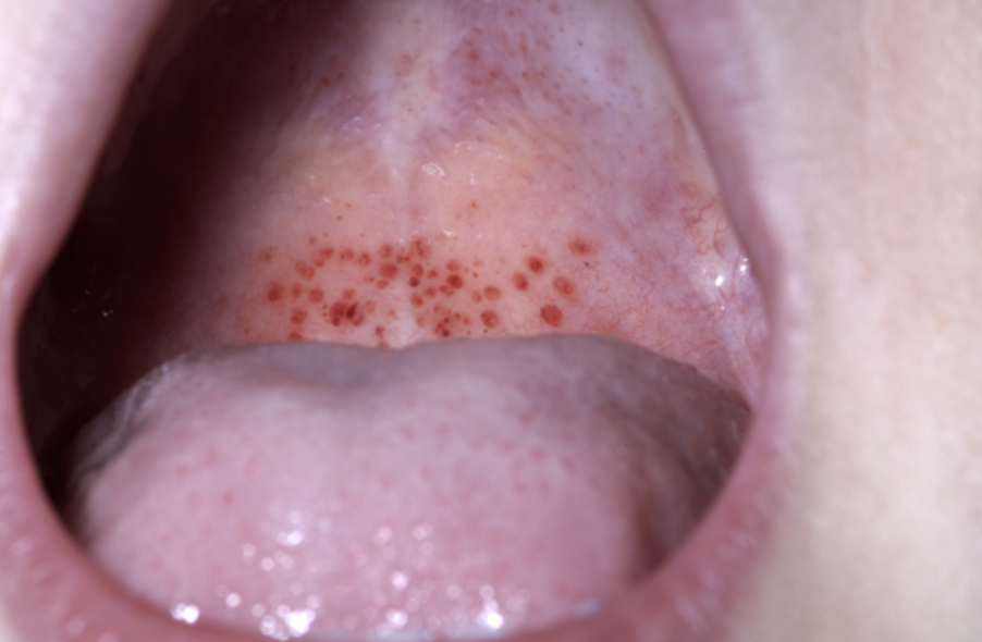
Petechiae on the lip in a case of ITP.
Image Source: Wikimedia Commons (2008). Oral petechiae, Licensed under a creative commons licence. Available for free at: https://commons.wikimedia.org/wiki/File:Oral_petechiae.JPG (Accessed on: 30 January 2023).

Investigations
The suspicion of ITP is raised by isolated thrombocytopenia with an otherwise normal FBC. A peripheral blood film should be visualised. Other causes of isolated thrombocytopenia should be excluded; the patient will therefore have bloods sent for U+Es, LFTs, a coagulation screen, a screen for Hepatitis B, C and HIV. A bone marrow biopsy is rarely performed but should be performed if the FBC or blood film reveals other abnormalities other than thrombocytopenia, if the clinical features are not typical, or if the patient is failing to respond to standard therapies.
Treatments
Patients with a platelet count of >30 x 10⁹/l with no symptoms and no bleeding can be monitored and treatment is not required. First line treatment for symptomatic patients/patients with a platelet count of <30 x 10⁹/l is oral prednisolone, usually given at a dose of 1mg/kg/day. Corticosteroids usually increase a patient’s platelet count (although this can take up to a month for some patients). When a response is achieved, corticosteroids should be weaned gradually. However, there is a risk of relapse of the ITP when corticosteroids are weaned. Other treatment options available for ITP include IV immunoglobulin (IVIG), rituximab, IV anti-D immune globulin and thrombopoietin receptor agonists (e.g., romiplostim, eltrombopag).
Splenectomy can be considered in patients with severe thrombocytopenia (platelet count <15 x 10⁹/l) who have failed medical therapies and the disease persists after a year. There can be potential complications for patients undergoing splenectomy, such as thrombosis or infection (especially infection with encapsulated bacteria). In patients undergoing splenectomy, vaccinations (to S. pneumoniae, H. influenzae, N. meningitidis) are advised at least 2 weeks prior to the procedure.
Thrombosis
Venous thrombosis
Venous thrombosis includes both deep vein thrombosis (DVTs) and pulmonary emboli (PEs). Treatment of confirmed PEs/proximal DVTs is with anticoagulation; the first-line choice in these cases is either apixaban or rivaroxaban. If these medications are not suitable for any reason then low molecular weight heparin (LMWH) should be given for 5 days, then replaced by dabigatran or edoxaban. Alternatively, LMWH can be given alongside warfarin for >5 days (or until the INR is >2 on 2 consecutive readings) and then warfarin can be continued alone.
NICE guidelines for the investigation of unprovoked DVT advise a full medical history and physical examination, and full blood tests should be sent for FBC, U+E, LFTS, PT and APTT. Patients should not be offered further investigations for cancer unless relevant symptoms or signs are apparent.
If a patient has an unprovoked PE/DVT, their ongoing risk of VTE versus their bleeding risk (e.g. using the HAS-BLED score) is assessed to decide whether anticoagulation should be continued for longer than 3 months (or 6 months for active cancer patients). If the DVT/PE was provoked, healthcare professionals can consider stopping anticoagulation once the patient has completed 3 months (for patients with active cancer, this decision should take place after 3-6 months). Again, the risk vs benefit of anticoagulation should be considered.
Risk factors
Strong risk factors include:
- Thrombophilia
- Trauma
- Following a period of immobility
- Malignancy
- Previous VTE
- Post-surgery
Weaker risk factors include:
- Myeloproliferative diseases, congestive heart failure, COPD, inflammatory bowel disease, some neurological diseases, obesity, increasing age, pregnancy/postnatal period, history of varicose veins, first degree relative with VTE, extended travel, HRT and oestrogen-containing contraceptive pills.
Thrombophilia
Thrombophilia refers to both congenital and acquired conditions, which increase susceptibility to thrombosis.
Congenital causes
- Proteins C and S deficiency
Proteins C and S prevent coagulation, as these proteins mainly inactivate factors V and VIII. Deficiencies in these proteins, therefore, lead to hypercoagulable states. If there is suspicion of Protein C and Protein S deficiencies following thrombosis, testing should only be offered to patients who are stopping anticoagulation as results can be affected by anticoagulants.
- Factor V Leiden
The genetic mutation, Factor V Leiden causes a defect in factor V, meaning that protein C cannot inactivate it. Factor V is then working to continuously activate downstream clotting factors, causing a hypercoagulable state. Following acute thrombosis, if there is suspicion of Factor V Leiden, testing should only be offered to patients who are stopping anticoagulation as results can be affected by anticoagulants.
Acquired causes
- Antiphospholipid syndrome
Antiphospholipid syndrome can present with arterial or venous thrombosis and/or recurrent miscarriages. The patient must have one of the clinical features to make a diagnosis alongside one of the laboratory markers. Detection of anticardiolipin antibody, lupus anticoagulant or anti-b2-glycoprotein I antibody on >2 occasions at least 12 months apart is diagnostic. The lupus anticoagulant activity may prolong the APTT, therefore prolonged APTT alongside thrombosis or recurrent miscarriages should raise the question of antiphospholipid syndrome. It is important to consider prophylactic treatments (warfarin or anti-platelets) for antiphospholipid syndrome to reduce the risk of complications. As soon as a pregnancy is confirmed and throughout the pregnancy, LMWH alongside low-dose aspirin is recommended for patients with antiphospholipid syndrome and recurrent miscarriages. When there is no history of recurrent miscarriages but a history of intrauterine growth restriction or pre-eclampsia, monotherapy with low-dose aspirin should be considered.
- Paroxysmal nocturnal haemoglobinuria (PNH)
The incidence of arterial and venous thrombosis in patients with PNH is approximately 10%. PNH can cause hemolysis, thrombosis, and haemoglobinuria as a result of stem cell malfunction. CD55 and CD59 on the surface of the cell (GPI-anchored proteins) are deficient due to a mutation in the PIGA gene. The deficiency of these proteins results in complement activation, haemolysis and thrombosis. PNH is diagnosed by flow cytometric analysis looking at antibodies to GPI-anchored proteins. Treatment is with eculizumab or ravulizumab.
- Myeloproliferative neoplasms
Myeloproliferative neoplasms include polycythaemia vera, essential thrombocythemia and myelofibrosis. These conditions can cause venous and arterial thrombosis. Other signs of the underlying disorder would be present on the FBC, for instance thrombocytosis in essential thrombocythemia. On a peripheral blood sample, testing for JAK2, CALR and MPL can be sent. Myeloproliferative neoplasms are often associated with these mutations and identifying if a mutation is present can also guide treatment, for instance, by using a JAK2 inhibitor. A bone marrow biopsy may be required.
Anticoagulant therapy
If a patient requires anticoagulation, consideration should be taken as to which anticoagulant to prescribe: warfarin, apixaban, dabigatran, rivaroxaban or edoxaban. Scores exist to help the prescriber decide whether anticoagulation is indicated, such as the CHA₂DS₂-VASc score for stroke risk in AF and the HAS-BLED score to assess the bleeding risk for patients with AF.
Warfarin
Warfarin inhibits clotting factors X, IX, VII, and II by inhibiting vitamin K (this can be remembered by the mnemonic 1972). Patients who have been recently started on warfarin should have their INR levels checked regularly at first. The INR should be checked daily initially / alternate days until the INR is in the target therapeutic range on a minimum of 2 consecutive times, then twice weekly for 1-2 weeks, then at longer intervals thereafter (up to every 12 weeks).
Patients have different target INRs depending on the reason for anticoagulation. The images below, taken from NICE guidelines, suggest the recommended INR for different indications:

INR targets.
Image Source: National Institute for Health and Care Excellence (2022). Scenario: Warfarin. Available for free at: https://cks.nice.org.uk/topics/anticoagulation-oral/management/warfarin/ (Accessed on: 30 January 2023).
Warfarin can interact with many drugs which are cytochrome P450 inducers or inhibitors, as warfarin is metabolised via the cytochrome P450 system by CYP 2C9, 1A2, and 3A4. After the initiation of one of the following drugs, the INR should be checked after 3-5 days. Drugs which are P450 inhibitors and may enhance the effect of warfarin include alcohol (acutely); amiodarone; certain antibiotics including sulfonamides, metronidazole, clarithromycin and ciprofloxacin; antifungal medications such as fluconazole and ketoconazole; antidepressants such as fluoxetine; aspirin; cranberry juice; clopidogrel; corticosteroids; protease inhibitors; fibrates; NSAIDS; tamoxifen; thyroxine. Drugs which are P450 inducers and therefore may reduce the effects of warfarin include St. John’s Wart, griseofulvin, rifampicin, carbamazepine, phenobarbital and phenytoin. There is a further list of drugs which require INR monitoring when used concurrently with warfarin on the NICE website.
A patient with an INR out of therapeutic range should be managed as per NICE guidelines:
| INR | No bleeding | Minor bleeding | Major bleeding |
|---|---|---|---|
| >8 | Stop warfarin and give oral vitamin K (if needed oral vitamin K can be given again after 24 hours). Warfarin can be restarted when the INR <5. | Stop warfarin and give IV vitamin K (if needed, IV Vitamin K can be given again after 24 hours). Warfarin can be restarted when the INR <5. | Stop warfarin, give urgent IV vitamin K and prothrombin complex concentrate (PCC) |
| 5 – 8 | Warfarin should be held for 1 or 2 doses and then the dose should subsequently be reduced. | Stop warfarin and give IV vitamin K. Restart warfarin when INR <5. | Stop warfarin, give urgent IV vitamin K and prothrombin complex concentrate (PCC) |
Patients who take warfarin and require elective invasive procedures may need to temporarily stop warfarin. If there is a risk of severe bleeding with the procedure, patients will need to stop the warfarin 3 – 5 days before. If it is necessary to continue anticoagulation (i.e. high thrombotic risk), when the INR <2.5, heparin therapy should be started. In an emergency, warfarin can be reversed by vitamin K. For minor surgeries with a minimal bleeding risk, the surgery can be performed if the INR <2.5, but local guidelines should be viewed.
For dental procedures, the INR should be checked before the procedure (if the INR is usually stable, <72 hours before is sufficient). If the INR is <4, then the dental procedure can go ahead with no interruption to warfarin therapy. If the INR is >4, it should be delayed until it is <4.
Direct oral anticoagulants
Apixaban, rivaroxaban, edoxaban and dabigatran are all examples of direct oral anticoagulants (DOACs). The first three of these medications directly and reversibly inhibit factor Xa. Dabigatran works by reversibly inhibiting the thrombin molecule.
Unlike warfarin, DOACs do not need regular INR monitoring. However, contraindications to DOACs, such as severe hepatic disease, mechanical heart valve and severe renal disease. In these cases, warfarin is the only recommended anticoagulant.
Before commencing a DOAC, the below factors need to be considered:
- Body weight
- Renal function
- Interacting medications
- Different dosing regimens
Therapeutic fibrinolysis
Fibrinolytic therapies result in the breakdown of plasminogen into active plasmin. Fibrinolytic therapies are used to dissolve life-threatening thrombi to improve blood flow to vital organs. Examples include streptokinase, alteplase, etc. These agents can be used in many medical emergencies caused by the formation of clots listed below, but contraindications and risks must be considered prior to use:
- Acute ischaemic stroke
IV alteplase (a tissue plasminogen activator) is mostly used as a fibrinolytic therapy within 4.5 hours of stroke onset if no contraindications exist.
- Acute myocardial infarction
Fibrinolytic therapy is used to treat ST elevation myocardial infarctions if percutaneous coronary intervention (PCI) is unavailable or during transport to a hospital which provides PCI.
- Acute limb ischaemia
For non-life-threatening ischaemic limbs of <14-day duration, fibrinolysis can be used to achieve clot lysis. This can take up to 72 hours and therefore, it would not be appropriate for ischaemia which threatens a limb.
- DVT
DVTs are usually managed with anticoagulants. However, patients with a <14-day history of DVT symptoms, upper-extremity DVTs or proximal DVTs and good functional status can be considered for thrombolysis.
- PE
Fibrinolytic therapy can be used to treat acute massive PE, causing sustained hypotension and signs of haemodynamic compromise.
Complete Guide to Clinical Genetics ST3 Application and Recruitment (2027)
Written by Medibuddy. Last updated: October 2026. Clinical Genetics is

Complete Guide to Clinical Radiology ST3 Application and Recruitment (2027)
Recruitment paused: NHS England is not running Clinical Radiology ST3+ recru

Complete Guide to Paediatric Surgery ST3 Application and Recruitment in United Kingdom (2026)
Written by Medibuddy. Last updated: October 2026. If you’re planning


Take your subscriptions with you
Our mobile app allows you to access your interview and exam question banks wherever you are.

