


 Close
Close
 1st June 2023
Disorders of haemostasis
Haemophilia
A deficiency of clotting factor VIII or IX can result in Haemophilia, an X-linked recessive bleeding disorder. Factor VIII or Factor XI deficiency results in Haemophilia A and B, respectively. Due to genetic inheritance, inherited haemophilia occurs almost entirely in males, but females can also be affected.
Acquired haemophilia is a much rarer, non-genetic bleeding disorder. It has an autoimmune component and is due to the presence of a coagulation factor inhibitor, such as a lupus anticoagulant.
Haemophilia A
Haemophilia A is caused by the deficiency of the coagulation factor VIII. This disorder encompasses three categories: mild, moderate, and severe; the severity is based on factor VIII levels. In the normal population, the factor VIII levels are between 50% and 100%. In mild, moderate, and severe Haemophilia A, the factor VIII levels are between 6 – 40%, 1 – 5% and <5%, respectively.
The symptoms of Haemophilia A depend on whether the disorder is mild, moderate, or severe. Symptoms range from instances where bleeding only occurs following an injury, procedure or childbirth to spontaneous bleeding such as into muscles and joints and intracranial bleeds.
Haemophilia B
Haemophilia B is caused by the deficiency of the coagulation factor IX, Haemophilia B is also known as Christmas Disease. Depending on the level of factor IX, it can be classified as mild, moderate, or severe. Again, the symptoms, like Haemophilia A, vary depending on the classification of the disorder.

Haemophilia inheritance
Image Source: Public Health Image Library (2022). Haemophilia. Available for free at: https://www.cdc.gov/ncbddd/hemophilia/facts.html (Accessed on: 30 January 2023).
Investigations
If a patient has a family history of haemophilia or signs and symptoms that could suggest haemophilia, then the investigations below should be performed:
- APTT – usually prolonged
- APTT mixing studies – APTT corrects
- Factor VIII or factor IX assay – decreased levels of VIII in haemophilia A or decreased levels of IX in haemophilia B
- FBC – usually normal, but anaemia can exist due to chronic bleeding
- PT – normal
- Von Willebrand factor assay – normal
- Bleeding time – normal
- Liver function tests – normal
Management
Broadly, management can be divided into management in the acute setting and prophylaxis.
In the acute setting of severe bleeding, patients should be managed with high-dose clotting factor concentrate of either factor VIII or factor IX (depending on whether they have haemophilia A or B). Some patients may require emergency surgery or procedures, for example if there is compartment syndrome caused by a severe muscular haematoma. Some patients can be treated with tranexamic acid. In mild Haemophilia A, desmopressin can be used to increase the factor VIII and von Willebrand factor (VWF) levels both as treatment and prophylactically prior to a procedure.
Occasionally patients who receive clotting factor concentrates can develop antibodies, called inhibitors, which make the factor concentrates less effective when they are next given. Patients should be regularly tested for inhibitors. If high-titre factor inhibitors are present, bypassing agents should be used instead of the clotting factor concentrates.
In the prophylactic setting, if the haemophilia is severe and there are no factor VIII/IX inhibitors, prophylactic recombinant or plasma-derived factor VIII or factor IX should be administered. If patients have factor VIII/IX inhibitors, then immune tolerance induction can be used to eliminate the inhibitor. Emicizumab, a monoclonal antibody, has now been approved for prophylaxis in haemophilia A (irrelevant of the inhibitor status).
Von Willebrand disease
In von Willebrand disease (VWD), the protein, von Willebrand factor, which is required for platelet adhesion is defective or diminished due to an autosomally inherited defect. VWD causes increased bleeding and can be divided into three types: type 1 (the mildest and commonest), 2 and 3 (the most severe and rarest).
| Type | Description | People with VWD (%) | Inheritance Pattern |
|---|---|---|---|
| Type 1 | Low levels of VWF cause mild bleeding | 60 – 80% | Inherited in a dominant pattern – only one parent has to have VWD for their child to inherit it. |
| Type 2 (includes 2A, 2B, 2M, 2N subtypes). |
VWF that doesn’t work properly causes mild to moderate bleeding
|
15 – 30% | Certain subtypes are inherited in a recessive pattern – both parents have to have VWD for their child to inherit it. |
| Type 3 | Almost no VWF causes severe bleeding | 5 – 10% | Inherited in a recessive pattern – both parents have to have VWD for their child to inherit it. |
Signs and symptoms
Some cases of VWD can be asymptomatic and are only diagnosed if a family member has the disease. Signs and symptoms depend on the type of VWD. Usually, Type 1, 2A, 2B and 2M VWD present with mucocutaneous bleeding, such as easy bruising, epistaxis, and menorrhagia. Type 2N WVD can present similarly to mild haemophilia and type 3 WVD can present with severe bleeding as patients have almost no von Willebrand’s factor.
Diagnosis
Screening tests include:
- FBC – may be normal, but can show microcytic anaemia due to ongoing blood loss or thrombocytopenia in some types of VWD
- APTT – can be normal or prolonged (if factor VIII levels are reduced)
- PT – usually normal
- Bleeding time – prolonged
Diagnostic tests include:
- VWF antigen levels – to measure the amount of VWF
- VWF ristocetin cofactor activity – to measure how well the VWF is working
- VWF multimers – this can help to diagnose the type of VWD
- Factor VIII clotting activity – to measure how well VWF is binding to factor VIII
Management
- Desmopressin – works by stimulating the body to release more von Willebrand factor (it can be used for mild/moderate type 1 VWD and it can occasionally be useful in type 2 VWD)
- VWF/FVIII concentrates – can be used to treat bleeding in patients with severe bleeding in type 1 VWD or type 1 WVD which is unresponsive to desmopressin, type 2 and type 3 VWD
- Fibrinolysis inhibitors, such as tranexamic acid
- COCP for menorrhagia
- Local measures to stop bleeding
Disseminated intravascular coagulation
Disseminated intravascular coagulation (DIC) is a life-threatening consumptive coagulopathy in which the coagulation and fibrinolytic cascades are systemically activated in an uncontrolled manner due to an underlying cause such as sepsis, trauma, surgery, or pregnancy complications. The activation of the coagulation pathway results in thrombus formation within the intravascular system, leading to depletion of coagulation factors and platelets and resulting in bleeding.
DIC is associated with a high mortality and results in severe, life-threatening complications. Early aggressive treatment is key to reducing mortality. Complications can include thrombosis, severe bleeding, acute renal failure, gangrene and digit loss, haemothorax, intracranial bleeds, cardiac tamponade and death.
The International Society of Thrombosis and Haemostasis composed a diagnostic algorithm for DIC to be used in patients with a clinical suspicion of DIC. A score is calculated using the table below; a score ≥5 suggests overt DIC.
| Test | Score 0 | Score 1 | Score 2 | Score 3 |
|---|---|---|---|---|
| Platelet Count | >100 x 10⁹/L | 50 – 100 x 10⁹/L | <50 x 10⁹/L | |
| PT Prolongation | <3 seconds | 3 – <6 seconds | >6 seconds | |
| Fibrinogen Level | >100 mg/dL | <100 mg/dL | ||
| D Dimer Level | No increase | Moderate increase | Strong increase |
International Society of Thrombosis and Haemostasis Criteria for DIC
Causes
An underlying disorder is required to cause DIC, and therefore, DIC can only be considered in the presence of an underlying disorder. For instance, in the acute setting: sepsis, severe obstetric complications, major trauma, severe burns, severe immunological reactions and malignancies can all lead to systemic activation of the coagulation pathway causing DIC.
Treatment
The management of DIC ultimately should focus on treatment of the underlying disorder causing the DIC. In the acute setting, if there is active bleeding or there is a high bleeding risk, transfusions can be considered. Platelet transfusion should be considered if the platelet count is <20 x 10⁹/L or <50 x 10⁹/L with active bleeding. If significant bleeding is present or the patient’s fibrinogen level is <100 mg/dL, fresh frozen plasma (FFP) may be administered. Second-line treatment would include cryoprecipitates or fibrinogen concentrates.
Thrombocytopenia
Normal platelet counts fall between 150 – 400 x 10⁹/L. The definition of thrombocytopenia is a platelet count of <150 x 10⁹/ l.
Causes
Thrombocytopenia can be classified into three main categories: a decrease in platelet production, an increase in platelet destruction and platelet sequestration.
Conditions which decrease platelet production include:
- Leukaemia, myelodysplastic syndrome (MDS), aplastic anaemia
- Paroxysmal nocturnal haematuria (PNH)
- B12 or folate deficiencies
- Liver failure
- Inherited thrombocytopenia disorders
- Systemic viral or bacterial infection
- Certain drugs can cause bone marrow suppression, for example sodium valproate, daptomycin, certain chemotherapy agents
Conditions which increase platelet destruction include:
- Immune thrombocytopenic purpura
- Thrombotic thrombocytopenic purpura
- Haemolytic uraemic syndrome
- Disseminated intravascular coagulation
- Systemic lupus erythematosus
Platelet sequestration:
- Conditions which cause splenomegaly, such as liver cirrhosis and portal hypertension, some leukaemias and lymphomas.

Blood film from a patient with TTP showing a lack of platelets, and fragmented red blood cells. TTP can cause a pentad of symptoms/signs: fever, renal failure, haemolytic anaemia, thrombocytopenia, and neurological symptoms. Caused by decreased activity of ADAMTS-13. Plasma exchange is the main treatment for TTP.
Image Source: Wikimedia Commons (2014). Thrombocytopenia 1, Licensed under a creative commons licence. Available for free at: https://commons.wikimedia.org/wiki/File:Thrombocytopenia_1.jpg (Accessed on: 30 January 2023).

Immune thrombocytopenia purpura
Immune thrombocytopenia purpura (ITP) usually occurs when autoantibodies are formed against platelets, leading to platelet destruction and inhibition of their production. The trigger of this process in adults is unknown.
Symptoms/Signs
Most patients are asymptomatic and thrombocytopenia is discovered on FBC performed for another reason. The most common signs/symptoms include petechiae (see image below of petechiae on the lip), mucosal bleeding, menorrhagia, epistaxis, and purpura. More rarely, patients can present with GI haemorrhage and intracranial haemorrhages.
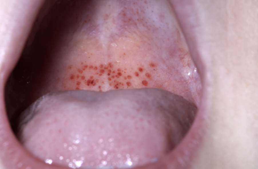
Petechiae on the lip in a case of ITP.
Image Source: Wikimedia Commons (2008). Oral petechiae, Licensed under a creative commons licence. Available for free at: https://commons.wikimedia.org/wiki/File:Oral_petechiae.JPG (Accessed on: 30 January 2023).

Investigations
The suspicion of ITP is raised by isolated thrombocytopenia with an otherwise normal FBC. A peripheral blood film should be visualised. Other causes of isolated thrombocytopenia should be excluded; the patient will therefore have bloods sent for U+Es, LFTs, a coagulation screen, a screen for Hepatitis B, C and HIV. A bone marrow biopsy is rarely performed but should be performed if the FBC or blood film reveals other abnormalities other than thrombocytopenia, if the clinical features are not typical, or if the patient is failing to respond to standard therapies.
Treatments
Patients with a platelet count of >30 x 10⁹/l with no symptoms and no bleeding can be monitored and treatment is not required. First line treatment for symptomatic patients/patients with a platelet count of <30 x 10⁹/l is oral prednisolone, usually given at a dose of 1mg/kg/day. Corticosteroids usually increase a patient’s platelet count (although this can take up to a month for some patients). When a response is achieved, corticosteroids should be weaned gradually. However, there is a risk of relapse of the ITP when corticosteroids are weaned. Other treatment options available for ITP include IV immunoglobulin (IVIG), rituximab, IV anti-D immune globulin and thrombopoietin receptor agonists (e.g., romiplostim, eltrombopag).
Splenectomy can be considered in patients with severe thrombocytopenia (platelet count <15 x 10⁹/l) who have failed medical therapies and the disease persists after a year. There can be potential complications for patients undergoing splenectomy, such as thrombosis or infection (especially infection with encapsulated bacteria). In patients undergoing splenectomy, vaccinations (to S. pneumoniae, H. influenzae, N. meningitidis) are advised at least 2 weeks prior to the procedure.
Complete Guide to Clinical Genetics ST3 Application and Recruitment (2027)
Written by Medibuddy. Last updated: October 2026. Clinical Genetics is

Complete Guide to Clinical Radiology ST3 Application and Recruitment (2027)
Recruitment paused: NHS England is not running Clinical Radiology ST3+ recru

Complete Guide to Paediatric Surgery ST3 Application and Recruitment in United Kingdom (2026)
Written by Medibuddy. Last updated: October 2026. If you’re planning


Take your subscriptions with you
Our mobile app allows you to access your interview and exam question banks wherever you are.

