


 Close
Close
 29th May 2023
Acute myeloblastic leukaemia (AML)
Overview
AML is a life-threatening malignancy in which a clonal population of early myeloid progenitors called blasts proliferate in the bone marrow, blood and sometimes extramedullary tissues. It is characterised particularly by cytogenetic abnormalities, but also genetic and epigenetic mutations. It is the most common type of acute leukaemia. The incidence in the UK is 4.05 per 100,000 people per year. It can present at any age, but incidence increases with age and the median age at diagnosis is 67.
As the blast population increases, there is progressively less room for healthy blood cell maturation, leading to a drop (usually) in red cells, normal white cells and platelets. Most symptoms of AML are attributable to these cytopenias. AML is a rapidly progressive condition, and the symptoms often develop quickly.
- Symptomatic anaemia
- Bleeding – from low platelets but also DIC
- Infection
- Hepatosplenomegaly (sometimes)
- Bone pain
- Gum infiltration
The white cell count (WCC) may be high or low at presentation. If the WCC is markedly raised (>100), this can cause hyperviscosity and leukostasis (sluggish blood flow). This tends to present with pulmonary infiltrates and hypoxia. This is a medical emergency and needs immediate action with leukapheresis and cytoreductive therapy.
Investigations
- Full panel of bloods, especially
- FBC – often anaemia and thrombocytopenia. WCC may be raised (due to blasts), normal or low. Neutropenia is common
- Coagulation screen – to look for DIC
- Blood film – often shows circulating blasts. Can be hard to distinguish from ALL but presence of Auer rods in blasts confirms AML
- It may be possible to perform flow cytometry (immunophenotyping) on peripheral blood to confirm the diagnosis, plus cytogenetics and molecular genetic analysis
- Bone marrow aspirate and trephine are essential to confirm the presence of blasts – diagnosis usually requires 20% blast percentage in the marrow. Flow, cytogenetics and molecular studies are also performed on this sample
- Further tests can include echo/multigated acquisition (MUGA) scan pre-chemo
Treatment
AML is potentially curable. However, the treatment is highly intense, with significant toxicity and so is largely delivered as an inpatient. Patients who are older in age (>65 as a general guide) or with significant comorbidities may not be fit enough for this treatment.
For patients treated with curative intent, this involves highly myelotoxic chemotherapy delivered in cycles – first in an induction phase and then a consolidation phase. Treatment and diagnostics have improved considerably in AML and patients are stratified into favourable, standard and unfavourable risk groups based on cytogenetic, genetic and epigenetic profile. This not only impacts prognosis but also treatment strategy. Most patients achieve remission with treatment, but patients with unfavourable risk profiles are much more likely to relapse. These patients may be offered allogeneic stem cell transplant after induction chemotherapy. Patients with a favourable risk profile would normally only be offered a transplant if they relapse, as the risks of a transplant outweigh the potential benefit given their lower chance of relapse.
Prognostic factors
The following are associated with a worse prognosis:
- Increasing age
- High risk features from cytogenetics or genetic analysis
- Progression to AML from pre-existing haematological disorder e.g. myelodysplastic syndrome (MDS), myeloproliferative neoplasm (MPN)
- Development of AML in context of previous chemotherapy (treatment associated)
Acute promyelocytic leukaemia
This is a variant of AML that is important to recognise as it commonly presents as an emergency with DIC and can lead to catastrophic bleeds. It is associated with a t(15;17) PML-RARA translocation gene, which fuses the promyelocytic leukaemia gene with the retinoic acid receptor alpha gene, leading to a block in cellular maturation and marked proliferation. Blasts in APML are promyelocytes and they have distinctive morphological features (pronounced cytoplasmic granulation and Auer rods with prominent nucleoli).
The treatment for APML is very different. It is based around all-trans retinoic acid (ATRA) and sometimes arsenic. It is imperative that treatment is started as soon as possible, and very close supportive care is necessary. If the diagnosis is suspected from morphology, treatment should be initiated without awaiting confirmation of genetics.
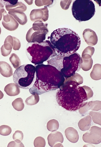
Blood film showing promyelocyte with Auer rods.
Image Source: Wikimedia Commons (2008). Faggot cell in AML-M3, Licensed under a creative commons licence. Available for free at: https://commons.wikimedia.org/wiki/File:Faggot_cell_in_AML-M3.jpg (Accessed on: 10 January 2023).

Complete Guide to Clinical Genetics ST3 Application and Recruitment (2027)
Written by Medibuddy. Last updated: October 2026. Clinical Genetics is

Complete Guide to Clinical Radiology ST3 Application and Recruitment (2027)
Recruitment paused: NHS England is not running Clinical Radiology ST3+ recru

Complete Guide to Paediatric Surgery ST3 Application and Recruitment in United Kingdom (2026)
Written by Medibuddy. Last updated: October 2026. If you’re planning


Take your subscriptions with you
Our mobile app allows you to access your interview and exam question banks wherever you are.

