


 Close
Close
 29th May 2023
Chronic leukaemia
Chronic myeloid leukaemia (CML)
Overview
CML is a type of (usually) slow-growing myeloproliferative neoplasm. The affected cells are haematological pluripotent stem cells, and these cells continue to undergo maturation into all three subsequent myeloid lineages – erythroid, platelet and granulocytes.
More than 90% of CML is associated with the Philadelphia chromosome BCR-ABL, which is caused by the gene translocation t(9;22). This fusion gene codes for the synthesis of a constitutively active tyrosine kinase signalling protein which leads to uncontrolled cell division and loss of cell cycle regulation in affected cells.
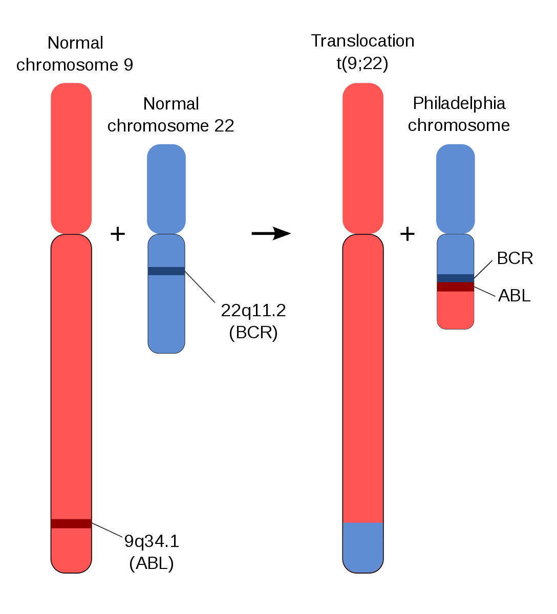
Schematic of Philadelphia chromosome translocation.
Image Source: Wikimedia Commons (2012). Schematic of the Philadelphia Chromosome, Licensed under a creative commons licence. Available for free at: https://commons.wikimedia.org/wiki/File:Schematic_of_the_Philadelphia_Chromosome.svg (Accessed on: 10 January 2023).

The incidence is 4.2 per 100,000 people per year. It can present at any age but the median age at diagnosis is 72 years.
Most cases are detected asymptomatically on routine bloods, but features can include:
- Constitutional symptoms
- Splenomegaly
- Symptomatic cytopenia – anaemia, bleeding, infections
- Gout (due to high cell turnover)
- Hyperviscosity (due to high WCC)
The following tests are usually performed during workup for suspected CML:
- Bloods – full panel, especially:
- FBC – leukocytosis is common with neutrophils, basophils and eosinophils. Thrombocytopenia and anaemia (normocytic) develop over time usually
- Blood film shows granulocytes of all stages of maturation
- BCR:ABL analysis can often be run on peripheral blood and is quantifiable – the percentage of BCR:ABL positive cells can be monitored as response to treatment
- Bone marrow is essential for evaluating blast percentages and staging disease and cytogenetics can also be sent
CML can be divided into three stages:
- Chronic phase – Most people are diagnosed in the chronic stage – counts are relatively well maintained and symptoms are minimal. This often lasts years.
- Accelerated phase – With rising blast counts (up to 29%), falling RBC and platelets and increasing symptoms.
- Blast phase – Finally in the blast phase the blast percentage rises to over 30% and the disease behaves like AML but with a very poor response to chemotherapy. The blast phase of CML is often rapidly fatal.
Treatment for CML has been revolutionised by the development of targeted therapy. The target is the tyrosine kinase protein produced by the BCR:ABL gene. These drugs are known as tyrosine kinase inhibitors; the first example of this class of drug was imatinib but there are now also second and third generation forms. They are generally well tolerated and have drastically changed prognosis for patients diagnosed with CML. The ten-year life expectancy has risen from around 11% to 84%, and patients diagnosed in the chronic phase have an almost normal life expectancy.
Chronic lymphocytic leukaemia (CLL)
Overview
Chronic lymphocytic leukaemia (CLL) is a malignant clonal expansion of immature B-lymphocytes. It is typically a slowly progressive condition and is increasingly common with age. The median age of diagnosis is 72 years. Only 10% are diagnosed under 55 years.
It is most commonly diagnosed on routine blood tests and is asymptomatic at presentation. Symptoms can include:
- Constitutional symptoms
- Enlarged lymph nodes or spleen
- Anaemia
- Bleeding symptoms from thrombocytopenia
- Increased susceptibility to infection
Diagnostic criteria:
- Persistent clonal lymphocytes >5 x 10⁹/L for more than 3 months
- Immunophenotype consistent with CLL
- Morphologically consistent with lymphocytes
Lymphadenopathy may or may not be present. There is an association of CLL with autoimmune phenomena such as Immune thrombocytopenia (ITP) and autoimmune haemolytic anaemia (AIHA).
Investigations for suspected CLL:
- Bloods – full panel, especially:
- FBC to look for cytopenia
- Blood film to evaluate morphology of lymphocytes
- Immunoglobulins, viral screen, DAT screen (AIHA)
- Flow cytometry to confirm CLL plus analysis for TP53 mutations and IGHV mutations (relevant for prognosis +/- treatment)
- CT scan – only if concerns about lymphadenopathy or splenomegaly
- Lymph node biopsy – only if large or rapidly growing lymph nodes to look for Richter’s transformation*
- Bone marrow biopsy – not always performed
*In a small proportion of cases, CLL can undergo a phenomenon known as Richter’s transformation. A proportion of clonal lymphocytes will mutate and take on the phenotype of a diffuse large B cell lymphoma. This carries a very poor prognosis.

Blood film showing typical appearance of CLL lymphocytes.
Image Source: Wikimedia Commons (2006). Chronic lymphocytic leukemia, Licensed under a creative commons licence. Available for free at: https://commons.wikimedia.org/wiki/File:Chronic_lymphocytic_leukemia.jpg (Accessed on: 10 January 2023).

Staging
Traditionally the Binet system was used in Europe, whereas the Rai system was used in the US. It is now more informative to use CLL-IPI, although the Rai-Binet system is still in use.
Binet A –Hb >100 g/L, Platelets >100 x 10⁹/L, <3 lymph node areas involved
Binet B – Hb and platelets as Binet A and >3 lymph node areas
Binet C – Hb <100 g/L or platelets <100 x 10⁹/L
Treatment
Not everyone needs treatment. CLL is a slow growing cancer and treatment carries significant morbidity risk. Most patients are managed with a ‘watch and wait’ approach to begin with. Treatment is not considered curative, and the disease course is of a relapsing and remitting nature.
Indications for treatment include:
- Progressive or troublesome lymphadenopathy
- Progressive splenomegaly
- Constitutional symptoms
- Cytopenias (Hb <100 g/L, Platelets <100 x 10⁹/L)
- Recurrent infections
- Associated immune phenomena e.g. AIHA not responsive to other treatment
- Lymphocyte doubling time < 6 months*
*This is a relative indication only
Treatment options themselves are beyond the scope of this course but include a combination of immunotherapy and conventional chemotherapy such as Fludarabine, Cyclophosphamide and Rituximab. Newer options such as Bruton Tyrosine Kinase inhibitors e.g. ibrutinib can also be used. The decision for treatment is determined by factors such as fitness for treatment, previous therapy, and mutation status of TP53. Some treatment courses can be quite intense (such as FCR) whereas ibrutinib is usually well tolerated.
Complete Guide to Clinical Genetics ST3 Application and Recruitment (2027)
Written by Medibuddy. Last updated: October 2026. Clinical Genetics is

Complete Guide to Clinical Radiology ST3 Application and Recruitment (2027)
Recruitment paused: NHS England is not running Clinical Radiology ST3+ recru

Complete Guide to Paediatric Surgery ST3 Application and Recruitment in United Kingdom (2026)
Written by Medibuddy. Last updated: October 2026. If you’re planning


Take your subscriptions with you
Our mobile app allows you to access your interview and exam question banks wherever you are.

