


 Close
Close
 25th June 2023
Cranial Nerve Disorders
Cranial Nerve Neuroanatomy
Rule of Four
The cranial nerves consist of twelve paired nerves that can be characterised as follows:
| Pure Sensory | Cranial nerves 1, 2, and 8 |
| Pure Motor | Cranial nerves 3, 4, 6, 11, and 12 |
| Mixed Sensorimotor | Cranial nerves 5, 7, 9, and 10 |
They can be structurally abstracted as part of the Rule of 4:
- 4 cranial nerves are situated above the pons: CN 1 and 2 in cerebrum, CN 3 and 4 in midbrain
- 4 cranial nerves are situated within the pons: CN 5, 6, 7, and 8
- 4 cranial nerves are situated below the pons: CN 9, 10, 11, and 12
- 4 motor cranial nerve nuclei are in the midline: CN 3, 4, 6 and 12, which are associated with three other important structures – the corticospinal tract (motor pathway), medial lemniscus (vibration and proprioception), and medial longitudinal fasciculus.
- 4 cranial nerve nuclei are in the lateral brainstem: CN 5, 7, 9 and 10, which are associated with three other important structures – the spinocerebellar pathway, spinothalamic pathway, and sympathetic pathway.
- 4 cranial nerves are closely related at the cerebellopontine angle – CN 5, 6, 7, 8, and the cerebellum. Tumours at that area, e.g. a vestibular schwannoma, are likely to cause ipsilateral CN 5,6,7 and 8 palsies with ataxia.
Decussations and bilateral innervation
A cranial nerve (i.e. the axon to and from its lower motor neuron nuclei) does not decussate, i.e. cross over to the opposite side, with the exceptions of the trochlear nerve (CN 4), and the medial fibres of the optic nerve (CN 2).
CN 2, 4, 7, and 12 cranial nerve nuclei receive bilateral innervation from upper motor neuron cortical inputs; therefore, cortical lesions can affect tracts to the cranial nuclei and subsequently result in an observed disorder of cranial nerve function, despite the lesion not affecting the cranial nerve directly.
Lower motor cranial nerve nuclei disorders will always result in an ipsilateral disorder except for CN 2 and CN 4, while lesions that affect upper motor cranial nuclei will result in bilateral or contralateral manifestations. The common examples of these will be discussed in the following sections.
Anatomical Relevance
In general, isolated disorders of the cranial nerve itself, i.e. lower motor neuron lesions of the cranial nerve, are less common than pathology affecting cortical/supranuclear pathways contributing to the relevant cranial nerves or pathology that will generally affect several nerves, e.g. generalised neuropathy. There are several common aetiologies that can affect any distribution of nerves and also affect supranuclear contributions to cranial nerves.
In general:
- Any physical impediment along the course of a cranial nerve can result in loss of function; this is commonly due to traumatic causes or neoplastic causes
- Neuroinflammatory conditions, e.g. multiple sclerosis, Lyme disease can affect any distribution of nerves and, while less common, can cause isolated cranial nerve palsies.
- Ischaemia and infarction; a macrovascular infarction, i.e. stroke, can cause supranuclear or lower motor nuclear manifestations.
- As per the rule of 4, infarctions above the level of the involved cranial nerve nuclei would result in an upper motor neuron manifestation of cranial nerve dysfunction, while lesions at the level of the nuclei cause lower motor neuron manifestations.
- For example, a pontine stroke due to posterior circulation infarction can cause an ipsilateral complete facial palsy (lesion at level of CN 7 nuclei ) with possible pseudobulbar palsy (above the level of CN 9, 10 and 12 nuclei), while a cortical anterior circulation infarction would cause a contralateral forehead sparing facial palsy (being above the level of CN 7 etc.)
- Microvascular changes, e.g. in diabetes mellitus, can cause ischaemia to any nerve, including cranial nerves.
False localising signs
A neurological sign is described as a false localising sign if it reflects dysfunction distant from the actual pathology. It commonly arises due to space-occupying lesions and diseases that cause raised intracranial pressure, including idiopathic intracranial hypertension. Extracranial false localising signs arise mainly due to spinal cord lesions.
Some anatomical considerations to consider are:
- Cranial nerve with the longest intracranial course is CN 4 – more vulnerable to head trauma
- Cranial nerve with the longest intradural course is CN 6 – most likely to be affected by raised intracranial pressure, vulnerable to stretching, classic example of false localising signs.
- CN 3 passes close to the tentorium and is vulnerable to uncal herniation/pressure effects at that level – vulnerable to false localisation
Not necessarily related to false localising signs but of interest :
- Cranial nerve with the longest intraosseous course is CN 7 – making it vulnerable to infection, trauma and congenital conditions with craniofacial craniosynostosis. Note its descent in the petrous part of the temporal bone which makes it vulnerable to facial fractures.
- Cranial nerve with longest extracranial course is CN 10 –vulnerable to traction and compression in the cervical area
- Cranial nerves in the cavernous sinus are CN 6, CN 3, CN 4, CN 5a – targets for cerebral venous sinus thrombosis.
Cranial nerves associated with vision – II, III, IV, VI
CNII – Optic Nerve
The optic nerve arises from the retina of each eye, passing through the optic canals and converging at the optic chiasm before continuing to the lateral geniculate body in the thalamus and into the cortex as optic radiations. There is bilateral innervation in that one half of each visual field is projected to the contralateral visual cortex, with the medial fibres of each optic nerve decussating at the optic chiasm to then travel to the contralateral cerebral hemisphere.
Relative Afferent Pupillary Defect – RAPD
Also known as a Marcus Gunn Pupil, a relative afferent pupillary defect (RAPD) is a condition where there is an asymmetric response to the same light stimuli as a result of unilateral damage to the optic afferent pathway involving the retina and optic nerve. This can be elicited by the swinging flashlight test (Marcus Gunn Test), where a pen torch is shone at one eye before moving to the other and alternating between the two to observe the pupils’ response. In RAPD, the affected optic afferent nerve would be less responsive to the light stimuli than the unaffected side and lead to a competition between the direct pupillary light reflex of that eye and the consensual pupillary light reflex of the contralateral eye. In the affected eye, the direct pupillary light reflex would be impaired by pathology, causing a paradoxical pupillary dilation in response to light stimuli.
Pathology that can cause RAPD can be divided into :
- Retinal causes
- Vascular causes –Ischaemic retinal disease and ocular disease
- Mechanical – Retinal detachment
- Infection
- Tumour
- Optic nerve causes
- Demyelination – Multiple sclerosis, neuromyelitis optica spectrum disorder
- Vascular –Ischaemia to the optic nerve, giant cell arteritis
- Inflammatory – Sarcoidosis, SLE
- Hereditary – Leber’s hereditary optic neuropathy
- Pressure – Glaucoma, Tumours
Optic Neuritis
Optic neuritis is an umbrella term for inflammation of the optic nerve, which can have many different aetiologies. A hallmark of optic neuritis is the presence of pain. It should be distinguished from optic neuropathy, which is a general term for any damage to the optic nerves. Optic neuritis is characterised by a rapid onset, painful ophthalmic condition associated with visual symptoms of dyschromatopsia, visual field defect or pupillary reflex abnormality.
There are several different conditions that can cause an optic neuritis:
- Demyelination:
- Multiple sclerosis
- Neuromyelitis optica spectrum disorders, which are a large spectrum of conditions exhibiting optic neuritis with myelitis, including:
- Anti-Aquaporin-4 IgG (Anti-AQP4) positive neuromyelitis optica
- Anti-AQP4 negative, Anti Myelin Oligodendrocyte Glycoprotein (anti-MOG) positive neuromyelitis optica.
- Anti-Glial Fibrillary Acidic protein positive neuromyelitis
- Autoimmune:
- Systemic Lupus Erythematosus
- Paraneoplastic
- Sarcoidosis
- Vasculitis, e.g. granulomatosis with polyangiitis
- Infective causes – Syphilis, Lyme disease
Treatment is with high dose IV methylprednisolone tapering onto oral prednisolone. As with all treatment with steroids, a PPI and bone protection should always be considered, and glycaemic control in patients with impaired glucose metabolism, e.g. diabetes, should also be considered. With antibody mediated optic neuritis, there is a role for plasma exchange (PLEX) or intravenous immunoglobulin (IVIg). Neuro-ophthalmology follow-up is essential.
Optic Neuropathy
Optic neuropathy is a very general term for any damage to the optic nerves, which also encompasses optic neuritis. The causes of optic neuropathy can broadly be divided into:
- Rapid onset – secondary to ischaemia, trauma, and autoimmune or inflammatory conditions.
- Gradual onset – secondary to toxins, e.g. lead toxicity-induced demyelination, drugs, e.g. ethambutol, compressive causes, e.g. glaucoma, metabolic, or mitochondrial causes.
Idiopathic Intracranial Hypertension – commonly affects the optic nerves
Idiopathic intracranial hypertension (IIH) commonly affects the optic nerves. IIH manifests as raised intracranial pressure without any obvious structural cause observed on imaging, with common associated clinical signs of vision loss and headache. Papilloedema can be found on fundoscopy.
Risk factors:
- Raised BMI
- Oral contraceptive pill use
- Tetracycline antibiotics
- Female
Treatment:
- Weight loss
- Acetazolamide
- Therapeutic lumbar puncture
Space occupying lesions can cause similar symptoms and should always be excluded before a diagnosis of IIH is made.
Leber’s hereditary optic neuropathy
Mitochondrially inherited defects in NADH-ubiquinone oxidoreductase chains interfere with glutamate transport and protection from reactive oxygen species, resulting in the loss of retinal ganglion cells and demyelination in the optic nerves, chiasm, and tracts. This manifests as acute progressive vision loss (both field and acuity) progressing from unilateral to bilateral, in young adults of 20 to 24 years old, with a preponderance for males. A key hallmark here is that it is painless and affects one eye before the other. It can be associated with extraocular neurological symptoms of dystonia and tremor and cardiac conduction defects, which if present, is termed LHON Plus.
Wolfram syndrome
Wolfram syndrome is a childhood onset autosomal-recessive genetic disorder that manifests as a combination of both diabetes insipidus and diabetes mellitus, with optic atrophy and deafness (the acronyms of which result in it being also known as DIDMOAD). It is caused by a mutation in the WFS-1 or WFS-2 gene which are responsible for the production of a protein called wolframin thought to regulate calcium metabolism. Not all the components of the syndrome may occur at once, but most sufferers of Wolfram syndrome will develop optic atrophy with diabetes mellitus.
CNIII – Oculomotor nerve
The oculomotor nerve extends from the ventral midbrain through the superior oblique fissure to travel to the extrinsic eye muscles. It has two nuclei in the midbrain, the motor nucleus which controls the extraocular muscles, and the parasympathetic nucleus, also known as the Edinger-Westphal nucleus, which controls the light reflex and accommodation reflex.
An oculomotor palsy consists of the following:
- Ptosis – paralysis of levator palpebrae superioris
- Lateral strabismus – unopposed CN 6 action
- Loss of pupillary light reflex
- Loss of accommodation
Care should be taken to distinguish the ptosis of an isolated CN 3 palsy from other causes of ptosis, including Horner’s Syndrome. The main differentiators are whether the ptosis is complete or partial, bilateral or unilateral, pupil size, along with other suggestive features.
| Laterality of ptosis | Pupil | Features | Condition |
|---|---|---|---|
| Unilateral | Small | Anhidrosis | Horner Syndrome – sympathetic interruption |
| Unilateral | Large | Pain is suggestive of sinister pathology | Isolated CN 3 palsy |
| Unilateral or Bilateral | Large | Hemiplegia contralateral to ophthalmoplegia | Midbrain lesion
Midbrain stroke syndrome (Weber Syndrome) |
| Bilateral | Normal | Fatigability | Myasthenia Gravis |
| Bilateral | Normal | Retinitis pigmentosa
Reduced eye movements |
Chronic Progressive External Ophthalmoplegia
Kearns – Sayre Syndrome |
| Bilateral | Normal | Frontal balding
Neck muscle wasting |
Dystrophica Myotonia |
| Bilateral | Normal | Areflexia, ataxia | Miller-Fisher Syndrome |
In the presence of a painful ophthalmoplegia, always be aware of intracavernous carotid artery dissections, particularly if the history involves trauma to the neck or head, or any Valsalva manoeuvre action, e.g. weightlifting, straining against a load. In a painful isolated CN 3 palsy, a posterior communicating artery dissection or aneurysm should always be excluded.
Other causes of a painful ophthalmoplegia include vascular causes, e.g. cerebral venous sinus thrombosis, neoplastic causes, infective causes, e.g. aspergillosis, inflammatory causes, e.g. sarcoidosis, and Tolosa Hunt syndrome, which is an idiopathic inflammatory diagnosis of exclusion that manifests as periorbital headache, ipsilateral painful ophthalmoplegia, and often pain in the distribution of the trigeminal nerve.
CN IV – Trochlear nerve
The trochlear nerve is a motor nerve that innervates the superior oblique muscle of the eye, allowing the eye to look down and outwards.
Palsy of the trochlear nerve primarily causes diplopia when looking downwards, e.g. when going downstairs. The trochlear nerve decussates in the brainstem after originating from its nuclei in the midbrain. A unilateral lesion to the trochlear nucleus affects the contralateral nerve and superior oblique muscle, whereas a lesion to the trochlear nerve itself results in ipsilateral signs. If there is evidence of involvement of other ocular motor nerves, a lesion in the cavernous sinus or midbrain is likely implicated as these nerves are in close proximity in this space and share blood supply.
CN VI – Abducens nerve
The abducens nerve is a motor nerve that innervates the lateral rectus muscle of the eye. Due to its length and course, it is most vulnerable to damage from raised intracranial pressure, such as idiopathic intracranial hypertension and is prone to producing a false localising sign. Palsy of the abducens nerve causes loss of function of the lateral rectus resulting in the inability to look laterally. This nerve is associated with CN 3 and 4 at points of its course and can be associated with painful ophthalmoplegia.
Internuclear Ophthalmoplegia
Internuclear ophthalmoplegia (INO) is the failure of adduction of the ipsilateral eye towards the midline and nystagmus in the contralateral eye as it looks away from the midline.

Schematic of right internuclear ophthalmoplegia.
Image Source: Wikimedia Commons (2006). Internuclear ophthalmoplegia, Licensed under a creative commons licence. Available for free at: https://commons.wikimedia.org/wiki/File:Internuclear_ophthalmoplegia.jpg (Accessed on: 11 January 2023).

INO results from a lesion in the median longitudinal fasciculus (MLF) which coordinates the oculomotor nerve of the ipsilateral eye with the contralateral trochlear and abducens nerves via the paramedian pontine reticular formation (PPRF). The PPRF is a supranuclear centre close to the ipsilateral abducens nucleus which controls horizontal conjugate movements.
An INO manifests as an inability to adduct the eye ipsilateral to the lesion of the MLF. A right INO is a disorder of the right MLF leading to an inability to adduct the right eye. Midbrain and pontine demyelinating lesions can cause a unilateral or bilateral INO.
Bilateral INO is very characteristic of multiple sclerosis and causes an inability to adduct either eye with resultant bilateral abduction nystagmus. If the ipsilateral PPRF or abducens nuclei are affected in addition to the ipsilateral MLF, this can cause a ‘One and a Half’ spectrum of syndromes, where an ipsilateral conjugate horizontal gaze palsy with an ipsilateral INO results in the ipsilateral eye being incapable of horizontal movement with varying degrees of response to horizontal vestibular reflexes depending on where the lesion is, while the contralateral eye also experiences the conjugate horizontal gaze palsy resulting inability to adduct the contralateral eye.
Vertical gaze palsy
A vertical gaze palsy, also known as a supranuclear gaze palsy, is a bilateral inability to make voluntary eye movements in the vertical plane, i.e. upward or downward gaze movements. Involuntary vertical eye movements would still be preserved, e.g. in reflex conjugate eye movements such as the vestibulo-ocular reflex, which can be demonstrated when an examiner passively moves the patient’s head up and down, leading to correction of the vertical paresis. On moving the head up while the patient’s gaze is fixed on a point, the eyes would reflexively move down as the head is moved up, a movement they would not be able to voluntarily do in a vertical gaze palsy.
This is a classic symptom in progressive supranuclear palsy and corticobasal degeneration but can also result from a vascular or infective insult to the structures involved – the interstitial nucleus of Cajal, rostral interstitial nucleus of medial longitudinal fasciculus, and cranial nerve 3 and 4 nuclei, and from mechanical causes like hydrocephalus or tumour. A caveat to this is if there is a bilateral infranuclear lesion, e.g. a neuromuscular junction or muscle lesion such as in myasthenia gravis, which would result in a gaze palsy that cannot be overcome by conjugate gaze testing.
Parinaud Syndrome
Parinaud syndrome, also known as dorsal midbrain syndrome, is a combination of a vertical gaze palsy with an Argyll-Robertson pupil with bilateral lid retraction (Collier sign). An Argyll-Robertson pupil is the manifestation of bilaterally small pupils that are not reactive to light but have a preserved accommodation reflex – i.e. are able to constrict when looking at near objects but not to light.
This sign is suggestive of damage to the midbrain involving damage of the pretectal nucleus or Edinger-Westphal nuclei, which results in loss of parasympathetic innervation to the iris sphincter muscles and an inability to constrict the pupil in response to light and is associated with diabetic neuropathy and neurosyphilis. As a component of Parinaud syndrome, dorsal midbrain lesions can result from a variety of aetiologies, including:
- vascular lesions, e.g. infarction, haemorrhage, arteriovenous malformations
- mechanical, e.g. tumours, hydrocephalus, trauma
- demyelinating and inflammatory conditions, e.g. neurosyphilis, multiple sclerosis and infection
The most common causes are pineal region tumours, and brainstem haemorrhage or infarction.
Kearns-Sayre syndrome
Kearns-Sayre syndrome, also known as oculocraniosomatic neuromuscular disorder with ragged red fibres, is a rare syndrome caused by mitochondrial DNA deletions resulting in defective oxidative phosphorylation and subsequent mitochondrial myopathy.
It manifests as a tetrad of:
- Chronic progressive external ophthalmoplegia – weakness and paralysis of eye muscles
- Pigmentary retinopathy leading to vision loss and night blindness
- Cardiomyopathy and cardiac conduction defects
- Cerebral folate deficiency leading to ataxia, sensorineural hearing loss, and cognitive impairment.
It typically manifests before the age of 20 and has no specific gender predilection. Treatment is supportive, including Co-enzyme Q supplementation and consideration of prophylactic pacemaker implantation.
Cranial nerves of the face – V, VII
CN V – Trigeminal nerve
The trigeminal nerve is divided into ophthalmic (V1), maxillary (V2) and mandibular (V3) divisions which convey sensory impulses in the distribution outlined in the image below.
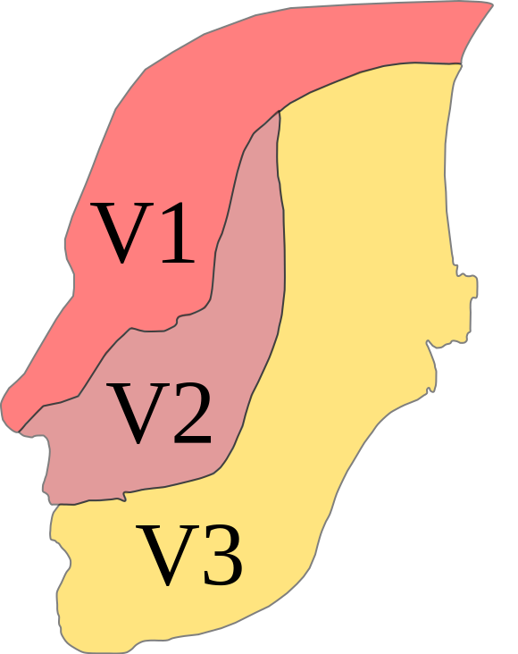
Dermatome distribution of the trigeminal nerve.
Image Source: Wikimedia Commons (2009). Trig innervation, Licensed under a creative commons licence. Available for free at: https://commons.wikimedia.org/wiki/File:Trig_innervation.svg (Accessed on: 11 January 2023).

The ophthalmic V1 branch supplies the scalp and forehead, conjunctiva, cornea, to the nose. The maxillary V2 supplies the lower eyelid, lower nose, maxilla, palate, and maxillary teeth, while the mandibular V3 branch supplies the oral cavity, mandibular teeth, and gums. Of note, all branches have some sensory supply to the commensurate parts of the meninges and sinuses in their distribution. The mandibular (V3) division also carries motor fibres for mastication, supplying the masseter, temporalis muscle, lateral and medial pterygoids, as well as supporting muscles that help with the process of mastication like the tensor tympani, which dampens loud noises including from chewing, and the mylohyoid, anterior belly of the digastric and tensor veli palatini which assist in biting and swallowing.
Hutchinson sign
Hutchinson sign refers to the involvement of the nasociliary nerve, a branch of V1 the ophthalmic nerve which innervates the cornea, eyelid, and lateral dorsum and tip of the nose in herpes zoster infections. It is relevant in the manifestation of herpes zoster ophthalmicus, which can present as periorbital and temporal pain before developing herpes zoster skin lesions in the distribution of the nasociliary nerve and can develop into ocular inflammation with corneal denervation. Care should be taken to differentiate this from Hutchinson’s pupil, which is unrelated and refers to an oculomotor nerve lesion causing an ipsilateral fixed dilated pupil.
Loss of corneal reflex
The corneal blink reflex is a loop between the trigeminal and the facial nerves, where contact with the cornea induces both the blink reflex and tear production. Disorder or disruption of the trigeminal nerve can cause the loss of this reflex, leading to loss of corneal health and damage. This can happen in pontine stroke or progressive supranuclear palsy.
Trigeminal neuralgia
Trigeminal neuralgia is an intense sporadic neuropathic pain confined to and following the distribution of the trigeminal nerve, manifesting with unilateral, severe, shooting or burning episodic pain with each episode lasting seconds to minutes, starting and stopping suddenly. Intervals between each episode are unpredictable and can happen multiple times a day, with episodes occurring over weeks, months to years. It can be triggered by touch (even that of a draught or wind), mastication, changes in temperature, and some types of food, including caffeine-containing, spicy, acidic, or strong flavoured foods.
Trigeminal neuralgia can be primary or secondary. The aetiology of primary trigeminal neuralgia is not clear but is strongly suspected to be related to compression of the trigeminal nerve divisions from contact with normal vascular structures, where the issue is intrinsic to the structure of the nerve with physiological variation. This accounts for 75% of cases and is the most common cause of trigeminal neuralgia. Secondary trigeminal neuralgia arises when the trigeminal nerve is affected by another medical condition, for example, demyelination from multiple sclerosis, invasion from a tumour or abnormal compression from masses like an arteriovenous malformation.
Trigeminal neuralgia needs to be distinguished from trigeminal autonomic cephalalgias such as cluster headaches, paroxysmal hemicrania, short-lasting unilateral neuralgiform headache attacks with conjunctival injection and tearing (SUNCT) and short-lasting unilateral neuralgiform headache attacks with autonomic symptoms (SUNA). The trigeminal autonomic cephalalgias share many features with trigeminal neuralgia, presenting as severe unilateral headache in a trigeminal distribution with ipsilateral cranial autonomic symptoms, such as ptosis, conjunctival tearing, lacrimation, and rhinorrhoea. While there is debate on whether these are distinct entities or a continuum of disorders on the same spectrum, these conditions can be distinguished by attack duration and frequency, triggers, as well as response to treatment and treatment types. Trigeminal neuralgia is characterised by a trigger area, with attacks that tend to be shorter (seconds to a few minutes) but which have a refractory period, with a high variability in frequency in attacks which can happen multiple times in the day, milder to non-existent autonomic symptoms, and with a lack of response to high dose oxygen and triptans which are useful in cluster headaches or lidocaine which is useful in SUNCT.
Treatment is primarily divided into medical and surgical options, with medical options focused on anticonvulsants and nerve modulating analgesic agents. Carbamazepine is the first line treatment for trigeminal neuralgia, with second line treatment being other anticonvulsants including oxcarbazepine which is a structural derivative of carbamazepine (reduced risk of the known adverse effects of agranulocytosis and anaemia of carbamazepine), lamotrigine, gabapentin, and phenytoin. Carbamazepine should be avoided in pregnancy due to the risk of spina bifida and cleft palate, while lamotrigine has the least reported severe effects in pregnancy. Nerve modulating analgesic agents aim to provide symptomatic relief and include amitriptyline and duloxetine.
Surgical treatments are generally reserved for patients who have severe pain with a debilitating impact on quality-of-life refractory to pharmacological treatments. These are categorised as destructive (i.e. lesioning techniques) or non-destructive depending on whether trigeminal nerve function and integrity is intentionally targeted. Non-destructive interventions include microvascular decompression, while destructive interventions like percutaneous radiofrequency rhizotomy, percutaneous balloon compression and stereotactic radiosurgery treat trigeminal neuralgia through the use of electrocoagulation, mechanical compression or ionising radiation respectively to selectively destroy the part of the nerve suspected to cause pain.
CN VII – Facial Nerve
The facial nerve is a mixed nerve that travels through the internal acoustic meatus and splits into five major branches (frontal, zygomatic, buccal, marginal mandibular, and cervical) within the parotid gland, which then go on to supply the motor functions of facial expression, autonomic impulses to lacrimal and salivary glands, stapedius muscle innervation that protects against loud noises and sensory function to the anterior two-thirds of the tongue.
Disorders of the CN VII lower motor nucleus will result in an ipsilateral facial asymmetry, with absence of transverse wrinkles of the forehead, loss of nasolabial fold, drooped corner of the mouth and loss of tone of the orbicularis oculi leading to eversion of the lower eyelid on the affected side. There is also loss of the efferent part of the conjunctival reflex, leading to uncontrolled tearing and inability to close the eye, which can result in eventual corneal ulceration. Bell’s Palsy is a classic example of an ipsilateral lower motor neuron CN 7 disorder.
Contralateral and bilateral innervation
CN VII demonstrates the effects of bilateral innervation from upper motor neuron sources as it receives stimulation from both the ipsilateral and the contralateral corticobulbar tracts.
The upper muscles of facial expression receive stimulation from both the ipsilateral and the contralateral corticobulbar tracts while the lower muscles receive stimulation only from the contralateral corticobulbar tract. A pathology disrupting this stimulation, e.g. an upper motor neuron lesion such as stroke, would cause loss of function of the contralateral lower muscles of facial expression but sparing of the upper muscles of facial expression as they would still be receiving stimulation from the ipsilateral upper motor neuron component.
Bell’s Palsy
A Bell’s Palsy is the classic example of an isolated lower motor neuron cranial nerve palsy. It manifests as a unilateral complete paralysis of the facial muscles of one side of the face. While the aetiology of a Bell’s Palsy is not completely clear, it has been associated with several conditions, specifically diabetes mellitus, herpes simplex type 1 viral infection, pregnancy, herpes zoster viral infection (shingles), hypertension, Lyme disease and autoimmune disease.
Due to the unilateral complete palsy of the facial muscles with no forehead sparing, this leads to ocular complications due to loss of the ability to blink and protect the exposed eye, dysarthria drooling, and difficulty eating and drinking due to the loss of facial muscle actions, as well as loss of ancillary functions of the facial nerve which include loss of function of the stapedius muscle in the ear leading to tinnitus and hyperacusis and loss of taste as a result of the facial nerve’s innervation to the anterior two thirds of the tongue.
During the recovery from Bell’s palsy, abnormal connections can form during nerve regeneration where efferent fibres from the superior salivary nucleus synapse with parasympathetic fibres of the facial nerve, causing tears to be produced during salivation in what is known as Bogorad syndrome or “Crocodile tears syndrome.”. This is similar to, but distinct from Frey syndrome, where sweating occurs with salivation due to abnormal connections between sympathetic and parasympathetic nerves associated with the auriculotemporal nerve (branch of mandibular division V3 of Trigeminal nerve), usually with an iatrogenic aetiology, e.g. parotid surgery.
Treatment of a Bell’s Palsy is with high dose oral prednisolone (a 10-day tapering course starting at 60mg Prednisolone) to reduce facial nerve inflammation. Investigations for conditions associated with Bell’s palsy should be considered where clinically suspected, e.g. Hba1c testing for diabetes, Lyme disease antibody titres. Antivirals such as aciclovir may be considered but should not be used in isolation. Eye protection is very important to replace the functions that serve to protect the cornea; lubricating eye drops and eye patches with ophthalmology follow-up is typically indicated.
Ramsay Hunt Syndrome
Ramsay Hunt syndrome is the manifestation of the reactivation of dormant varicella-zoster virus (shingles) with spread to the facial nerve. It is more common in immunocompromised individuals, e.g. patients with HIV, patients on immunotherapy, chemotherapy, or radiotherapy. Clinically, it manifests as a vesicular, erythematous, painful rash, in or around one ear with an ipsilateral ‘Bell’s palsy’ and can affect CN 5 and CN 8 with symptoms such as vertigo, tinnitus and hearing loss. The vesicles can be confined only to the auditory canal with facial pain and otalgia; all patients with facial pain and palsy should have an otoscopic exam.
Treatment is focused on the following:
- Disease modifying: Antivirals, e.g. acyclovir, famciclovir, together with corticosteroids.
- Symptomatic: carbamazepine for neuralgia, antihistamines, and anticholinergics for vertigo
- Eye care as in the treatment of a Bell’s palsy
Parotid tumour
The extracranial motor branch of the facial nerve splits into 5 branches in the parotid gland – the temporal branch, zygomatic branch, buccal branch, marginal mandibular branch, and cervical branch. A lesion in the parotid gland, e.g. trauma/iatrogenic during surgery or a tumour, can result in a lower motor neuron facial nerve palsy.
Vestibular schwannomas
A vestibular schwannoma, also known as an acoustic neuroma, is a benign tumour developing from the vestibulocochlear nerve. It is the most common cerebellopontine angle tumour (85%).
Cerebellopontine angle (CPA) tumours are the most common tumours in the posterior cranial fossa, specifically in the triangular space in the posterior cranial fossa, which is bound by the tentorium superiorly, medulla posteromedially and petrous part of temporal bone posterolaterally, through which run the anterior inferior cerebellar artery, CNs 5, 6, 7, and 8. Tumours in this area can cause ipsilateral LMN signs in CN 7, with overlapping CN5, 8, and 9 palsies. Thus a patient presenting with such a collection of ipsilateral signs, including the loss of corneal reflex, should prompt suspicion of a CPA lesion. Bilateral vestibular schwannomas are associated with neurofibromatosis type 2.
Pontine lesions
The facial cranial nerve nuclei of CN7 lies in the pons and is closely associated with the nuclei of the abducens nerve (CN6). A lesion in the pons, e.g. infarct or tumour, can cause an ipsilateral CN 7 and CN 6 palsy, with contralateral hemiplegia due to being above the decussation of the corticospinal tracts.
Bilateral facial palsy
A rare manifestation of facial palsy, these can be sequential, i.e. unilateral progressing to bilateral facial palsy or concurrent. A wide range of causes of unilateral facial palsy can have bilateral facial palsy, albeit rarely. As noted above, bilateral CPA tumours, or large pontine lesions (stroke/tumour) or systemic disease with multi nerve affecting pathologies such as Lyme disease, syphilis, sarcoidosis, demyelinating disease like Guillain Barre syndrome, vasculitis, and diabetes mellitus can be a cause. Rarer causes include hereditary congenital facial palsy, Melkersson-Rosenthal Syndrome (recurrent antihistamine resistant oedema of lips, cheeks, and eyelids with recurrent facial palsy and cheilitis) and Moebius-Poland syndrome (congenital facial and abducens palsy with pectoral and limb deformities).
Cranial nerves of the oropharynx and of smell – I, X, XII
CN1 – Olfactory nerve
The olfactory nerve arises from the olfactory epithelium, passing through the cribriform plate of the ethmoid bone and running through the olfactory bulb to terminate in the primary olfactory cortex. It carries afferent impulses for the sense of smell and is commonly affected by trauma and infection.
Problems with olfaction can also be a sentinel sign of neurodegenerative disorders which affect the olfactory bulb. Hyposmia is recognised as an early ‘pre-clinical’ sign of Parkinson’s disease, present in 75% of patients with idiopathic Parkinson’s disease, higher than the prevalence of resting tremor. Other α-synuclein and tau-related disorders, e.g. Alzheimer’s dementia, also have associations with olfactory dysfunction.
Kallmann syndrome
Kallmann syndrome is a rare form of hypogonadotropic hypogonadism. It is associated with anosmia or hyposmia as a result of failure of production or activity of gonadotropin-releasing hormone (GnRH) by the hypothalamus. This defect in GnRH producing neurons is also associated with the loss of olfactory neurons, resulting in the syndrome of anosmia and hypogonadism, failure to achieve puberty and cranio-facial defects.
CN X – Vagus nerve
The vagus nerve is a mixed nerve that extends beyond the cranial and cervical area, travelling via the jugular foramen to provide motor parasympathetic function to the cardiac and respiratory systems, and all visceral organs except the adrenal glands. It also innervates part of the pharynx and is responsible for the gag reflex.
Disorders of the vagus nerve can result in dysphagia as part of bulbar and pseudobulbar palsy, loss of the gag reflex which can contribute to aspiration pneumonia and brain-gut axis disorders causing gastroparesis and gastroesophageal reflux disease. Isolated disorder of the vagal nerve is very rare, and it is more likely to be involved as part of the manifestation of systemic pathology like stroke and multiple sclerosis.
CN XII – Hypoglossal nerve
The hypoglossal nerve arises from the medulla and travels through the hypoglossal canal, innervating the intrinsic and extrinsic muscles of the tongue. Disorders of the nerve cause problems with speech and swallowing.
Damage to the hypoglossal nerve itself (a lower motor neuron lesion) is very rare, mostly due to trauma and will cause an ipsilateral weakness of the tongue muscles, manifesting as deviation of the tongue to the ipsilateral side, due to ipsilateral tongue muscles being unable to balance the contraction of the genioglossus from the contralateral side.
Unilateral upper motor neuron lesions disrupting pathways to the hypoglossal nerve nuclei are more common and will result in contralateral weakness of the tongue, and therefore deviate to the contralateral side, as the contralateral muscles are affected and would be unable to balance the action of the unaffected side. Both unilateral and bilateral damage can occur, with bilateral damage resulting in a complete inability of the tongue to function.
Pseudobulbar palsy and bulbar palsy
Pseudobulbar palsy results from a lesion that is supranuclear to the hypoglossal nerve (i.e. an upper motor neuron lesion). Causes can be traumatic, metabolic, vascular, neoplastic, or autoimmune. In this instance, the corticobulbar tracts are most likely to be involved, and the manifestation is similar to that of a true bulbar palsy with impairment of articulation, mastication, and swallowing, but as the lesion is above the bulbar areas, it is termed ‘pseudo’ bulbar. Features include slow dysarthric speech, stiff and spastic tongue without fasciculations, dysphagia, and brisk jaw jerk. Due to the bilateral innervation of CN XII from the cortex, unilateral corticobulbar tract involvement would result in relatively preserved function.
True bulbar palsy is usually caused by a neurodegenerative disease that affects the bulbar areas, in particular, motor neuron disease with bulbar involvement such as progressive bulbar palsy, bulbar amyotrophic lateral sclerosis, and spinal and bulbar muscular atrophy (Kennedy’s disease – an X-linked recessive, CAG repeat mutation of the androgen receptor causing neurological, muscular, and endocrine loss of function.)
It is possible for other general causes of nerve disorders to have bulbar involvement and cause a bulbar palsy, e.g. a medullary brainstem stroke, or tumour specifically involving the brainstem, or neuroinflammatory conditions that can affect any nerve, e.g. Lyme disease, neurosarcoidosis, the acute bulbar palsy plus variant of Guillain Barré syndrome, but these are much rarer manifestations.
Vestibulocochlear Nerve and Spinal Accessory Nerve – VIII, XI
CN VIII – Vestibulocochlear nerve
The vestibulocochlear nerve arises from the inner ear apparatus for hearing and equilibrium, passing through the internal acoustic meatus. It has two divisions:
- The cochlear division – for hearing
- The vestibular division – for balance
Labyrinthitis and vestibular neuritis
Vestibular neuritis occurs when only the vestibular division of the vestibulocochlear nerve is inflamed, resulting in continuous dizziness and vertigo but no change in hearing. There is also an association with viral infections, usually upper respiratory tract infections.
Labyrinthitis occurs when both the cochlear and vestibular divisions of the nerve are affected, resulting in hearing changes in addition to continuous dizziness and vertigo. There is also an association with upper respiratory tract infections as with vestibular neuritis. Treatment for vestibular neuritis and labyrinthitis is with antihistamines like betahistine and meclizine.
It should be noted that Ménière’s disease with its presentation of vertigo, tinnitus and hearing loss is a disorder of the inner ear rather than of the vestibulocochlear nerve itself. The difference is that symptoms of Ménière’s disease are chronic and episodic, and there is no association with infection.
Vestibular schwannoma (acoustic neuroma)
As covered earlier in the section, a vestibular schwannoma (or acoustic neuroma) is a benign tumour of the vestibulocochlear nerve (CN 8) that can also affect the facial nerve (CN 7).
CN XI – Spinal accessory nerve
The spinal accessory nerve is formed from a cranial root from the medulla and a spinal root from the superior spinal cord which passes into the cranium via the foramen magnum. The combined accessory nerve leaves the cranium via the jugular foramen. The spinal accessory nerve supplies motor fibres to the larynx, pharynx, and soft palate, and innervates the sternocleidomastoid and the trapezius muscles.
Disorder of CN XI manifests primarily as weakness of the trapezius and sternocleidomastoid with signs of:
- Asymmetric neckline
- Drooping shoulder ipsilaterally
- Winged scapula
- Weakness of elevation of the shoulder ipsilaterally
- Weakness of head turning to the contralateral side – this is because the ipsilateral sternocleidomastoid muscle, which functions to push the head to the contralateral side, is affected
The aetiology is primarily due to trauma and more commonly iatrogenic trauma, e.g. neck surgery, lymph node biopsy.
Complete Guide to Clinical Genetics ST3 Application and Recruitment (2027)
Written by Medibuddy. Last updated: October 2026. Clinical Genetics is

Complete Guide to Clinical Radiology ST3 Application and Recruitment (2027)
Recruitment paused: NHS England is not running Clinical Radiology ST3+ recru

Complete Guide to Paediatric Surgery ST3 Application and Recruitment in United Kingdom (2026)
Written by Medibuddy. Last updated: October 2026. If you’re planning


Take your subscriptions with you
Our mobile app allows you to access your interview and exam question banks wherever you are.

