


 Close
Close
 31st January 2024
Crystal Arthropathy
Background
Crystal arthropathy is the deposition of crystals in one or more joints, resulting in joint destruction and soft tissue inflammatory reaction.
The two most common types of crystal arthropathy are gout and pseudogout. Gout is due to the deposition of monosodium urate crystals, and pseudogout is due to crystal deposition other than monosodium urate. Commonly, these crystals are calcium pyrophosphate dihydrate, but pseudogout can also occur due to deposition of the following:
- Calcium phosphate hydroxyapatite
- Calcium oxalate aluminium phosphate (deposited in end-stage renal failure patients on dialysis)
- Cholesterol deposited in rheumatoid arthritis and osteoarthritis
Prevalence
Crystal arthritis is more common in men, with a 20:1 male-to-female ratio. It is associated with increasing age, co-morbidities, and certain medical therapies. Importantly, it can be seen in tumour lysis syndrome in cancer patients receiving chemotherapy.
Tumour lysis syndrome occurs commonly in non-Hodgkin’s lymphoma and acute leukaemia but can also arise in other forms of cancer. It can occur spontaneously but is also commonly due to intracellular components being released during cell death in those receiving chemotherapy. The contents released into the bloodstream results in hyperuricaemia, hyperkalaemia, hypocalcaemia and hyperphosphataemia. The accumulation of urate can precipitate in joint spaces, causing gout.
Risk factors
Dietary factors associated with gout
- Meat consumption, specifically beef, pork, and lamb
- Alcohol: beer has a higher association with gout than spirits. There is no specific association between wine consumption and gout
- Sugary drinks- Consuming 2 or more sugar-sweetened soft drinks daily is strongly associated with gout
- There is an association between coffee consumption and gout but not with tea.
Medications
- Diuretics- thiazide and loop diuretics. Thiazide and loop diuretics lead to urate reabsorption at the proximal convoluted tubule (PCT) via the URAT1 transporter. It is thought to be due to salt and water loss promoting urate reabsorption, but the mechanism is still unclear.
- Cyclosporin when compared to tacrolimus and azathioprine in transplant recipients. This is due to reduced urate excretion and, therefore, a risk of gout.
Co-morbidities
- Obesity
- Hypertension
- Diabetes mellitus
- Hypercholesterolaemia and hypertriglyceridemia
- Cancers
- Increased age
- Osteoarthritis
- Previous joint trauma
- Metabolic disease such as hyperparathyroidism, hemochromatosis and hypomagnesaemia are less common.
Pathophysiology
In gout and pseudogout, crystal deposition in the joint occurs, which can be asymptomatic despite a trigger for the inflammatory process. An acute gout flare results in a significant degree of pain; however, the specific trigger of this is still unknown. During the inflammatory process, neutrophils cause phagocytosis of the crystals and release pro-inflammatory cytokines.
Gout is a problem with either purine metabolism or urate excretion. Adenine and guanine, 2 purine nucleic acids are metabolised into uric acid. Approximately two-thirds of uric acid is eliminated by the kidneys, and the remaining third by the gastrointestinal tract.
Uric acid has low water solubility and is mostly circulated in the plasma bound to albumin. In the renal tract, it is filtered via 4 channels: the urate transporter (UAT), 2 organic anion transporters (OAT1 and OAT3) and URAT1, which is responsible for tubular reabsorption of urate located at the apical membrane of the PCT. The URAT1 channel is thought to be key in the association of gout and diuretic use. With increasing diuresis, there is an increase in urate reabsorption; this, in combination with dehydration, provokes a gout attack. Alterations in these channels increases the risk of gout.
The normal level of urate is 1.5-6.0mg/dL in women and 2.5- 7.0 mg/dL in men. High urate levels, typically >7mg/dL can result in gout. Please note if the urate level is measured during an acute attack, the level may be below 7mg/dL- this does not exclude gout as a cause. This is thought to be due to the presence of tophi or a uric acid pool related to the acute flare; therefore, urate levels may be normal. If measured during intercritical phase (between acute painful attacks), the urate level will likely be elevated.
Under excretion can occur at the level of the kidneys due to altered transportation through the renal tubules or under excretion at the level of the gut (extra renal). This triggers an acute inflammatory reaction and the innate immune system.
There are five recognised stages of gout:
- Asymptomatic hyperuricaemia
- Acute gouty arthritis
- Intercritical gout (between acute painful attacks)
- Chronic tophaceous gout- tophi are urate crystal deposition in soft tissue. They are often asymptomatic.
- Gouty nephropathy
Although an acute attack of gout is self-limiting, the crystals remain within the joint space and, with time, can cause local erosion and destruction of the joint and surrounding soft tissue. The only way to definitively treat gout is by lowering the serum urate level. While elevated serum urate levels predispose the patient to developing gout, it does not automatically guarantee that gout will arise.
The following are contributing factors to an acute gout flare:
- Trauma
- Cold temperatures
- Previous disease
- Diet
- Dehydration
Pseudogout belongs to a group of diseases known as calcium pyrophosphate deposition (CPPD) diseases, to which chondrocalcinosis also belongs. It is also recognised to have different stages:
- CPPD: occurrence of calcium pyrophosphate crystals, with or without symptoms
- asymptomatic CPPD: chondrocalcinosis +/- changes of osteoarthritis, but clinically asymptomatic
- acute CPPD crystal arthritis (formerly pseudogout): self-limiting synovitis in the setting of CPPD
- osteoarthritis with CPPD: typical changes of osteoarthritis in the setting of CPPD
- chronic CPPD crystal inflammatory arthritis
References
- Feger J, Hacking C. Crystal arthropathy. Radiopaedia.org [Internet]. 2015 Aug 3 [cited 2022 May 9]; Available from: https://radiopaedia.org/articles/crystal-arthropathy?lang=gb
- C Ronco, Rodeghiero F. Hyperuricemic syndromes : pathophysiology and therapy. Basel ; New York: Karger; 2005.
- Howard SC, Jones DP, Pui C-H. The Tumor Lysis Syndrome. The New England journal of medicine [Internet]. 2011 May 12;364(19):1844–54. Available from: https://www.ncbi.nlm.nih.gov/pmc/articles/PMC3437249/
- Pascual E. Gout, diuretics and the kidney. Annals of the Rheumatic Diseases. 2006 Jan 13;65(8):981–2.
- Lin H-Y, Rocher LL, McQuillan MA, Schmaltz S, Palella TD, Fox IH. Cyclosporine-Induced Hyperuricemia and Gout. New England Journal of Medicine. 1989 Aug 3;321(5):287–92.
- Singh JA, Reddy SG, Kundukulam J. Risk factors for gout and prevention: a systematic review of the literature. Current Opinion in Rheumatology. 2011 Feb;23(2):1.
- Rho YH, Zhu Y, Zhang Y, Reginato AM, Choi HK. Risk factors for pseudogout in the general population. Rheumatology. 2012 Aug 11;51(11):2070–4.8. Furuhashi M. New insights into purine metabolism in metabolic diseases: role of xanthine oxidoreductase activity. American Journal of Physiology-Endocrinology and Metabolism. 2020 Nov 1;319(5):E827–34.
- Dalbeth N. Pathophysiology of gout. Oxford Medicine Online. Oxford University Press; 2016.
- Harris MD, Siegel LB, Alloway JA. Gout and Hyperuricemia. American Family Physician [Internet]. 1999 Feb 15 [cited 2022 May 9];59(4):925. Available from: https://www.aafp.org/afp/1999/0215/p925.html#:~:text=Gout%20progresses%20through%20four%20clinical
- Schumacher HR. The pathogenesis of gout. Cleveland Clinic Journal of Medicine [Internet]. 2008 Jul 1;75 Suppl 5:S2-4. Available from: https://www.ncbi.nlm.nih.gov/pubmed/18822468
- Zhang W, Doherty M, Bardin T, Barskova V, Guerne P-A, Jansen TL, et al. European League Against Rheumatism recommendations for calcium pyrophosphate deposition. Part I: terminology and diagnosis. Annals of the Rheumatic Diseases. 2011 Jan 7;70(4):563–70.
- Maiuolo J, Oppedisano F, Gratteri S, Muscoli C, Mollace V. Regulation of uric acid metabolism and excretion. International Journal of Cardiology [Internet]. 2016 Jun [cited 2022 May 24];213(213):8–14. Available from: https://www.sciencedirect.com/science/article/pii/S0167527315303429
- Schlesinger N, Norquist J M, Watson D J. Serum Urate During Acute Gout. The Journal of Rheumatology. 2009 Apr 15;36(6):1287–9.
- Torres RJ, Puig JG. Hypoxanthine-guanine phosophoribosyltransferase (HPRT) deficiency: Lesch-Nyhan syndrome. Orphanet Journal of Rare Diseases. 2007 Dec;2(1)
Presentation
Crystal arthropathy usually causes monoarthropathy. Typical joints involved in gout are the first metatarsophalangeal joint, ankle, knee, fingers, wrist, and elbow. If up to 4 joints are affected, this is oligoarthropathy, and it is polyarthropathy when more than 5 joints are affected.
Signs and Symptoms
Patients usually present with sudden onset pain. The associated signs on examination include erythema, swelling, tenderness and warmth of the affected joint. Generally, the patient would be otherwise systemically well. The pain is severe and often affects activities of daily living- this can be demonstrated by asking the patient to perform certain tasks (please see the examination section below). The acute joint pain can be associated with mild fever and rise in inflammatory markers, so it is essential to rule out septic arthritis; in septic arthritis, the patient would be acutely unwell.
Articular vs Extra-articular Manifestations
Extra-articular manifestations of gout present as gouty tophi. These are subcutaneous concentrations of urate crystals. These appear as white deposits of a chalk-like substance beneath the skin. Tophi can occur without previously having an acute attack of gout and do not typically cause pain. Common sites for tophaceous deposits include:
- The pinna of the ear.
- The Achilles tendon
- The pulp of the fingers
- The olecranon bursa. This can give the appearance of rheumatoid nodules, so careful history and examination of the patient are needed.
Another extra-articular feature of gout are renal calculi. These form due to uric acid salt deposition within the renal matrix. Low urinary pH increases the risk of uric acid stones and nephrolithiasis. Stone formation is an important cause of renal failure and should be considered and the risk reduced.
Complications of Hyperuricaemia
In addition to renal impairment, studies have shown that uric acid levels are independent risk factors for cardiovascular disease and associated mortality, particularly in those with hypertension and diabetes mellitus. This is related to oxidative stress caused by raised urate levels and inflammatory changes in the vasculature and is supported by urate-lowering therapies, such as allopurinol, resulting in improved cardiovascular outcomes.
The inflammatory process, when triggered within blood vessels, causes damage to the endothelium. Attempts to repair the damage results in fatty plaque deposits within the blood vessels, causing atheroma formation. This contributes to hypertension, which in turn increases blood vessel damage. Studies have shown that individuals with obesity but without metabolic syndrome have increased risk of diabetes and hypertension with raised serum urate levels. This is also true of lean individuals.

Image Source: Wikimedia Commons (2014). Tophaceous gout, Licensed under a creative commons licence. Available for free at: https://commons.wikimedia.org/wiki/File:Tophaceous_gout.jpg (Accessed: 30 April 2022).
Pseudogout often has a more insidious course, and symptoms may take longer to reach maximum debilitation during each flare, and there is often a longer recovery period. It is not uncommon for larger joints to be affected by pseudogout, such as knees.
References
- Harris MD, Siegel LB, Alloway JA. Gout and Hyperuricemia. American Family Physician [Internet]. 1999 Feb 15 [cited 2022 May 9];59(4):925. Available from: https://www.aafp.org/afp/1999/0215/p925.html#:~:text=Gout%20progresses%20through%20four%20clinical
- Society PCD. Gouty tophi [Internet]. Primary Care Dermatology Society. [cited 2022 Apr 30]. Available from: https://www.pcds.org.uk/clinical-guidance/gouty-tophi
- e.roddy@cphc.keele.ac.uk ARUPCC Primary Care Sciences, Keele University, Keele, UK. English: Tophaceous gout affecting the right great toe and finger interphalangeal joints. Note the asymmetrical swelling and yellow-white discolouration. [Internet]. Wikimedia Commons. 2014 [cited 2022 Apr 30]. Available from: https://commons.wikimedia.org/wiki/File:Tophaceous_gout.jpg
- MacMullan P, McCarthy G. Treatment and management of pseudogout: insights for the clinician. Therapeutic Advances in Musculoskeletal Disease. 2011 Dec 21;4(2):121–31.
- Arthritis Foundation. Juvenile Idiopathic Arthritis [Internet]. Arthritis.org. 2020 [cited 2022 Feb 5]. Available from: https://www.arthritis.org/diseases/juvenile-idiopathic-arthritis
- Lillicrap M. Crystal arthritis: contemporary approaches to diseases of antiquity. Clinical Medicine. 2007 Jan 1;7(1):60–4.
- Gupta MK, Singh JA. Cardiovascular Disease in Gout and the Protective Effect of Treatments Including Urate-Lowering Therapy. Drugs. 2019 Mar 13;79(5):531–41.
- Kuwabara M, Kuwabara R, Hisatome I, Niwa K, Roncal-Jimenez CA, Bjornstad P, et al. “Metabolically Healthy” Obesity and Hyperuricemia Increase Risk for Hypertension and Diabetes: 5-year Japanese Cohort Study. Obesity. 2017 Sep 18;25(11):1997–2008.
Diagnosis
History
Often, the diagnosis is clinical and based on a clear history; however, tests may be needed if there is doubt or atypical presentation. A good history should include some of the following questions:
- When did the pain start?
- Over what time frame did the pain reach its maximal intensity?
- Is there any association with the time of day?
- Does the pain get better/ worse with movement?
- Are there any exacerbating or relieving factors?
- Have there been any previous flares of this type of joint pain?
- Is the pain associated with systemic illness- fever, dysuria, rigors, etc.? If the answer to these questions is ‘yes,’ it is essential to consider septic arthritis as a cause for the presentation and treat it appropriately- septic arthritis should be treated promptly with input from the orthopaedic team- if missed septic arthritis can lead to death.
- Are there any other painful sites, e.g. tendons, eyes in uveitis? If uveitis is suspected, slit lamp examination should be performed and treated urgently to avoid visual loss.
In the case of an inflammatory joint pain, in addition to taking a full family and past medical history, an accurate sexual history helps to rule in or rule out reactive arthritis. Taking a sexual history requires a non-judgemental and sensitive approach. The following questions are important to ask:
- Are you sexually active?
- Do you practise genital/ anal/ oral sex or a combination of these? This will aid sites for testing if needed.
- Ask about genital symptoms:
- Do you have any burning or stinging on passing urine?
- Have you noticed any unusual discharge from the penis or any change to your normal discharge (in women)?
- Have you noticed any testicular pain?
- Is there pain on intercourse (dyspareunia)? If so, where is the pain?
- Have you noticed any bleeding between your periods (intermenstrual bleed)?
- Have you ever been tested and/ or treated for a sexually transmitted infection in the past? If so, when?
- Do you have a regular sexual partner? If not, then further questions related to contacts are needed, which can be done at GUM clinic.
- Do you use barrier contraception- i.e. condoms or diaphragms?
Examination
Examine the joint for warmth, erythema, swelling and injury. Assess for impairment in function- depending on the joint involved; it can affect walking, writing, etc. Perform a systemic examination to assess for infection, which could point to septic arthritis or signs to suggest an autoimmune or metabolic-related cause.
When examining the patient, tell the patient to show the affected joint- they will often be handling it with extreme care and may not wish to be touched immediately due to fear of pain. Be patient and offer to help the patient if they are in discomfort and struggling to expose the joint.
Start with inspection of the joint and compare it to the unaffected joint on the left/ right side. Ensure you carefully inspect both joints at all angles accessible- anterior, lateral, posterior and medial. Inspect the area immediately above and below the joint. Take note of scars, deformities, colour change, swelling or masses and where these are in relation to the joint- are they immediately overlying the joint, or do they appear to be related to the muscle or ligaments, for example. If a swelling is present, ask the patient how long it has been there for and if it has changed- the patient may say it has been there for 10 years, so may be irrelevant to the current presentation.
Following inspection comes palpation. Be mindful of the patient when palpating the joint. Explain clearly what you will do and tell the patient to mention if pain arises. Start with the “normal” side first- this will not only prepare the patient for what is to come for the affected side but will also give you information about the normal range of movement and function of the patient. Palpate above and below the joint. Feel the joint itself- you are trying to elicit any areas of swelling which are not obvious on initial inspection. Active synovitis is an inflammatory change in the joint space itself- it is felt as a fluctuant boggy pocket of fluid. Feel for temperature change- gentle palpation of both joints at the same time to identify temperature discrepancy- use the back of the hand and move proximal to distal.
Next, assess movement- active movement first, then passive movement. Depending on the joint involved, assessment of flexion, extension, abduction, adduction, internal and external rotation. Assessment of the degrees of movement actively (the patient doing it for themselves) and then passively (the examiner doing the movement for the patient) in comparison to the unaffected side. Whilst moving the joint, the examiner should also comment on evidence of crepitation of the joint and any clear difficulty expressed by the patient in doing these movements.
Examination of the joint above and below should be performed in thorough assessment of the joint and general physical examination of the individual. General examination can help rule in or rule out differential diagnoses or additional diagnoses that were not elicited on history taking.
General inspection can give clues about other co-morbidities relevant to joint pathology:
- Plaques seen on the extensor surfaces of the elbows, shins and near the hairline can indicate psoriasis
- Symmetrical deformity of the fingers and toes, as well as nodules near the elbows, can indicate a diagnosis of rheumatoid arthritis.
- Asymmetrical deformity of the distal interphalangeal joints and large joints such as knees can indicate a diagnosis of osteoarthritis.
Finally, assessing the function of the joint involved is key to assessing the effect the problem has on the patient. If the lower limb joints are involved, you may wish to watch the patient walk or take off their shoes and socks. If the upper limb is involved, you may wish to observe the patient pick up a coin, undo and do up buttons of a shirt or watch the patient pick up a pen and write. These demonstrate how the problem is affecting daily living activities. Above all else, an understanding of how the patients’ life is affected by the pain shows a holistic approach to patient care. It can lead to improved management of the patient’s expectations and consideration of prophylactic treatment.
Blood tests
- Full blood count- look for rise in white cells in infection, anaemia in autoimmune conditions such as rheumatoid arthritis, and platelet count-this is important if considering joint aspiration.
- Urea and electrolytes- deranged renal function can impact treatment choice- many drugs are renally excreted for dose adjustments and need to be considered.
- Urate- can be raised in gout but can be normal in acute flares
- CRP- raised in inflammatory conditions and infection
- Lipids profile and glucose- raised in metabolic conditions such as diabetes mellitus and hypercholesterolaemia. Elevated serum insulin levels affect the renal excretion of uric acid, therefore increasing urate concentration in the blood. Some studies demonstrated an association between high plasma triglycerides and elevated urate levels. This is thought to be related to triglyceride synthesis and de novo synthesis of uric acid by the liver.
Imaging
- X-rays in gout: asymmetrical punched-out erosions, tophi in chronic gout (occurs years after the initial attack). Preservation of joint space. Punched-out lytic lesions are also seen which can be large. During an acute flare, inflammatory changes can also be seen on radiographs but are non-specific.
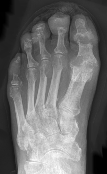
Image Source: Wikimedia Commons (2013). Gicht-Roe-multiple Arrosionen Destruktionen, Licensed under a creative commons licence. Available for free at: https://commons.wikimedia.org/wiki/File:Gicht_-_Roe_-_multiple_Arrosionen_Destruktionen.png (Accessed: 30 April 2022).
- X-rays in pseudogout: chondrocalcinosis. Chondrocalcinosis is not specific to pseudogout and is often present asymptomatically.

Image Source: Wikimedia Commons (2016). Chondrocalcinosis-annotated, Licensed under a creative commons licence. Available for free at: https://commons.wikimedia.org/wiki/File:Chondrocalcinosis_-_annotated.jpg (Accessed: 30 April 2022).
- NICE do not recommend further imaging if gout or pseudogout is suspected, as it is often a clinical diagnosis. However, if there is doubt and further imaging is recommended, then modalities such as ultrasound, MRI and CT scan can be considered.
Joint fluid aspiration
Request MC&S Turbid fluid should always be sent for MC&S, and aspirate should be viewed under plain polarised light:

Image Source: Wikimedia Commons (2008). Gout-monosodium-urate crystals in joint fluid (2), Licensed under a creative commons licence. Available for free at: https://commons.wikimedia.org/wiki/File:Gout_-_monosodium_urate_crystals_in_joint_fluid_(2).jpg (Accessed: 30 April 2022).

Image Source: Wikimedia Commons (2020). Birefringence microscopy of pseudogou, Licensed under a creative commons licence. Available for free at: https://commons.wikimedia.org/wiki/File:Birefringence_microscopy_of_pseudogout.jpg (Accessed: 30 April 2022).
Prior to joint aspiration, ensure the patient is not on any anticoagulant and has no history of clotting or bleeding disorders. If there is doubt, wait for formal clotting factors and full blood count results to come back before proceeding.
Bacteraemia is also a relative contraindication- however, if the source is thought to be due to a septic joint, then it should be performed preferably by an orthopaedic doctor.
Severe overlying infection, such as cellulitis over the swollen joint, is a contraindication to joint aspiration as this may introduce infection into a sterile joint.
References
- De Oliveira EP, Burini RC. High plasma uric acid concentration: causes and consequences. Diabetology & Metabolic Syndrome. 2012 Apr 4;4(1).
- Gentili A. The advanced imaging of gouty tophi. Current Rheumatology Reports. 2006 Jun;8(3):231–5.
- Hellerhoff. Deutsch: Gicht im Röntgenbild des Fußes mit multiplen Destruktionen. [Internet]. Wikimedia Commons. 2013 [cited 2022 Apr 30]. Available from: https://commons.wikimedia.org/wiki/File:Gicht_-_Roe_-_multiple_Arrosionen_Destruktionen.png
- Taokinesis. English: X-ray of a knee with chondrocalcinosis. [Internet]. Wikimedia Commons. 2016 [cited 2022 Apr 30]. Available from: https://commons.wikimedia.org/wiki/File:Chondrocalcinosis_-_annotated.jpg
- USA EU from H TX. Gout: monosodium urate crystals in joint fluid [Internet]. Wikimedia Commons. 2007 [cited 2022 Apr 30]. Available from: https://commons.wikimedia.org/wiki/File:Gout_-_monosodium_urate_crystals_in_joint_fluid_(2).jpg
- features MH M. D.-Author info- Reusing images- Conflicts of interest: None Mikael Häggström Consent note: Consent from the patient or patient’s relatives is regarded as redundant, because of absence of identifiable. English: Microscopy with polarised light of tissue by a metatarsal joint, showing crystals of which some have rhomboid shape and weak positive birefringence, consistent with calcium pyrophosphate dihydrate crystal deposition disease (pseudogout). [Internet]. Wikimedia Commons. 2020 [cited 2022 Apr 30]. Available from: https://commons.wikimedia.org/wiki/File:Birefringence_microscopy_of_pseudogout.jpg
- Thomsen TW, Shen S, Shaffer RW, Setnik GS. Arthrocentesis of the Knee. New England Journal of Medicine [Internet]. 2006 May 11 [cited 2020 Jan 29];354(19):e19. Available from: https://www.nejm.org/doi/full/10.1056/NEJMvcm05191440. Lillicrap M. Crystal arthritis: contemporary approaches to diseases of antiquity. Clinical Medicine. 2007 Jan 1;7(1):60–4.
Differential diagnoses
Septic arthritis
Septic arthritis is a potentially life-threatening condition. It should be suspected in anyone who is systemically unwell with or without fever and has an acutely hot, swollen, painful joint. The sepsis 6 guidelines should be followed, and local guidelines of antibiotic choice used. Once clinically suspected, septic arthritis should be investigated and treated urgently, as there is a ~10% mortality rate in septic arthritis.
Treatment with intravenous antibiotics and fluids should commence within 1 hour of clinical suspicion. Culture sample should be taken in the same way as for treatment of sepsis; however, importantly, joint aspiration samples should be obtained using an antiseptic non-touch technique. If confirmed, discussion with the Orthopaedics team is often needed for consideration of joint washout. If there is a suspected joint prosthesis infection, discussion with the orthopaedic team is needed promptly, and joint aspiration should be performed by them.
Joints are commonly infected with bacteria, but viral and fungal infections should be considered in the differential diagnoses as treatment will differ. Microbiology advice should be sought if there is doubt.
Delayed or poorly treated septic arthritis can result in reduced joint functioning and possibly joint loss, and therefore, long-term disability.
Haemarthrosis
Haemarthrosis is due to bleeding into a joint. It typically results following trauma and affects 1 joint- therefore, it can present as a painful, swollen single joint. A clear history is important to diagnose the condition, as well as clinical examination and imaging. Atraumatic haemarthrosis can occur in people with bleeding disorders, for example, haemophilia. A definitive diagnosis will involve joint aspiration- where clotting or bleeding disorders are of concern, this should be done by Orthopaedic colleagues and should be discussed with the Haematology team as consideration of blood products may be needed to avoid life-threatening bleeding.
Reactive arthritis
Reactive arthritis is a seronegative arthritis resulting from an inflammatory response to an extra-articular infection (rather than intra-articular). It commonly presents with urogenital infection (such as Chlamydia) or gastrointestinal infection (such as Campylobacter, Shigella, Salmonella and Yersinia). There is a delay in joint manifestation following an acute infection, and aspiration of the joint will not yield a micro-organism, unlike in septic arthritis. There is a typical triad of symptoms- conjunctivitis, urethritis and arthritis, although other manifestations can also be present. It is also common for only 2 parts of the triad to be present in reactive arthritis.
Reactive arthritis can present as a monoarthritis or a polyarthritis. It is typically asymmetrical, and a distant site of infection can be determined on thorough history taking and systems examination, which can include examination of the external genitalia. A detailed but sensitive sexual history is important here.
Monoarticular manifestation of inflammatory arthritis
Inflammatory arthritis, such as rheumatoid arthritis, psoriatic arthritis, and other connective tissue diseases with joint manifestations, can all result in monoarticular pathology. However, inflammatory arthritis is usually polyarticular, and some are typically symmetrical in pattern. The type of joint involved can also vary, from small joints to large joints.
The pathophysiology of arthritis is similar in crystal arthropathy- an influx of inflammatory cells from the circulation to the joint space and synovium, triggering the inflammatory reaction. However, inflammatory arthritis typically follows a more chronic course and has different extra-articular manifestations. The pain and loss of function of the joints involved are associated with disuse of the joint and improvement on movement- therefore, time of day and exacerbating factors are important to ask about. The treatment options for inflammatory arthritis also differ with consideration of disease-modifying anti-rheumatoid drugs, DMARDs, and immunosuppressive therapy.
Traumatic synovitis
Traumatic synovitis is an inflammatory process occurring after a traumatic insult. The inflammatory reaction occurs as part of the healing process. Traumatic synovitis is associated with osteoarthritis.
References
- Taylor-Robinson D, Keat A. Septic and aseptic arthritis: a continuum? Best Practice & Research Clinical Rheumatology. 1999 Mar;13(1):179–92.
- Nigrovic P. UpToDate [Internet]. www.uptodate.com. 2022 [cited 2022 May 7]. Available from: https://www.uptodate.com/contents/overview-of-hemarthrosis
- Yu D, Tubergen A. UpToDate [Internet]. www.uptodate.com. 2021 [cited 2022 May 16]. Available from: https://www.uptodate.com/contents/reactive-arthritis
- Stavropoulos PG, Soura E, Kanelleas A, Katsambas A, Antoniou C. Reactive Arthritis. Journal of the European Academy of Dermatology and Venereology. 2014 Sep 8;29(3):415–24.
- Smolen J. UpToDate [Internet]. www.uptodate.com. 2020 [cited 2022 May 16]. Available from: https://www.uptodate.com/contents/undifferentiated-early-inflammatory-arthritis-in-adults
- Liao L, Zhang S, Zhao L, Chang X, Han L, Huang J, et al. Acute Synovitis after Trauma Precedes and is Associated with Osteoarthritis Onset and Progression. International Journal of Biological Sciences. 2020;16(6):970–80.
Management
The approach to management of patients with gout and pseudogout are initially similar- symptom control and aim to enable return of joint use and independence. However, the management of pseudogout is more restricted- there are no specific agents that can be used to reduce the crystal deposition in pseudogout, whereas multiple agents are currently in use for the management and prevention of gout.
Conservative
- Rest, ice and elevate
- Avoid trauma
- Lifestyle changes- encourage exercise and healthy diet
As mentioned previously, gout is linked to purine levels. Increased consumption of food and drink with high levels of purine increases the risk of gout flares occurring.
Alcohol consumption increases the risk of suffering from gout. Beers and lagers have higher levels of purine than spirits, whereas consuming moderate levels of wine does not increase the risk of gout. The consumption of 2 or more sugar-sweetened drinks in a day also increases the incidence of gout. Consuming high quantities of meats, notably beef, pork, lamb, and seafood increases this risk of gout due to the levels of purine consumption. Reducing the consumption of these products can aid in reducing gout attacks. By contrast, consuming vegetables with high purine content has not demonstrated an increased risk of gout. In addition, people who consume high quantities of dairy products have a lower incidence of gout.
Pharmacological
Consider contraindications to proposed pharmacological therapy prior to initiation. In particular, renal function needs to be considered. Other contraindications include intolerance and, therefore, non-compliance, severe side effects such as peptic ulceration and gastrointestinal bleeding, allergic reaction, previous failure with this agent and medication interactions. Drug-drug interactions are numerous depending on what the patient is taking and should be considered individually.
During an acute flare
- Non-steroidal anti-inflammatory (NSAID) agents- to be continued until 1-2 days after cessation of the attack. (NB Aspirin is not indicated in gout. In fact, low-dose aspirin can affect renal excretion of uric acid and increase the risk of a gout flare)
- Consider co-prescribing a proton pump inhibitor with NSAIDs, particularly if there is a risk of gastric ulceration and GI bleed.
- Prednisolone is considered for those with an acute flare with difficult symptom control and affecting quality of life. Not all patients with a flare of crystal arthropathy require steroids, and this should be considered on an individual basis. Co-morbidities are also important to consider; for example, those with diabetes will likely suffer from labile blood sugar levels. If steroids are needed, a short course of approximately 5 days only should be used; therefore, weaning is usually not needed. The use of steroids is aimed at the inflammatory response- to dampen this and aid the symptoms of an attack.
- Oral colchicine- be aware of the common side effects of these medications. The main common side effects affect the gastrointestinal system. Colchicine is often used in gout management but the evidence for use in pseudogout is variable. Colchicine is increasingly used in prophylaxis of gout with gradual dose increase as tolerated by the patient.
- Colchicine is an alkaloid used in the symptomatic relief of pain in attacks of gout and to treat the inflammatory symptoms of Familial Mediterranean Fever.
- Colchicine works by blocking the mitotic activity during metaphase; it binds to tubulin, which, by forming complexes, prevents elongation, thus impairing the inflammatory process.
Prophylaxis
- Xanthine oxidase inhibitors, such as allopurinol or febuxostat, are used for prevention of gout attacks. Initiation should be avoided during an acute flare, but once initiated, can be continued during flares.
- Allopurinol: allopurinol undergoes first-pass metabolism and is converted to its active form Oxypurinol (Alloxanthine), which acts as an inhibitor of xanthine oxidase enzyme. A starting dose of between 50mg-100mg daily is recommended, with gradual dose increase to a maximum of 900mg in daily divided doses with the aim to reach a target serum urate level <6mg/dL. During an acute gout flare, allopurinol should be continued.
- Febuxostat is a non-purine selective xanthine oxidase inhibitor recommended in the lowering of uric acid levels in a dose-dependent manner in those who are not responsive or intolerant of allopurinol. It has been shown to lower serum urate levels to <6mg/ dL better than allopurinol.
- Uricase agents, e.g. Pegloticase, can be used to reduce urate levels to prevent further acute flares.
Surgical intervention
Tophi can be surgically excised, but this will not prevent further tophi from appearing. Patients should be counselled on the risks vs. benefits of surgical intervention, particularly as this is not a definitive solution. Risks include infection, structural damage and failure. People with co-morbidities, such as diabetes mellitus, are at greater risk of these complications.
Patients sometimes request surgery for joint remodelling or replacement, but again, this does not treat the underlying condition and is not a long-term treatment option.
References
- MacMullan P, McCarthy G. Treatment and management of pseudogout: insights for the clinician. Therapeutic Advances in Musculoskeletal Disease. 2011 Dec 21;4(2):121–31.
- Lillicrap M. Crystal arthritis: contemporary approaches to diseases of antiquity. Clinical Medicine. 2007 Jan 1;7(1):60–4.
- CKS is only available in the UK [Internet]. NICE. 2018 [cited 2022 Apr 30]. Available from: https://cks.nice.org.uk/topics/gout/prescribing-information/allopurinol/
- Choi HK, Atkinson K, Karlson EW, Willett W, Curhan G. Alcohol intake and risk of incident gout in men: a prospective study. The Lancet. 2004 Apr;363(9417):1277–81.
- Leung YY, Yao Hui LL, Kraus VB. Colchicine—Update on mechanisms of action and therapeutic uses. Seminars in Arthritis and Rheumatism. 2015 Dec;45(3):341–50.
- Hainer BL, Matheson E, R. Travis Wilkes. Diagnosis, Treatment, and Prevention of Gout. American Family Physician [Internet]. 2014 [cited 2022 May 9];90(12):831–6. Available from: https://www.aafp.org/afp/2014/1215/p831.html
- Qurie A, Pankaj Bansal, Amandeep Goyal, Musa R. Allopurinol [Internet]. Nih.gov. StatPearls Publishing; 2019 [cited 2022 Apr 30]. Available from: https://www.ncbi.nlm.nih.gov/books/NBK499942/
- Gerriets V, Ishwarlal Jialal. Febuxostat [Internet]. Nih.gov. StatPearls Publishing; 2021 [cited 2022 Apr 30]. Available from: https://www.ncbi.nlm.nih.gov/books/NBK544239/
- Neogi T. Gout. New England Journal of Medicine. 2011 Feb 3;364(5):443–52.
Complete Guide to Clinical Genetics ST3 Application and Recruitment (2027)
Written by Medibuddy. Last updated: October 2026. Clinical Genetics is

Complete Guide to Clinical Radiology ST3 Application and Recruitment (2027)
Recruitment paused: NHS England is not running Clinical Radiology ST3+ recru

Complete Guide to Paediatric Surgery ST3 Application and Recruitment in United Kingdom (2026)
Written by Medibuddy. Last updated: October 2026. If you’re planning


Take your subscriptions with you
Our mobile app allows you to access your interview and exam question banks wherever you are.

