


 Close
Close
 31st January 2024
Diagnosis
History
Often, the diagnosis is clinical and based on a clear history; however, tests may be needed if there is doubt or atypical presentation. A good history should include some of the following questions:
- When did the pain start?
- Over what time frame did the pain reach its maximal intensity?
- Is there any association with the time of day?
- Does the pain get better/ worse with movement?
- Are there any exacerbating or relieving factors?
- Have there been any previous flares of this type of joint pain?
- Is the pain associated with systemic illness- fever, dysuria, rigors, etc.? If the answer to these questions is ‘yes,’ it is essential to consider septic arthritis as a cause for the presentation and treat it appropriately- septic arthritis should be treated promptly with input from the orthopaedic team- if missed septic arthritis can lead to death.
- Are there any other painful sites, e.g. tendons, eyes in uveitis? If uveitis is suspected, slit lamp examination should be performed and treated urgently to avoid visual loss.
In the case of an inflammatory joint pain, in addition to taking a full family and past medical history, an accurate sexual history helps to rule in or rule out reactive arthritis. Taking a sexual history requires a non-judgemental and sensitive approach. The following questions are important to ask:
- Are you sexually active?
- Do you practise genital/ anal/ oral sex or a combination of these? This will aid sites for testing if needed.
- Ask about genital symptoms:
- Do you have any burning or stinging on passing urine?
- Have you noticed any unusual discharge from the penis or any change to your normal discharge (in women)?
- Have you noticed any testicular pain?
- Is there pain on intercourse (dyspareunia)? If so, where is the pain?
- Have you noticed any bleeding between your periods (intermenstrual bleed)?
- Have you ever been tested and/ or treated for a sexually transmitted infection in the past? If so, when?
- Do you have a regular sexual partner? If not, then further questions related to contacts are needed, which can be done at GUM clinic.
- Do you use barrier contraception- i.e. condoms or diaphragms?
Examination
Examine the joint for warmth, erythema, swelling and injury. Assess for impairment in function- depending on the joint involved; it can affect walking, writing, etc. Perform a systemic examination to assess for infection, which could point to septic arthritis or signs to suggest an autoimmune or metabolic-related cause.
When examining the patient, tell the patient to show the affected joint- they will often be handling it with extreme care and may not wish to be touched immediately due to fear of pain. Be patient and offer to help the patient if they are in discomfort and struggling to expose the joint.
Start with inspection of the joint and compare it to the unaffected joint on the left/ right side. Ensure you carefully inspect both joints at all angles accessible- anterior, lateral, posterior and medial. Inspect the area immediately above and below the joint. Take note of scars, deformities, colour change, swelling or masses and where these are in relation to the joint- are they immediately overlying the joint, or do they appear to be related to the muscle or ligaments, for example. If a swelling is present, ask the patient how long it has been there for and if it has changed- the patient may say it has been there for 10 years, so may be irrelevant to the current presentation.
Following inspection comes palpation. Be mindful of the patient when palpating the joint. Explain clearly what you will do and tell the patient to mention if pain arises. Start with the “normal” side first- this will not only prepare the patient for what is to come for the affected side but will also give you information about the normal range of movement and function of the patient. Palpate above and below the joint. Feel the joint itself- you are trying to elicit any areas of swelling which are not obvious on initial inspection. Active synovitis is an inflammatory change in the joint space itself- it is felt as a fluctuant boggy pocket of fluid. Feel for temperature change- gentle palpation of both joints at the same time to identify temperature discrepancy- use the back of the hand and move proximal to distal.
Next, assess movement- active movement first, then passive movement. Depending on the joint involved, assessment of flexion, extension, abduction, adduction, internal and external rotation. Assessment of the degrees of movement actively (the patient doing it for themselves) and then passively (the examiner doing the movement for the patient) in comparison to the unaffected side. Whilst moving the joint, the examiner should also comment on evidence of crepitation of the joint and any clear difficulty expressed by the patient in doing these movements.
Examination of the joint above and below should be performed in thorough assessment of the joint and general physical examination of the individual. General examination can help rule in or rule out differential diagnoses or additional diagnoses that were not elicited on history taking.
General inspection can give clues about other co-morbidities relevant to joint pathology:
- Plaques seen on the extensor surfaces of the elbows, shins and near the hairline can indicate psoriasis
- Symmetrical deformity of the fingers and toes, as well as nodules near the elbows, can indicate a diagnosis of rheumatoid arthritis.
- Asymmetrical deformity of the distal interphalangeal joints and large joints such as knees can indicate a diagnosis of osteoarthritis.
Finally, assessing the function of the joint involved is key to assessing the effect the problem has on the patient. If the lower limb joints are involved, you may wish to watch the patient walk or take off their shoes and socks. If the upper limb is involved, you may wish to observe the patient pick up a coin, undo and do up buttons of a shirt or watch the patient pick up a pen and write. These demonstrate how the problem is affecting daily living activities. Above all else, an understanding of how the patients’ life is affected by the pain shows a holistic approach to patient care. It can lead to improved management of the patient’s expectations and consideration of prophylactic treatment.
Blood tests
- Full blood count- look for rise in white cells in infection, anaemia in autoimmune conditions such as rheumatoid arthritis, and platelet count-this is important if considering joint aspiration.
- Urea and electrolytes- deranged renal function can impact treatment choice- many drugs are renally excreted for dose adjustments and need to be considered.
- Urate- can be raised in gout but can be normal in acute flares
- CRP- raised in inflammatory conditions and infection
- Lipids profile and glucose- raised in metabolic conditions such as diabetes mellitus and hypercholesterolaemia. Elevated serum insulin levels affect the renal excretion of uric acid, therefore increasing urate concentration in the blood. Some studies demonstrated an association between high plasma triglycerides and elevated urate levels. This is thought to be related to triglyceride synthesis and de novo synthesis of uric acid by the liver.
Imaging
- X-rays in gout: asymmetrical punched-out erosions, tophi in chronic gout (occurs years after the initial attack). Preservation of joint space. Punched-out lytic lesions are also seen which can be large. During an acute flare, inflammatory changes can also be seen on radiographs but are non-specific.
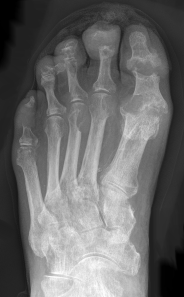
Image Source: Wikimedia Commons (2013). Gicht-Roe-multiple Arrosionen Destruktionen, Licensed under a creative commons licence. Available for free at: https://commons.wikimedia.org/wiki/File:Gicht_-_Roe_-_multiple_Arrosionen_Destruktionen.png (Accessed: 30 April 2022).
- X-rays in pseudogout: chondrocalcinosis. Chondrocalcinosis is not specific to pseudogout and is often present asymptomatically.

Image Source: Wikimedia Commons (2016). Chondrocalcinosis-annotated, Licensed under a creative commons licence. Available for free at: https://commons.wikimedia.org/wiki/File:Chondrocalcinosis_-_annotated.jpg (Accessed: 30 April 2022).
- NICE do not recommend further imaging if gout or pseudogout is suspected, as it is often a clinical diagnosis. However, if there is doubt and further imaging is recommended, then modalities such as ultrasound, MRI and CT scan can be considered.
Joint fluid aspiration
Request MC&S Turbid fluid should always be sent for MC&S, and aspirate should be viewed under plain polarised light:

Image Source: Wikimedia Commons (2008). Gout-monosodium-urate crystals in joint fluid (2), Licensed under a creative commons licence. Available for free at: https://commons.wikimedia.org/wiki/File:Gout_-_monosodium_urate_crystals_in_joint_fluid_(2).jpg (Accessed: 30 April 2022).

Image Source: Wikimedia Commons (2020). Birefringence microscopy of pseudogou, Licensed under a creative commons licence. Available for free at: https://commons.wikimedia.org/wiki/File:Birefringence_microscopy_of_pseudogout.jpg (Accessed: 30 April 2022).
Prior to joint aspiration, ensure the patient is not on any anticoagulant and has no history of clotting or bleeding disorders. If there is doubt, wait for formal clotting factors and full blood count results to come back before proceeding.
Bacteraemia is also a relative contraindication- however, if the source is thought to be due to a septic joint, then it should be performed preferably by an orthopaedic doctor.
Severe overlying infection, such as cellulitis over the swollen joint, is a contraindication to joint aspiration as this may introduce infection into a sterile joint.
References
- De Oliveira EP, Burini RC. High plasma uric acid concentration: causes and consequences. Diabetology & Metabolic Syndrome. 2012 Apr 4;4(1).
- Gentili A. The advanced imaging of gouty tophi. Current Rheumatology Reports. 2006 Jun;8(3):231–5.
- Hellerhoff. Deutsch: Gicht im Röntgenbild des Fußes mit multiplen Destruktionen. [Internet]. Wikimedia Commons. 2013 [cited 2022 Apr 30]. Available from: https://commons.wikimedia.org/wiki/File:Gicht_-_Roe_-_multiple_Arrosionen_Destruktionen.png
- Taokinesis. English: X-ray of a knee with chondrocalcinosis. [Internet]. Wikimedia Commons. 2016 [cited 2022 Apr 30]. Available from: https://commons.wikimedia.org/wiki/File:Chondrocalcinosis_-_annotated.jpg
- USA EU from H TX. Gout: monosodium urate crystals in joint fluid [Internet]. Wikimedia Commons. 2007 [cited 2022 Apr 30]. Available from: https://commons.wikimedia.org/wiki/File:Gout_-_monosodium_urate_crystals_in_joint_fluid_(2).jpg
- features MH M. D.-Author info- Reusing images- Conflicts of interest: None Mikael Häggström Consent note: Consent from the patient or patient’s relatives is regarded as redundant, because of absence of identifiable. English: Microscopy with polarised light of tissue by a metatarsal joint, showing crystals of which some have rhomboid shape and weak positive birefringence, consistent with calcium pyrophosphate dihydrate crystal deposition disease (pseudogout). [Internet]. Wikimedia Commons. 2020 [cited 2022 Apr 30]. Available from: https://commons.wikimedia.org/wiki/File:Birefringence_microscopy_of_pseudogout.jpg
- Thomsen TW, Shen S, Shaffer RW, Setnik GS. Arthrocentesis of the Knee. New England Journal of Medicine [Internet]. 2006 May 11 [cited 2020 Jan 29];354(19):e19. Available from: https://www.nejm.org/doi/full/10.1056/NEJMvcm05191440. Lillicrap M. Crystal arthritis: contemporary approaches to diseases of antiquity. Clinical Medicine. 2007 Jan 1;7(1):60–4.
Complete Guide to Clinical Genetics ST3 Application and Recruitment (2027)
Written by Medibuddy. Last updated: October 2026. Clinical Genetics is

Complete Guide to Clinical Radiology ST3 Application and Recruitment (2027)
Recruitment paused: NHS England is not running Clinical Radiology ST3+ recru

Complete Guide to Paediatric Surgery ST3 Application and Recruitment in United Kingdom (2026)
Written by Medibuddy. Last updated: October 2026. If you’re planning


Take your subscriptions with you
Our mobile app allows you to access your interview and exam question banks wherever you are.

