


 Close
Close
 31st January 2024
Imaging
- X-rays in gout: asymmetrical punched-out erosions, tophi in chronic gout (occurs years after the initial attack). Preservation of joint space. Punched-out lytic lesions are also seen which can be large. During an acute flare, inflammatory changes can also be seen on radiographs but are non-specific.
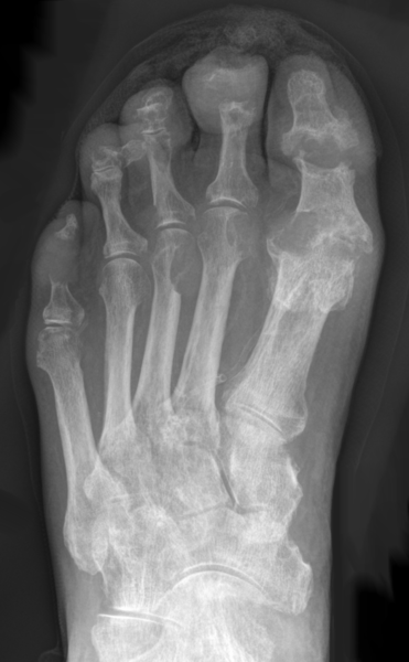
Image Source: Wikimedia Commons (2013). Gicht-Roe-multiple Arrosionen Destruktionen, Licensed under a creative commons licence. Available for free at: https://commons.wikimedia.org/wiki/File:Gicht_-_Roe_-_multiple_Arrosionen_Destruktionen.png (Accessed: 30 April 2022).
- X-rays in pseudogout: chondrocalcinosis. Chondrocalcinosis is not specific to pseudogout and is often present asymptomatically.

Image Source: Wikimedia Commons (2016). Chondrocalcinosis-annotated, Licensed under a creative commons licence. Available for free at: https://commons.wikimedia.org/wiki/File:Chondrocalcinosis_-_annotated.jpg (Accessed: 30 April 2022).
- NICE do not recommend further imaging if gout or pseudogout is suspected, as it is often a clinical diagnosis. However, if there is doubt and further imaging is recommended, then modalities such as ultrasound, MRI and CT scan can be considered.
Complete Guide to Clinical Genetics ST3 Application and Recruitment (2027)
Written by Medibuddy. Last updated: October 2026. Clinical Genetics is

Complete Guide to Clinical Radiology ST3 Application and Recruitment (2027)
Recruitment paused: NHS England is not running Clinical Radiology ST3+ recru

Complete Guide to Paediatric Surgery ST3 Application and Recruitment in United Kingdom (2026)
Written by Medibuddy. Last updated: October 2026. If you’re planning


Take your subscriptions with you
Our mobile app allows you to access your interview and exam question banks wherever you are.

