


 Close
Close
 29th December 2023
Differential Diagnoses
Parkinson’s Disease
Ophthalmological signs in Parkinson’s can arise from efferent or afferent dysfunction due to the decreased levels of dopamine in the CNS. In normal physiology, dopamine is a neurotransmitter in the retina and is theorised to control the activity of several cell types. Ocular symptoms can include horizontal diplopia (mono or binocular), convergence insufficiency where a patient struggles to read or clearly see items at close distance, photosensitivity, and subjective reductions in visual quality.
Skew deviation
This is a vertical and oblique diplopia which cannot be localised to a single extraocular muscle or cranial nerve and is, therefore, a diagnosis of exclusion. Suspicion for skew deviation should be raised if a 3-step test does not highlight a problem with the 4th cranial nerve, i.e. the problematic muscle is not superior oblique. Further testing can be performed via Maddox Rod; however, this requires specialist input and interpretation.
6ᵗʰ nerve palsies
The abducens nerve has a long course from its source in the pons to the terminus at the lateral rectus muscle. Innervation of the lateral rectus allows for abduction (lateral gaze) of the eye. Understanding the anatomy of the abducens nerve helps in highlighting the key pathologies which can result in 6ᵗʰ nerve palsies.
Nuclear – a nuclear sixth nerve palsy can present as a horizontal gaze palsy where the patient is unable to abduct the ipsilateral eye and adduct the contralateral eye. The 6ᵗʰ nerve nucleus intermixes with the contralateral 3ʳᵈ nerve nucleus via the medial longitudinal fasciculus, allowing for gaze to one direction or another (i.e. both eyes moving in the same direction at the same time.) When the 6ᵗʰ nerve nucleus has been affected by such processes as brainstem demyelination, compression secondary to a neoplasm or micro-ischaemia, then the contralateral 3ʳᵈ nerve can also be affected. Cranial nerve VII may also be affected as the nerve fascicle loops around the nucleus of cranial nerve 6. The medial longitudinal fasciculus can be damaged by any lesion or ischaemia as it is supplied by branches of the basilar artery; this can cause internuclear ophthalmoplegia (INO). INO is an ocular movement disorder characterised by partial or complete loss of adduction on the same side as the lesion/ischaemia and contralateral nystagmus on abduction. The MLF passes through the pons into the midbrain, meaning the location of the lesion can be in both. If the patient is able to converge their eyes (i.e. become cross-eyed), then that means the 3ʳᵈ nerve nucleus is functioning via communications from the convergence centre in the midbrain, meaning the lesion is likely in the pons.
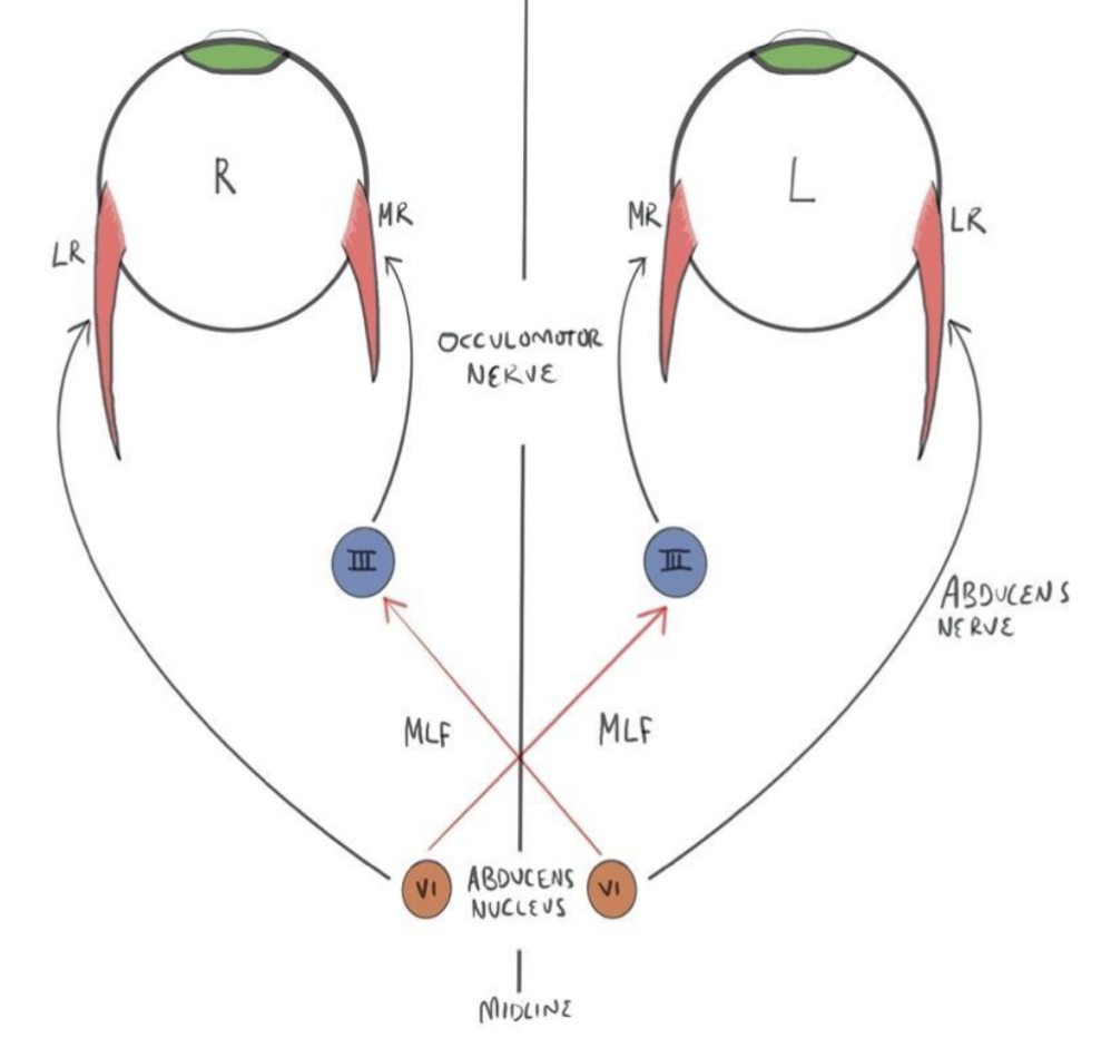
Fascicle – The nerve fascicle can be affected as it travels towards its exit point at the pontomedullary junction. This often presents with contralateral hemiparesis due to involvement of the pyramidal tract.
The subarachnoid space – the abducens nerve is anchored at its exit at the pontomedullary junction and to the petroclinoid ligament, where the nerve turns 90 degrees after following the slope of the clivus. Between these two anchoring points, the nerve exists in the subarachnoid space, and changes in intracranial pressure (ICP), either increased or decreased, may result in downward displacement of the brainstem, causing stretching of the nerve.
Petrous – as it passes under the petroclinoid ligament, the nerve contacts the tip of the petrous pyramid, making it susceptible to pathologies related to the temporal bone. These can be neoplastic, inflammatory, via compression and often involve the 8ᵗʰ, 7ᵗʰ or 5ᵗʰ nerves due to their close proximity.
Cavernous sinus – as the nerve passes through the cavernous sinus, it can be associated with the 3ʳᵈ, 4ᵗʰ and V1/2 branches of the 5ᵗʰ cranial nerve. Cavernous sinus thrombosis, most commonly due to infection, can produce cranial nerve palsies, the most affected being the abducens nerve. The internal carotid artery also passes through the cavernous sinus, giving rise to the possibility of aneurysms and fistulas, which again can lead to cranial nerve palsies.
Superior orbital fissure – pathologies involved in the cavernous sinus can also affect the superior orbital fissure due to its mixed contents of cranial nerves and vessels. Problems at this point of the abducens nerve length are often associated with proptosis and decreasing vision.
The most likely aetiology of isolated six nerve palsy in adults is ischaemia. This can be due to a host of comorbidities, including diabetes, hypertension, temple arthritis or anaemia.
3ʳᵈ nerve palsies
The oculomotor nerve has three main functions: innervation to the pupil and lens, innervation to the upper eyelid and innervation of the extraocular muscles (SR, MR, IR, IO). Complete third nerve palsies are likely to present with ptosis, a dilated pupil with sluggish to no reactions and a down-and-out eye with an inability to adduct, infraduct or supraduct. Partial third nerve palsies are more common, and these can present with a mix of the previously mentioned signs to varying degrees of severity.
The third nerve originates from two nuclei in the midbrain and exits near the midline at the base of the midbrain. From here, the nerve passes through the cavernous sinus towards and through the superior orbital fissure and to the orbit of the eye. Therefore, the same pathologies which can affect the 6ᵗʰ cranial nerve in the cavernous sinus or orbital fissure can also affect the 3ʳᵈ nerve. Patients presenting with new signs and symptoms of 3ʳᵈ nerve palsy raise suspicion for aneurysms, which may be compressing the nerve between it exiting the midbrain and entering the cavernous sinus, mainly an aneurysm of the posterior communicating artery. Also, CN III has parasympathetic fibres controlling pupil constriction running along the outside of the nerve. Therefore, external compression of the 3ʳᵈ nerve may lead to pupil dilation. If any ocular motility examination suggests involvement of CN III + deficits of duction with or without a dilated pupil, urgent non-contrast CT head with possibility of additional CT angiogram should be discussed. MRA should be discussed for patients who are pregnant, with poor renal function or allergy to contrast agents.
Lesions affecting the 3ʳᵈ nerve nucleus in the midbrain can present with bilateral complete ptosis due to there being only one sub-nucleus innervating both levator palpebrae superioris muscles. If the corticospinal tract has been affected, patients can present with contralateral hemiplegia or ataxia if the superior cerebellar peduncle is involved. The 3rd nerve partly exists within the subarachnoid space and is therefore at risk of damage secondary to infections, inflammation or masses within the subarachnoid space. In patients presenting and not requiring urgent CT imaging of the brain, it is not unreasonable to consider an MRI with contrast to evaluate the entire path of the 3rd nerve to rule out lesions/processes along its course.
4ᵗʰ nerve palsies
As mentioned earlier, a palsy of the trochlear nerve will result in vertical diplopia. Superior oblique intorts and depresses the eye in the adducted position. So, clinical examination of a patient with a 4ᵗʰ nerve palsy may reveal abnormal head posture (torticollis) in an attempt to compensate for the diplopia, strabismus in the affected eye where there would be a hypertropia and in some cases an esotropia. The trochlear nerve is the only cranial nerve which crosses the midline to innervate the contralateral side and exits the brainstem dorsally. This means that in cases of trauma to the dorsal aspect of the brainstem, a patient can have a bilateral 4th nerve palsy, which presents with vertical diplopia and strabismus.
The Parks 3 Step test can be utilised as a gross bedside test to help determine which extraocular muscle is causing a vertical diplopia. Understanding why the test works requires an in-depth understanding of how the extraocular muscles rotate the eye on 3 axes, which is not covered in this section. The test will not be diagnostic if more than one extraocular muscle is affected.
- Identify the hypertrophic eye in primary position – ask the patient to gaze ahead and see which eye is hypertrophic. If, for example, the right eye is higher, then the muscles affected are narrowed to either the depressors of the right eye (IR + SO) or the elevators (SR + IO) of the left eye.
- Identify if the hypertropia worsens in left or right gaze – in a trochlear nerve palsy, hypertropia of the paretic eye will increase on the opposite gaze.
- Identify if the hypertropia is worse on head tilt to the left or right – in a trochlear nerve palsy, the hypertropia will worsen on tilting to the same side.
There is a very quick and easy diagram you can utilise when identifying which extraocular muscle may be the culprit in presentations of vertical diplopia.

In this example, the patient presents with vertical diplopia and a right-sided hypertropia on examination. Step 1 is to circle the muscles on the bottom of the eye, which is deviated, and circle the muscles on the top on the contralateral side (highlighted in green). The next step of the test is to examine when the deviation worsens in left or right gaze. In this example, the patient’s vision worsens in left gaze, so you circle the muscles utilised for left gaze (amber). The final step is to identify which direction of head tilt worsens the deviation; in this case, it is right-sided head tilt. Highlight the muscles on the diagonal, which corresponds to whichever direction the deviation is greatest. The extraocular muscle, which has been circled 3 times, is the muscle causing the deviation, which in this example is the right superior oblique.
Thyroid eye disease
The position of the lid must be observed when diagnosing thyroid eye disease. Pathological signs include lid lag, where the upper lid lags behind the limbus when the patient rapidly diverts their eye from superior to inferior gaze, and lid retraction, where the sclera is visible above and below the iris. Thyroid eye disease causes enlargement of the extraocular muscles, which restricts their movement and causes diplopia.
Myasthenia gravis
Patients may complain of associated symptoms such as dysphagia, dysarthria and dyspnoea, which worsen as the day goes on and improve with rest. Ocular signs include ptosis, sometimes beginning as unilateral, which frequently shifts from one eye to the other to eventually become bilateral. Pupils are never involved in myasthenia.
References
- Margolin E. Approach to patients with diplopia. Journal of the Neurological Sciences. 2020 Oct 15;417:117055.
- Crumbie L. Diencephalon [Internet]. Kenhub. 2022. Available from: https://www.kenhub.com/en/library/anatomy/diencephalon
- Kanukollu VM, Sood G. Strabismus.
- Danchaivijitr C, Kennard C. Diplopia and eye movement disorders. Journal of Neurology, Neurosurgery & Psychiatry. 2004 Dec 1;75(suppl 4):iv24-31.
- The Cavernous Sinus – Contents – Borders – Thrombosis – TeachMeAnatomy [Internet]. Available from: https://teachmeanatomy.info/neuroanatomy/vessels/cavernous-sinus/
- Nguyen V, Reddy V, Varacallo M. Neuroanatomy, cranial nerve 6 (abducens). InStatPearls [Internet] 2021 Nov 29. StatPearls Publishing.
- Azarmina M, Azarmina H. The six syndromes of the sixth cranial nerve. Journal of ophthalmic & vision research. 2013 Apr;8(2):160.
- Joyce C, Le PH, Peterson DC. Neuroanatomy, Cranial Nerve 3 (Oculomotor) [Updated 2022 Mar 28]. In: StatPearls [Internet]. Treasure Island (FL): StatPearls Publishing; 2022 Jan-.
- Elkington AR, Frank HJ. 2nd ed. Oxford: Blackwell Science; 1991. Clinical Optics p.94-98
- Tan AK, Faridah HA. The two-minute approach to monocular diplopia. Malaysian Family Physician: the official journal of the Academy of Family Physicians of Malaysia. 2010;5(3):115.
Complete Guide to Clinical Genetics ST3 Application and Recruitment (2027)
Written by Medibuddy. Last updated: October 2026. Clinical Genetics is

Complete Guide to Clinical Radiology ST3 Application and Recruitment (2027)
Recruitment paused: NHS England is not running Clinical Radiology ST3+ recru

Complete Guide to Paediatric Surgery ST3 Application and Recruitment in United Kingdom (2026)
Written by Medibuddy. Last updated: October 2026. If you’re planning


Take your subscriptions with you
Our mobile app allows you to access your interview and exam question banks wherever you are.

