


 Close
Close
 29th December 2023
6ᵗʰ nerve palsies
The abducens nerve has a long course from its source in the pons to the terminus at the lateral rectus muscle. Innervation of the lateral rectus allows for abduction (lateral gaze) of the eye. Understanding the anatomy of the abducens nerve helps in highlighting the key pathologies which can result in 6ᵗʰ nerve palsies.
Nuclear – a nuclear sixth nerve palsy can present as a horizontal gaze palsy where the patient is unable to abduct the ipsilateral eye and adduct the contralateral eye. The 6ᵗʰ nerve nucleus intermixes with the contralateral 3ʳᵈ nerve nucleus via the medial longitudinal fasciculus, allowing for gaze to one direction or another (i.e. both eyes moving in the same direction at the same time.) When the 6ᵗʰ nerve nucleus has been affected by such processes as brainstem demyelination, compression secondary to a neoplasm or micro-ischaemia, then the contralateral 3ʳᵈ nerve can also be affected. Cranial nerve VII may also be affected as the nerve fascicle loops around the nucleus of cranial nerve 6. The medial longitudinal fasciculus can be damaged by any lesion or ischaemia as it is supplied by branches of the basilar artery; this can cause internuclear ophthalmoplegia (INO). INO is an ocular movement disorder characterised by partial or complete loss of adduction on the same side as the lesion/ischaemia and contralateral nystagmus on abduction. The MLF passes through the pons into the midbrain, meaning the location of the lesion can be in both. If the patient is able to converge their eyes (i.e. become cross-eyed), then that means the 3ʳᵈ nerve nucleus is functioning via communications from the convergence centre in the midbrain, meaning the lesion is likely in the pons.
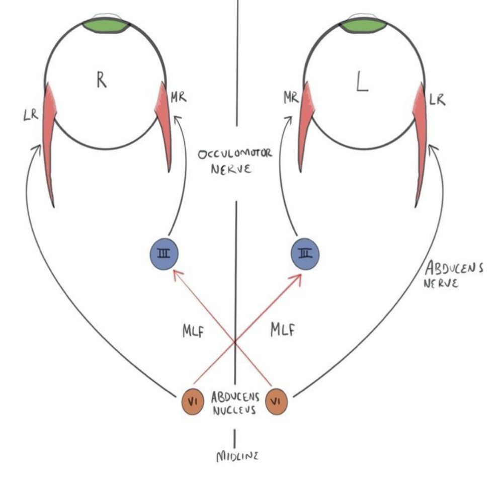
Fascicle – The nerve fascicle can be affected as it travels towards its exit point at the pontomedullary junction. This often presents with contralateral hemiparesis due to involvement of the pyramidal tract.
The subarachnoid space – the abducens nerve is anchored at its exit at the pontomedullary junction and to the petroclinoid ligament, where the nerve turns 90 degrees after following the slope of the clivus. Between these two anchoring points, the nerve exists in the subarachnoid space, and changes in intracranial pressure (ICP), either increased or decreased, may result in downward displacement of the brainstem, causing stretching of the nerve.
Petrous – as it passes under the petroclinoid ligament, the nerve contacts the tip of the petrous pyramid, making it susceptible to pathologies related to the temporal bone. These can be neoplastic, inflammatory, via compression and often involve the 8ᵗʰ, 7ᵗʰ or 5ᵗʰ nerves due to their close proximity.
Cavernous sinus – as the nerve passes through the cavernous sinus, it can be associated with the 3ʳᵈ, 4ᵗʰ and V1/2 branches of the 5ᵗʰ cranial nerve. Cavernous sinus thrombosis, most commonly due to infection, can produce cranial nerve palsies, the most affected being the abducens nerve. The internal carotid artery also passes through the cavernous sinus, giving rise to the possibility of aneurysms and fistulas, which again can lead to cranial nerve palsies.
Superior orbital fissure – pathologies involved in the cavernous sinus can also affect the superior orbital fissure due to its mixed contents of cranial nerves and vessels. Problems at this point of the abducens nerve length are often associated with proptosis and decreasing vision.
The most likely aetiology of isolated six nerve palsy in adults is ischaemia. This can be due to a host of comorbidities, including diabetes, hypertension, temple arthritis or anaemia.
Complete Guide to Clinical Genetics ST3 Application and Recruitment (2027)
Written by Medibuddy. Last updated: October 2026. Clinical Genetics is

Complete Guide to Clinical Radiology ST3 Application and Recruitment (2027)
Recruitment paused: NHS England is not running Clinical Radiology ST3+ recru

Complete Guide to Paediatric Surgery ST3 Application and Recruitment in United Kingdom (2026)
Written by Medibuddy. Last updated: October 2026. If you’re planning


Take your subscriptions with you
Our mobile app allows you to access your interview and exam question banks wherever you are.

