


 Close
Close
 10th March 2024
Diseases of the Arteries
Arterial Waveform
The arterial pulse results from the abrupt expansion of an artery caused by the sudden ejection of blood into the aorta and resulting transmission throughout the arterial system. A typical pulse wave usually has an upstroke and a downstroke with a dicrotic notch, which is not palpable but only visible on a direct arterial trace (see image below). Pulses should be examined bilaterally to identify any differences in amplitude, contour, or upstroke, which could indicate the presence of cardiovascular pathology.
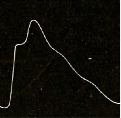
Normal arterial pulse waveform. Image source: Wikimedia Commons (2015). Hand-book of physiology (1892) (14742233966), Licensed under a Creative Commons licence. Available for free at: https://en.wikipedia.org/wiki/Pulse (Accessed: 25 January 2024).
Arterial Pulse Abnormalities
Unequal arterial pulses may occur when there is obstructive arterial disease, aortic dissection, aortic aneurysm, Takayasu disease, coarctation of the aorta, or supravalvular aortic stenosis. The arterial abnormalities caused by these conditions cause a delayed expansion of the arteries. If simultaneous palpation of the radial and femoral pulses reveals a delay, coarctation of the aorta should be considered. Below are some common arterial pulse abnormalities and associated cardiac conditions.
Pulsus alternans
The pulse amplitude varies between strong and weak beats due to changing systolic pressure (See image below). This is typically due to structural or functional abnormalities of the left ventricle, e.g., left heart failure, hypertrophic cardiomyopathy, cardiac tamponade, severe aortic regurgitation ectopic beats.

Image source: Wikimedia Commons (2007). Pulsus alternans trace, Licensed under a Creative Commons licence. Available for free at: https://en.wikipedia.org/wiki/Pulsus_alternans#/media/File:Pulsus_alternans_trace.jpg (Accessed: 25 January 2024).
Pulsus paradoxus
There is an exaggerated fall in BP >10 mmHg on inspiration, which is caused by a decrease in arterial pressure due to a decline in left ventricular stroke volume. The decline happens when there is an increase in right ventricular end-diastolic volume and decreased left ventricular end-diastolic volume secondary to cardiac tamponade.
Pulsus bisferiens
A single central pulse wave with two systolic peaks separated by a mid-systolic dip. The first peak is caused by rapid LV ejection, and the second peak is caused by the arterial recoil effect. Commonly seen in mixed aortic valve disease, HOCM and aortic regurgitation.
Dicrotic Pulse
Although clinically similar to the pulsus bisferiens, the arterial pulse waveform differs. In this case, an accentuated diastolic dicrotic wave follows the dicrotic notch. Dicrotic pulse can be found in patients with severe heart failure, hypovolemic shock, cardiac tamponade, and during the postoperative period following aortic valve replacement.
Water Hammer/Collapsing pulse
Characterised by an abrupt and very rapid upstroke followed by a rapid collapse. During the examination, it is best appreciated palpating the radial pulse while raising the arm abruptly. Commonly found in patients with significant aortic regurgitation but also in other increased stroke volume-associated conditions like patent ductus arteriosus, large arteriovenous fistulas, hyperkinetic states, thyrotoxicosis anaemia, and extreme bradycardia.
Additionally, the mean arterial pressure (MAP) can be calculated with the following formula: DP + 1/3 (SP-DP) (DP: diastolic pressure; SP: systolic pressure). The MAP can help monitor the average arterial pressure throughout one cardiac cycle, representing the overall perfusion of organs. A normal MAP is considered between 60 and 100, meaning that a MAP below 60 represents poor tissue perfusion and vice versa.
References
- DeMers, D. and Wachs, D., 2021. Physiology mean arterial pressure. In StatPearls [Internet]. StatPearls Publishing.
- Gersh, B. (2021) Examination of the arterial pulse’. In: UpToDate. (Accessed: April 2nd, 2022).
- Levine, J. (2010) ‘Peripheral Arterial Disease is Underdiagnosed in the Elderly’, Available at: http://jmlevinemd.com/peripheral-arterial-disease-is-underdiagnosed/ (Accessed: April 2nd, 2022).
- Nirmalan, M. and Dark, P.M., 2014. Broader applications of arterial pressure waveform analysis. Continuing Education in Anaesthesia, Critical Care & Pain, 14(6), pp.285-290.
- Singh, A. (no date) ‘Pulse Measurement – Normal and Abnormal’. Available at: https://medcaretips.com/pulse/ (Accessed: April 2nd, 2022).
Conditions of the Aorta
Aortic Dissection
Background
Aortic syndromes refer to life-threatening conditions, including aortic dissection, intramural haematoma and penetrating atherosclerotic ulcer. Aortic dissection is defined as the separation of the aortic wall layers due to an intimal tear. The incidence of aortic dissection is 5 to 30 cases per 1 million people per year and carries a mortality of 1 to 2% per hour after the onset of symptoms in untreated patients, with an overall in-hospital mortality rate of 27%. Aortic dissection can be spontaneous, iatrogenic, or traumatic.
Risk factors for aortic dissection include:
- Hypertension
- Trauma
- Connective tissue disorders (Marfan’s and Ehlers-Danlos syndrome
- Aortic aneurysm
- Previous aortic dissection
- Bicuspid aortic valve (Turner’s syndrome)
- Pregnancy
- Aortic instrumentation or surgery
- Coarctation of the aorta
- Inflammatory disease (vasculitis, rheumatoid arthritis, giant cell arteritis, Takayasu’s arteritis
- Fluoroquinolone use
Classification
Aortic dissection can be classified according to the anatomical location of the lesion, location of the initial tear and onset of presentation. The Stanford (most used) and DeBakey classifications describe aortic dissection according to the location of the lesion (See Table 1). Here the Stanford classification will be used to understand clinical presentation and management. According to time, aortic dissection can be classified as:
- Hyperacute: <24 hours
- Acute: 1 to 14 days
- Subacute: >14 to 90 days
- Chronic: >90 days
| The Stanford Classification | |
| Type A | Involves any part of the aorta proximal to the origin of the left subclavian artery
|
| Type B | Arises distal to the left subclavian artery origin
|


Table 1. Aortic dissection classification systems. Image source: https://en.wikipedia.org/wiki/Aortic_dissection
Clinical presentation
Aortic dissection commonly presents with acute severe chest or back pain. Most patients will describe the pain as sharp, stabbing or tearing. Pulse deficit (e.g., weak or absent carotid, brachial, or femoral pulse), unequal blood pressure in both arms, aortic regurgitation murmur, hypotension, syncope, or neurological symptoms can often present in type A dissection. In contrast, abdominal pain and hypertension are more common in type B dissection. It is worth noting that depending on the type of dissection, and hypotension can represent a rupture of the abdominal aorta (type B) or secondary severe aortic regurgitation (resulting in a new diastolic murmur) and/or extravasation into the pericardial space leading to cardiac tamponade (type A). Additionally, type A dissection can cause acute myocardial ischemia due to coronary occlusion, haemothorax or hemoperitoneum and compression of the surrounding structure, causing focal neurological deficit (e.g. stroke or altered consciousness, Horner syndrome, hoarseness, acute paraplegia, etc.). Due to the clinical presentation, other conditions should be considered when assessing patients with suspected aortic dissection (e.g., myocardial infarction or pulmonary embolism).
Diagnosis
Initial investigations include chest x-ray, which may reveal mediastinal widening of the aortic silhouette (60-90% of cases). ECG can differentiate between aortic dissection and acute coronary syndromes but is less valuable when coronary occlusion is secondary to aortic dissection. A D-dimer can rule out aortic dissection with levels below 500 ng/mL, indicating that a patient does not have acute aortic dissection.
Aortic dissection should be considered in patients presenting with any high-risk features (see Table 2). Diagnosis is confirmed with a CT Aortogram. This modality has similar accuracy to transoesophageal echo and magnetic resonance aortogram.
| Conditions | Pain Features | Clinical Findings |
|---|---|---|
| Marfan Syndrome or connective Tissue Disease
Family History Aortic Disease Aortic Valve Disease Recent Aortic Manipulation Known Thoracic Aortic Aneurysm |
Chest, back or abdominal pain which is severe in intensity/ abrupt onset
Or tearing/sharp/stabbing pain radiating to the back |
Pulse deficit
Systolic BP deficit of >20mmHg between arms New AR murmur with pain Focal neurological deficit with pain |
Table 2. High-risk features associated with aortic dissection
Management
Patients should ideally be managed in an intensive care unit for anti-impulse therapy aimed at reducing the velocity of left ventricular contraction and reducing the shear stress and progression of the lesion. Anti-impulse therapy is accomplished using IV beta-blockers (Labetalol or Esmolol) to reduce heart rate below 60 bpm and blood pressure range between 100 and 120 mmHg. It should be noted that blood pressure should be lower, preserving mental state and urine output. Where beta-blockers are not tolerated, diltiazem or verapamil may be used as an alternative. IV morphine and anxiolytics are used to control pain and anxiety.
Initial management in aortic dissection type A focuses on aggressive blood pressure control and cardiac surgical consultation. For type B dissections, management is aimed at blood pressure control and only requires surgery if there is a rupture or worsening pain. Chronic aortic dissection is usually managed medically.
Surgical management of aortic dissection type A may involve replacing the ascending aorta, resectioning intimal tears and aneurysmal aorta, and aortic valve repair or replacement. Surgical intervention should be considered in type B aortic dissection where there is persistent severe hypertension or pain, lesion progression, aneurysmal expansion, poor perfusion leading to end-organ ischemia, or rupture.
Abdominal Aortic Aneurysm
Background
An abdominal aortic aneurysm (AAA) is a segmental, full-thickness dilation of the abdominal aorta 50 per cent greater than the normal aortic diameter and is the most common true arterial aneurysm. In adults, a diameter greater than 3 cm is considered an aneurysm. An abdominal aortic aneurysm requires timely management; if not, it can progress to further expansion, rupture, haemorrhage, and death. AAA has a 50 – 80% mortality, and the most common risk factors are the following:
- Older age
- Male sex
- Cigarette smoking
- Positive family history of AAA
- Other large artery aneurysms (e.g., iliac, femoral, popliteal)
- Atherosclerosis
- Hypertension
Additionally, most family-based studies found a predisposition for a patient to have an abdominal aortic aneurysm if a relative had one but not if they had a thoracic aortic aneurysm.
Classification
Although an infrarenal aorta with a maximum diameter ≥3.0 cm is considered aneurysmal, the following classification based on diameter does not predict clinical events.
- Small aneurysms have a diameter of <4.0 cm
- Medium aneurysms have a diameter between 4.0 and 5.5 cm
- Large aneurysms have a diameter >5.5 cm
- Very large aneurysms have a diameter ≥6.0 cm
Clinical presentation
AAA can be asymptomatic, and despite advances in screening, it continues to be a significant cause of death in the elderly. When symptomatic, it usually means that there has been a rapid change in the aneurysm’s configuration. Patients will often complain of abdominal, back, or flank pain. Sometimes, patients can present signs of limb ischemia or other systemic manifestations like fever or malaise. The most common signs and symptoms of AAA rupture are severe pain, hypotension and a pulsatile abdominal mass. In some cases, symptoms might be unspecific when a retroperitoneal rupture occurs.
Diagnosis
AAA diagnosis can be made clinically when the aneurysm is greater than 5.5 cm; however, a CT is considered the test of choice for symptomatic AAA. When asymptomatic, abdominal ultrasonography is recommended for follow-up in patients with small and medium-sized aneurysms when managed conservatively. When considering a surgical approach, contrast-enhanced CT aortography is used.
Management
Once AAA is confirmed, the management will be based on the diameter and whether the presentation was symptomatic or asymptomatic. If the AAA ruptures, surgical management can be offered, and imaging is ideal but not required, especially when patients are hemodynamically unstable. Additionally, resuscitation fluids should be aimed to maintain mentation and stabilise blood pressure and heart rate. When patients are stable, imaging can be performed urgently to determine if endovascular repair is possible.
Thoracic Aneurysm
Like AAA, thoracic aneurysms affect 5 to 10 patients per million population each year. Presentation may be asymptomatic, but where symptoms are present, the patient may report chest pain and tenderness, back pain, cough, hoarseness and shortness of breath. Many thoracic aneurysms are first present in the context of a rupture. This should be suspected if the patient has sharp, sudden pain in the upper back, which spreads downward; chest, jaw, neck or arm pain, dysphagia, shortness of breath, hypotension and altered consciousness state. A CT aortogram remains the best diagnostic tool but should not delay management if the patient is unstable.
References
- Ayala, IADL and Chen, Y.F. (2012). Acute aortic dissection: an update – The Kaohsiung Journal of Medical Sciences. Available at: https://doi.org/10.1016/j.kjms.2011.11.010
- Black, J. & Manning, W. (2021). Overview of acute aortic dissection and other acute aortic syndromes UpToDate.
- Cantrill et al (2015). Clinical policy: critical issues in the evaluation and management of adult patients with suspected acute nontraumatic thoracic aortic dissection. Annals of Emergency Medicine. Available at: https://www.acep.org/globalassets/new-pdfs/clinical-policies/tad1-2015.pdf
- Callaway et al (2021). Diagnosis of Thoracic Aortic Dissection in the Emergency Department. RCEM. Available at: https://rcem.ac.uk/wp-content/uploads/2021/12/Diagnosis_of_Thoracic_Aortic_dissection.pdf
- Czerny, M. and Beyersdorf, F., 2020. Abdominal Aortic Aneurysm: Treatment Choice and Volume Effects. Deutsches Ärzteblatt International. Available at: https://doi.org/10.3238/arztebl.2020.0811
- Dalman, R. & Mell, M. (2021) Overview of abdominal aortic aneurysm In UpToDate. (Accessed: March 23rd, 2022).
- Melo, R.G., Duarte, G.S., Lopes, A., Alves, M., Caldeira, D., e Fernandes, R.F. and Pedro, L.M., 2021, March. Incidence and prevalence of thoracic aortic aneurysms: A systematic review and meta-analysis of population-based studies. In Seminars in Thoracic and Cardiovascular Surgery. WB Saunders.
Peripheral Vascular Disease
Acute Limb Ischaemia
Acute limb ischaemia is a life-limb-threatening condition caused by a rapid or sudden decrease in limb perfusion, endangering tissue viability. Two possible events are implicated in the cause. Arterial thrombosis originating from a pre-damaged atherosclerotic site represents 85% of causes, and non-atherosclerotic peripheral embolism accounts for 10-15%. The incidence is approximately 1.5 cases per 10,000 persons per year. The most common aetiologies are:
- Native arterial thrombosis: atherosclerotic plaque, aneurysm, dissection, hypercoagulability.
- Injury: iatrogenic or traumatic
- Thrombosis following intervention
- Embolism: arterial or paradoxical
- Peripheral vasoconstriction/ vasospasm
Most patients with acute limb ischaemia will have a background of peripheral artery disease (PAD) and will experience intermittent claudication caused by a mismatch between active muscle demand and blood supply; and atypical lower extremity pain. A threatened limb is referred to those patients whose arterial blood flow is insufficient to meet the demand of resting muscle or tissue.
Clinical presentation
Presentation may be acute or acute-on-chronic limb ischaemia, depending on the onset, location and the possible collateral vessels available.
Acute-on-chronic limb ischaemia is common in patients with background of peripheral artery occlusive disease or previous revascularisation. Symptoms will have an increasing course and are similar to those of PAD. Acute limb ischemia, on the other hand, will present with rapidly evolving symptoms which can be remembered as the six Ps:
- Pain
- Pallor
- Poikilothermia (inability to maintain a constant core temperature)
- Pulseless
- Paraesthesia
- Paralysis
Diagnosis
Diagnosis can be made using clinical categories or with the use of ankle-brachial pressure index (see Table 3). After categorising based on cause and severity, consideration can be made whether to proceed with additional investigations. The clinical categories used are:
- Viable or marginally threatened: no sensory loss or muscle weakness, and both arterial and venous Doppler signals are audible.
- Immediately threatened: Sensory loss involves more than the toes; rest pain and mild-to-moderate muscle weakness are present. Arterial Doppler signals are usually inaudible, and venous Doppler signals are audible.
- Irreversibly ischemic: profound sensory loss and muscle weakness (paralysis and possible rigor). Arterial and venous Doppler signals are inaudible.
CT angiography or catheter-based arteriography are the investigations of choice, the second one providing a therapeutic option.
| ABPI value | Interpretation | Recommendation |
|---|---|---|
| >1.4 | Calcification/vessel hardening | Refer to vascular specialist |
| 1.0-1.4 | Normal | None |
| 0.9-1.0 | Acceptable | |
| 0.8-0.9 | Some arterial disease | Treat risk factors |
| 0.5-0.8 | Moderate arterial disease | Refer to vascular specialist |
| >0.5 | Severe arterial disease | Refer to vascular specialist |
Table 3. Ankle-Brachial Pressure Index interpretation.
Management
The success of treatment depends on several factors, including the aetiology, location, duration of symptoms, availability of autologous vein for bypass grafting and suitability of the patient for surgery or intervention. The management of acute limb ischaemia is done by catheter-directed thrombolysis (with or without endovascular intervention) or surgical revascularisation.
Raynaud’s Phenomenon
Raynaud’s phenomenon (RP) is defined as a vascular response to cold temperature or emotional stress that is above the normal physiological response. The pathogenesis of RP is a complex interplay of different factors, including genetic, neural, vascular and intravascular factors. This vasospastic condition affects around 5% of the population, is more common in women and can be classified as primary or secondary. Primary RP is more common before the age of 30 in contrast to secondary RP, which usually presents after the age of 40. Secondary causes include:
- Vascular
- Occupational
- Autoimmune
- Drug/chemical-related
- Conditions associated with increased plasma viscosity and reduced digital perfusion
- Other causes, e.g., carpal tunnel syndrome, frostbite, and hypothyroidism
Clinical presentation
RP presents as a characteristic colour change in hands and sometimes feet associated with cold temperatures or emotional stress. Most patients will not present to a healthcare provider during the episodes due to the regular short durations but will capture the episodes on their mobile phone or describe the typical clinical picture. The classical triad of colour change will traverse from white (pallor) to blue (cyanosis) to red before turning back to the patient’s regular skin colour.
Diagnosis
RP is considered a symptom, not a diagnosis; however, the cause should be investigated to identify whether this is primary or secondary. The burden of digital complications should be considered to aid the decision on whether drug or surgical intervention is needed. In primary RP nailfold capillaroscopy, FBC, ANA and ESR should be normal. In secondary RP or systemic sclerosis, these will be abnormal. Once the cause is established, the management will be tailored to the underlying cause and consideration for symptom control with drugs or surgical treatment.
Management
Initial management of primary RP involves conservative measures such as patient education to avoid the cold, hand-warming gloves and smoking cessation. After this, first-line medications such as calcium channel blockers, phosphodiesterase inhibitors, ARBs, SSRIs, alpha-blockers, ACE-I and topical nitrates can be used. In refractory RP or where there is digital ulceration, intravenous prostanoid can be used. When all medical treatments fail to control RP and ulceration and critical ischemia begins to occur, a digital sympathectomy should be considered.
References
- Fluck, F. et al. 2020. Current treatment options in acute limb ischemia. In RöFo-Fortschritte auf dem Gebiet der Röntgenstrahlen und der bildgebenden Verfahren. Vol. 192, No. 04, pp. 319-326.
- Mitchell, M. and Carpenter, J. 2020. Clinical features and diagnosis of acute lower extremity ischemia. In UpToDate.
- Van, D. et al. 2018. Acute limb ischemia.Revue medicale de Liege. 73(5-6), pp.304-311.
- Haque, A. and Hughes, M. 2020. Raynaud’s phenomenon. Clinical Medicine. Available at https://doi.org/10.7861/clinmed.2020-0754
- Herrick, A.L et al. 2020. New perspectives in the imaging of Raynaud’s phenomenon. European Journal of Rheumatology. Available at https://doi.org/10.1177/2397198319826467
Complete Guide to Clinical Genetics ST3 Application and Recruitment (2027)
Written by Medibuddy. Last updated: October 2026. Clinical Genetics is

Complete Guide to Clinical Radiology ST3 Application and Recruitment (2027)
Recruitment paused: NHS England is not running Clinical Radiology ST3+ recru

Complete Guide to Paediatric Surgery ST3 Application and Recruitment in United Kingdom (2026)
Written by Medibuddy. Last updated: October 2026. If you’re planning


Take your subscriptions with you
Our mobile app allows you to access your interview and exam question banks wherever you are.

