


 Close
Close
 10th March 2024
Arterial Waveform
The arterial pulse results from the abrupt expansion of an artery caused by the sudden ejection of blood into the aorta and resulting transmission throughout the arterial system. A typical pulse wave usually has an upstroke and a downstroke with a dicrotic notch, which is not palpable but only visible on a direct arterial trace (see image below). Pulses should be examined bilaterally to identify any differences in amplitude, contour, or upstroke, which could indicate the presence of cardiovascular pathology.
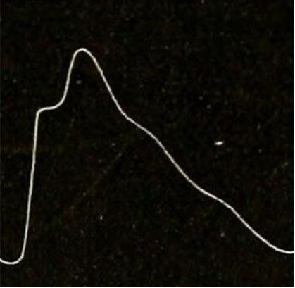
(14742233966), Licensed under a Creative Commons licence. Available for free at: https://en.wikipedia.org/wiki/Pulse (Accessed: 25 January 2024).
Arterial Pulse Abnormalities
Unequal arterial pulses may occur when there is obstructive arterial disease, aortic dissection, aortic aneurysm, Takayasu disease, coarctation of the aorta, or supravalvular aortic stenosis. The arterial abnormalities caused by these conditions cause a delayed expansion of the arteries. If simultaneous palpation of the radial and femoral pulses reveals a delay, coarctation of the aorta should be considered. Below are some common arterial pulse abnormalities and associated cardiac conditions.
Pulsus alternans
The pulse amplitude varies between strong and weak beats due to changing systolic pressure (See image below). This is typically due to structural or functional abnormalities of the left ventricle, e.g., left heart failure, hypertrophic cardiomyopathy, cardiac tamponade, severe aortic regurgitation ectopic beats.

Image source: Wikimedia Commons (2007). Pulsus alternans trace, Licensed under a Creative Commons licence. Available for free at: https://en.wikipedia.org/wiki/Pulsus_alternans#/media/File:Pulsus_alternans_trace.jpg (Accessed: 25 January 2024).
Pulsus paradoxus
There is an exaggerated fall in BP >10 mmHg on inspiration, which is caused by a decrease in arterial pressure due to a decline in left ventricular stroke volume. The decline happens when there is an increase in right ventricular end-diastolic volume and decreased left ventricular end-diastolic volume secondary to cardiac tamponade.
Pulsus bisferiens
A single central pulse wave with two systolic peaks separated by a mid-systolic dip. The first peak is caused by rapid LV ejection, and the second peak is caused by the arterial recoil effect. Commonly seen in mixed aortic valve disease, HOCM and aortic regurgitation.
Dicrotic Pulse
Although clinically similar to the pulsus bisferiens, the arterial pulse waveform differs. In this case, an accentuated diastolic dicrotic wave follows the dicrotic notch. Dicrotic pulse can be found in patients with severe heart failure, hypovolemic shock, cardiac tamponade, and during the postoperative period following aortic valve replacement.
Water Hammer/Collapsing pulse
Characterised by an abrupt and very rapid upstroke followed by a rapid collapse. During the examination, it is best appreciated palpating the radial pulse while raising the arm abruptly. Commonly found in patients with significant aortic regurgitation but also in other increased stroke volume-associated conditions like patent ductus arteriosus, large arteriovenous fistulas, hyperkinetic states, thyrotoxicosis anaemia, and extreme bradycardia.
Additionally, the mean arterial pressure (MAP) can be calculated with the following formula: DP + 1/3 (SP-DP) (DP: diastolic pressure; SP: systolic pressure). The MAP can help monitor the average arterial pressure throughout one cardiac cycle, representing the overall perfusion of organs. A normal MAP is considered between 60 and 100, meaning that a MAP below 60 represents poor tissue perfusion and vice versa.
References
- DeMers, D. and Wachs, D., 2021. Physiology mean arterial pressure. In StatPearls [Internet]. StatPearls Publishing.
- Gersh, B. (2021) Examination of the arterial pulse’. In: UpToDate. (Accessed: April 2nd, 2022).
- Levine, J. (2010) ‘Peripheral Arterial Disease is Underdiagnosed in the Elderly’, Available at: http://jmlevinemd.com/peripheral-arterial-disease-is-underdiagnosed/ (Accessed: April 2nd, 2022).
- Nirmalan, M. and Dark, P.M., 2014. Broader applications of arterial pressure waveform analysis. Continuing Education in Anaesthesia, Critical Care & Pain, 14(6), pp.285-290.
- Singh, A. (no date) ‘Pulse Measurement – Normal and Abnormal’. Available at: https://medcaretips.com/pulse/ (Accessed: April 2nd, 2022).
Complete Guide to Clinical Genetics ST3 Application and Recruitment (2027)
Written by Medibuddy. Last updated: October 2026. Clinical Genetics is

Complete Guide to Clinical Radiology ST3 Application and Recruitment (2027)
Recruitment paused: NHS England is not running Clinical Radiology ST3+ recru

Complete Guide to Paediatric Surgery ST3 Application and Recruitment in United Kingdom (2026)
Written by Medibuddy. Last updated: October 2026. If you’re planning


Take your subscriptions with you
Our mobile app allows you to access your interview and exam question banks wherever you are.

