


 Close
Close
 11th April 2024
Disorders of Female Reproduction
Physiology of the menstrual cycle
Several hormones tightly regulate the mechanisms underpinning fertility and pregnancy. Gonadotrophin-releasing hormone (GnRH) from the hypothalamus stimulates the release of luteinising hormone (LH) and follicle-stimulating hormone (FSH) from the anterior pituitary. This begins the follicular phase, where a steady rise in FSH acts on granulosa cell receptors to cause the conversion of androgens into oestrogen and trigger oocyte development. Androgens are produced by theca cells in response to LH. FSH also promotes follicular development until mid-cycle, where a spike in LH causes ovulation. The residual follicle then becomes the corpus luteum, which produces primarily progesterone, oestrogen, and inhibin A.
Fertilisation of the ovum prevents regression of the corpus luteum. This is due to the production of β-hCG by the trophoblast cells that surround the embryo. β-hCG stimulates the corpus luteum to continue producing hormones which maintain the endometrial lining. However, involution of the corpus luteum occurs in the absence of fertilisation as high β-hCG levels are not sustained. The decrease in progesterone and oestrogen leads to endometrial shedding (menstruation), and the cycle resets with FSH increasing again to develop new follicles.
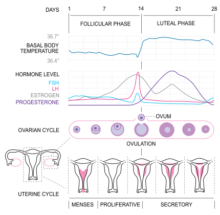
Image source: Wikimedia Commons (2010). MenstrualCycle2 en, Licensed under a Creative Commons licence. Available for free at: https://commons.wikimedia.org/wiki/File:MenstrualCycle2_en.svg (Accessed: 07 February 2024).
Table 1 Overview of the main hormones involved in the menstrual cycle
| Hormone | Produced where | Function |
|---|---|---|
| GnRH | Hypothalamus | Stimulates the release of FSH and LH |
| FSH | Anterior pituitary | Stimulates the development of a follicle |
| LH | Anterior pituitary | Spike causes ovulation
Development of the endometrium Negative feedback control of FSH and LH |
| Oestrogen | Corpus luteum
Placenta (>5 weeks) |
Stimulates growth of myometrium of the uterus
Mammary gland development |
| Progesterone | Corpus luteum
Placenta (>5 weeks) |
Prepares endometrium for implantation
Mammary gland development Maintains pregnancy and increases threshold for contractions |
| β-hCG | Placenta after implantation | Promotes maintenance of the corpus luteum |
| Prolactin | Anterior pituitary | Milk production and development of mammary glands |
| Inhibin A | Corpus luteum | Inhibits FSH |
References
- The European Society of Human Reproduction and Embryology. POI Guideline Development Group.
Hypogonadism
Physiology
The normal functioning of the Hypothalamic-Pituitary-Adrenal axis (HPA axis) consists of a series of negative feedback loops that regulate the release of hormones from endocrine glands around the body. The HPA regulation of reproductive hormone release involves the secretion of GnRH from the hypothalamus. GnRH acts on anterior pituitary gonadotrophs to release LH and FSH. LH and FSH then act on cells in the gonads to produce sex hormones, predominantly testosterone in males and oestrogen in females. Testosterone and oestrogen have an inhibitory effect on the release of GnRH from the hypothalamus and FSH and LH from the Anterior Pituitary.
Pathophysiology
Defined as failure of the gonads to produce adequate sex hormones, hypogonadism can be classified by aetiology into primary (gonadal) or secondary (central/extra-gonadal). Primary hypogonadism results from pathology of the sex glands, while secondary hypogonadism is caused by disruption of the HPA axis. In females, this results in ovarian dysfunction with a degree of impaired oestrogen production and ovulatory failure.
Aetiology
Primary hypogonadism
Primary hypogonadism (hypergonadotropic hypogonadism) in a woman is a pathology of the ovaries resulting in failure of normal ovulation and production of adequate oestrogen. It is characterised by disruption to the menstrual cycle (amenorrhoea/oligomenorrhoea) as well as biochemical features such as raised FSH and LH with low oestradiol. Congenital causes of gonadal dysgenesis include Turner (45X) and Fragile X syndrome. Streak ovaries are seen when there is abnormality or absence of the X chromosome and are associated with Turner syndrome. Acquired primary hypogonadism can be due to iatrogenic causes such as chemotherapeutic agents, therapeutic oophorectomy, as well as infections and autoimmune diseases, including haemochromatosis.
Secondary hypogonadism
Secondary hypogonadism is caused by pathology of the HPA axis (hypothalamus and/or pituitary), resulting in a downstream reduction in the production of gonadotrophins. Congenital causes include Kallmann syndrome, which is the most common genetic cause and is seen more often in men (3.6:1). Space occupying lesions, infiltrative diseases such as sarcoidosis and amyloidosis, meningitis, pituitary apoplexy and Sheehan’s syndrome are examples of secondary causes.
Clinical Features
In pre-pubertal females, primary hypogonadism is more common and diagnosed at the time of expected puberty with primary amenorrhoea and a lack of secondary sexual characteristics. In post-pubertal females’ secondary hypogonadism is more common, and women present with secondary amenorrhoea/oligomenorrhoea as well as features of oestrogen deficiency.
Investigations
In women presenting with menstrual disturbance, clinicians should enquire about symptoms of oestrogen deficiency and other systemic features.
Initial biochemical tests include human chorionic gonadotrophin (hCG) and FSH. LH, Oestradiol, testosterone, prolactin, thyroid stimulating hormone, and other pituitary function tests may be useful in aiding diagnosis.
In pre-pubertal females, screening for genetic causes of primary hypogonadism should be considered, such as a pelvic ultrasound scan and karyotyping if the uterus is absent.
Additional imaging, such as an MRI brain for patients with hypogonadotropic hypogonadism, may be considered if an intracranial cause is suspected based on clinical presentation. Abdominal imaging may reveal a local cause or horseshoe kidneys, which are associated with Turner syndrome.
Management
Hormone replacement therapy is the mainstay of treatment for hypogonadism. Treatment should be individualised to the patient, specific to the underlying aetiology and should take a holistic approach with consideration of psychosocial implications. As well as the physiological development of secondary sexual characteristics and fertility if desired, the main aim for treatment is to prevent long-term complications of low oestrogen, such as cardiovascular disease and osteoporosis.
References
- Dzemaili S, Tiemensma J, Quinton R, et al. Beyond hormone replacement: Quality of life in women with congenital hypogonadotropic hypogonadism. Endocrine Connections 2017; 6: 404–412.
Amenorrhoea
Physiology
The International Federation of Gynaecology and Obstetrics (FIGO) defines the parameters for normal uterine bleeding.
| Frequency: Days between cycles | ≥24 to ≤38 days |
| Duration: Duration of menses | ≤8 days |
| Regularity: Cycle variation dependent on age | ≤7 to 9 days |
| Flow Volume | Normal as determined by the patient |
Primary Amenorrhoea
Primary Amenorrhoea is defined as the failure of the onset of menses by the age of 15 in the presence of secondary sexual characteristics or by the age of 13 in the absence of secondary sexual characteristics.
| Causes of primary amenorrhoea | Features |
|---|---|
| Anatomical abnormalities in Mullerian development | Normal secondary sexual characteristics
Examples include: imperforate hymen; Mayer-Rokitansky-Kuster-Hauser syndrome; |
| 5-alpha reductase deficiency | Genetically male with female-appearing genitalia or genital ambiguity |
| 17-hydroxylase deficiency | In females, this is a non-virilising congenital adrenal hyperplasia (CAH) |
| Partial 21-hyrdroxylase deficiency | The most common cause of CAH and results in virilisation. |
| Flow Volume | Normal as determined by the patient |
| Genetic primary hypogonadism | Distinguished by the absence of or poorly defined secondary sexual characteristics
Turner syndrome, Klinefelter syndrome (XY, distinguished by anosmia), Kallman syndrome |
| Type 1 autoimmune polyglandular syndrome (APS-1) | Rare, recessively inherited auto-immune condition characterised by the triad of chronic mucocutaneous moniliasis, hypoparathyroidism, and Addison’s disease.
Sequelae include ovarian failure, which may present as primary amenorrhoea or infertility. |
| Complete androgen insensitivity syndrome | XY, phenotypically female
Usually diagnosed after puberty, though may be diagnosed in those with prepubescent bilateral inguinal hernias (due to testes in the inguinal canal) |
| Non-classic congenital adrenal hyperplasia | Clitoromegaly and clinical hyperandrogenism |
Secondary Amenorrhoea
Secondary Amenorrhoea is defined as the absence of menstruation for 3-6 months in a patient with previous regular menses or 6-12 months in patients with oligomenorrhoea.
| Causes of secondary amenorrhoea | Features |
|---|---|
| Pregnancy | Physiological androgen levels, confirmed with β-hCG test |
| Premature ovarian insufficiency (POI) | Females under 40 years old with premature menopausal symptoms (such as amenorrhoea or oligomenorrhoea) with elevated FSH levels on two samples 4-6 weeks apart |
| Functional hypothalamic cause | Physiological androgen levels
Leads to suppression of the hypothalamic-pituitary-ovarian axis and suppression of gonadotrophin-releasing hormone Risk factors include stress and over-exercise |
| Cushing’s syndrome and exogenous androgen-producing tumours | Secondary amenorrhoea results from hypercortisolism. Negative feedback to the hypothalamus leads to decreased GnRH release. |
| Prolactinoma | Presents with galactorrhoea due to prolactin’s suppressive effect on GnRH production from the hypothalamus. Microprolactinomas are more common than macroprolactinomas. The latter may be suspected if clinical signs of raised intracranial pressure are present. Diagnosis is confirmed with an MRI pituitary. |
| Hyperprolactinaemia secondary to medication | Presents with galactorrhoea due to prolactin’s suppressive effect on GnRH production from the hypothalamus. First-generation antipsychotics are the most common cause, though second-generation antipsychotics may also cause hyperprolactinaemia. Other causes include oral contraceptives, metoclopramide, verapamil, and opiates. |
| Asherman’s syndrome | Characterised by scar tissue in the uterine cavity and may present as oligomenorrhoea or amenorrhoea and infertility
History of dilatation and curettage and normal gonadotrophins |
Investigations
After excluding pregnancy, initial biochemical tests include FSH, LH, TSH and prolactin. If there are systemic features of androgen excess, total testosterone levels could be considered. A pelvic ultrasound should be considered if an anatomical abnormality of the genital tract is suspected. Diagnosis of POI in women <40 years is based on clinical features such as oligomenorrhoea/amenorrhoea >4 months and biochemical confirmation of hypergonadotropic hypogonadism (two raised FSH (>30) levels taken at a 4–6-week interval). Hysteroscopy is the gold standard for diagnosing Asherman’s syndrome.
Management
Management is specific to the underlying condition and includes specialist referral to a gynaecologist and/or an endocrinologist as appropriate. The main aims for treatment are to restore fertility if desired, treat the underlying pathology and prevent complications of hormone deficiency. In POI, either HRT or a combined hormonal contraceptive) reduces the risk of chronic diseases, including cardiovascular disease and osteoporosis. For symptomatic prolactinoma, cabergoline is the first-line treatment. If pregnant, it is important to review medications as many are contraindicated, such as lithium, which is teratogenic and may cause hypothyroidism.
References
- Management of spontaneous primary ovarian insufficiency (premature ovarian failure) – UpToDate, https://www.uptodate.com/contents/management-of-spontaneous-primary-ovarian-insufficiency-premature-ovarian-failure?search=hypogonadism women&source=search_result&selectedTitle=8~150&usage_type=default&display_rank=8 (accessed 20 July 2022).
- Munro MG, Critchley HOD, Fraser IS, et al. The two FIGO systems for normal and abnormal uterine bleeding symptoms and classification of causes of abnormal uterine bleeding in the reproductive years: 2018 revisions. Int J Gynecol Obstet 2018; 143: 393–408.
- Amenorrhoea | Health topics A to Z | CKS | NICE, https://cks.nice.org.uk/topics/amenorrhoea/
- Evaluation and management of primary amenorrhea – UpToDate, https://www.uptodate.com/contents/evaluation-and-management-of-primary-amenorrhea?search=hypogonadotropic hypogonadism female&source=search_result&selectedTitle=4~95&usage_type=default&display_rank=4 (accessed 20 July 2022).
- Evaluation and management of secondary amenorrhea – UpToDate, https://www.uptodate.com/contents/evaluation-and-management-of-secondary-amenorrhea?search=hypogonadotropic hypogonadism female&source=search_result&selectedTitle=1~95&usage_type=default&display_rank=1#H180195634.
- Diagnosis of premature ovarian insufficiency | Diagnosis | Menopause | CKS | NICE, https://cks.nice.org.uk/topics/menopause/diagnosis/diagnosis-of-premature-ovarian-insufficiency/ (accessed 21 June 2022).
- Polycystic ovary syndrome – NICE CKS, https://cks.nice.org.uk/topics/polycystic-ovary-syndrome/background-information/prevalence/ (accessed 28 July 2022).
Complete Guide to Clinical Genetics ST3 Application and Recruitment (2027)
Written by Medibuddy. Last updated: October 2026. Clinical Genetics is

Complete Guide to Clinical Radiology ST3 Application and Recruitment (2027)
Recruitment paused: NHS England is not running Clinical Radiology ST3+ recru

Complete Guide to Paediatric Surgery ST3 Application and Recruitment in United Kingdom (2026)
Written by Medibuddy. Last updated: October 2026. If you’re planning


Take your subscriptions with you
Our mobile app allows you to access your interview and exam question banks wherever you are.

