


 Close
Close
 6th February 2024
Disorders of Metalloproteins and Porphyrias
An Introduction to Metalloproteins
Metalloproteins
Metalloprotein is a broad term applied to any protein which contains a metal ion cofactor. A large number of proteins fall under this category. For example, around 1,000 out of 20,000 human proteins contain zinc-binding domains.
Many physiological processes rely on metalloproteins, such as insulin biosynthesis, respiration, and nerve transmission. A particularly important metalloprotein is haemoglobin, which contains iron.
Haem Synthesis
Haem synthesis takes place in the mitochondria of liver cells and bone marrow. It is a complex pathway, which is outlined below:
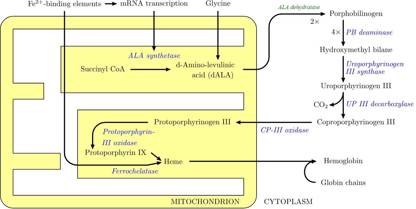
Image Source: Wikimedia Commons (2023). Heme synthesis, Licensed under a creative commons licence. Available for free at: https://upload.wikimedia.org/wikipedia/commons/a/a2/Heme_synthesis.svg (Accessed 4 January 2022).
Porphyria and Metalloproteins
As we have already mentioned, haemoglobin is a vital metalloprotein. Therefore, disorders relating to the synthesis of this protein can lead to clinical abnormalities. One prominent group of such disorders are termed porphyria. These are a group of inherited disorders of haem synthesis resulting in the accumulation of porphyrin and its precursors.
Depending on which enzyme involved in haem synthesis is deficient, there are seven main types of porphyria. This range of possible aetiologies leads to many clinical and biochemical features. The full list comprises
| Porphyria | Enzyme | Inheritance Pattern |
|---|---|---|
| Aminolaevulinic acid dehydratase porphyria (ADP) | Aminolaevulinic acid dehydratase | Autosomal recessive |
| Acute intermittent porphyria (AIP) | Porphobilinogen deaminase | Autosomal dominant |
| Congenital erythropoietic porphyria (CEP) | Uroporphyrinogen III synthase | Autosomal recessive |
| Porphyria cutanea tarda (PCT) | Uroporphyrinogen decarboxylase | Usually, autosomal dominant |
| Hereditary coproporphyria (HCP) | Coproporphyrinogen oxidase | Autosomal dominant |
| Variegate porphyria (VP) | Protoporphyrinogen oxidase | Autosomal dominant |
| Erythropoietic protoporphyria (EPP) | Ferrochelatase | Autosomal dominant |
There are numerous ways of classifying porphyria – most commonly by their clinical features:
- Acute porphyria featuring neuropsychiatric features: ADP and AIP.
- Cutaneous porphyria primarily affects the skin: PCT, EPP, and CEP.
- Mixed porphyria with features of both: VP and HCP.
However, we will focus on the three most clinically relevant porphyria: AIP, VP, and PCT.
References
- Fu, D., Finney, L. (2015). Metalloproteomics: challenges and prospective for clinical research applications. Expert Rey Proteomics, 11(1): 13–19. doi: 10.1586/14789450.2014.876365
- The BMJ (2023, Apr 5). Acute Intermittent Porphyria. https://bestpractice.bmj.com/topics/en-gb/235/treatment-algorithm#
- The BMJ (2023, Apr 5). Acute Porphyrias. https://bnf.nice.org.uk/treatment-summaries/acute-porphyrias/
Porphyria in clinical practice
Summary
Acute intermittent porphyria
AIP has a prevalence of around 1 in 20,000 people and is the commonest acute porphyria. It most commonly presents between the ages of 20-40.
As with other porphyrias, the presence of risk factors is one of the main signs to be aware of. These include family history, female sex (5:1 ratio female to male), nutritional changes such as fasting, intercurrent illness, and exposure to certain drugs or hormones.
Exposure to drugs such as barbiturates, phenytoin, and metoclopramide can precipitate an attack. Elevated progesterone levels have also been linked with attacks. Therefore, females may experience cyclical attacks in the luteal phase of the menstrual cycle.
AIP is not a mixed porphyria and will not result in any cutaneous manifestation.
Patients experiencing an acute intermittent porphyria attack are likely to experience the following:
- Abdominal pain that is out of proportion to physical exam findings with no apparent or common cause identified.
- Muscle weakness.
- Tachycardia.
- Hypertension: acutely but may progress to become chronic.
- Dark or red urine: due to the excretion of porphyrins and degradation products of porphobilinogen.
- Nausea and vomiting.
- Abdominal distension.
- Muscle weakness or sensory loss in a ‘bathing suit’ distribution.
- Neuropsychiatric symptoms include behavioural changes, insomnia, depression, confusion, and seizures.
- Rarely, in severe and prolonged attacks, there may be quadriparesis or respiratory failure.
Differential diagnoses to consider include:
- Guillain-Barre syndrome.
- Epilepsy.
- Psychiatric illnesses, particularly psychosis.
- Neurological deficit.
Diagnostic investigations to be considered are:
- Urinary and serum porphobilinogen. These are typically raised but may be normal outside acute episodes.
- DNA analysis, mainly if there is a strong family history.
Early diagnosis and treatment are vital in preventing neurological damage or fatal outcomes. Immediate management of AIP includes:
- Removal of any precipitating factors.
- Analgesia = usually opiates due to the level of pain experienced.
- Antiemetics = phenothiazines may also aid in calming anxiety.
- Severe attacks warrant intravenous haem arginate therapy.
- Glucose (at least 300g of 10% glucose) can benefit some patients with milder attacks. However, it is essential to note that large volumes may cause or exacerbate hyponatraemia.
- Long-term management:
- Patient education on identifying precipitating factors is a mainstay of long-term management.
- Givosiran and aminolevulinate synthase 1-directed small interfering RNA can be used in patients where this proves ineffective.
- Regular haem infusion may prevent recurrent or severe attacks.
- Screening of family members.
- GnRH analogues such as leuprorelin may effectively prevent cyclical attacks in women.
- Patients who are non-responsive to the above management may ultimately require a liver transplant, as chronic liver damage can occur due to the build-up of haem precursors, with approximately 10% of patients with AIP developing hepatocellular carcinoma.
Variegate porphyria
VP is most prevalent in people of South African origin. This porphyria is mixed, meaning there are cutaneous and systemic features. However, symptoms are predominantly cutaneous and will occur in 50% of people with VP. This is due to an accumulation of light-sensitive porphyrins (C and D.)
The most significant features are the cutaneous manifestations:
- Photosensitive blistering.
- Hyperpigmentation.
Other features that VP has in common with other porphyrias are:
- Neuropsychiatric symptoms.
- Dark urine.
- Abdominal pain.
The investigation and treatment of VP are like those of other porphyrias.
Porphyria cutanea tarda
This is the commonest porphyria with a prevalence of 1 in 10,000. There are two forms: sporadic (90%) and familial (10%.)
PCT is a cutaneous porphyria and presents with lesions similar to VP.
Other symptoms/signs include:
- Varying degree of iron overload, which may require management with venesection.
- Hypertrichosis (excess hair growth.)
This porphyria is associated with:
- Hepatitis C and alcoholic liver disease.
- Haemochromatosis.
- HIV.
- Systemic lupus erythematosus.
Attacks are often precipitated by alcohol.
Again, the diagnosis and management are like those of other porphyrias.
References
- National Organisation of Rare Diseases (NORD). (2022, Jun 1). Variegate Porphyria. https://rarediseases.org/rare-diseases/variegate-porphyria/
- National Institute for Health and Care Excellence (NICE). (2023, Apr 5). Treatment Summaries – Acute Porphyrias. https://bnf.nice.org.uk/treatment-summaries/acute-porphyrias/
Complete Guide to Clinical Genetics ST3 Application and Recruitment (2027)
Written by Medibuddy. Last updated: October 2026. Clinical Genetics is

Complete Guide to Clinical Radiology ST3 Application and Recruitment (2027)
Recruitment paused: NHS England is not running Clinical Radiology ST3+ recru

Complete Guide to Paediatric Surgery ST3 Application and Recruitment in United Kingdom (2026)
Written by Medibuddy. Last updated: October 2026. If you’re planning


Take your subscriptions with you
Our mobile app allows you to access your interview and exam question banks wherever you are.

