


 Close
Close
 12th April 2024
Disorders of Puberty
Pubertal development
Puberty begins when the hypothalamus starts releasing gonadotrophin-releasing hormone (GnRH) in pulses. GnRH travels into a network of capillaries called the hypophyseal portal system, which then stimulates cells known as gonadotrophs in the anterior pituitary gland to promote the production and release of gonadotrophins of luteinising hormone (LH) and follicle-stimulating hormone (FSH). LH and FSH stimulate the sex organs to produce sex hormones, which act via a negative feedback mechanism to inhibit the secretion of GnRH in the hypothalamus. This is known as the hypothalamus-pituitary-adrenal axis (HPA). Below is an image that illustrates the HPA axis.
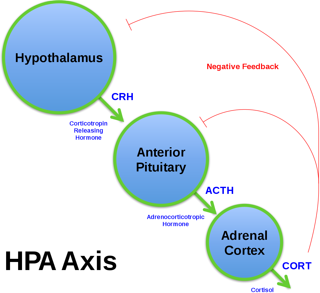
Image source: Wikimedia Commons (2016). HPA Axis Diagram (Brian M Sweis 2012), Licensed under a Creative Commons licence. Available for free at: https://commons.wikimedia.org/wiki/File:HPA_Axis_Diagram_(Brian_M_Sweis_2012).svg (Accessed: 06 February 2024).
In a male XY genotype, LH acts on Leydig cells in the interstitium of the testes to signal the conversion of cholesterol into testosterone. FSH act on the Sertoli cells in the seminiferous tubules of the testes to increase the production of sperm. Testosterone inhibits the production of GnRH and LH via a negative feedback mechanism, while the Sertoli cells produce inhibin to inhibit the production of FSH. Testosterone is important in promoting the development and growth of primary sexual characteristics.
In the female XX genotype, LH acts on the follicles of the ovaries to produce oestrogen and progesterone. The cells of the primary oocyte include theca cells and granulosa cells. Theca cells produce androstenedione in response to LH during puberty. Granulosa cells, in response to FSH, convert androstenedione into oestrogen and progesterone. Both oestrogen and progesterone work to regulate the monthly changes to ovary maturation and changes to the uterine wall, which is what causes the menstrual cycle. Oestrogen acts on the hypothalamus via a negative feedback mechanism to suppress further secretion of GnRH.
The stages and sequence in puberty are described by Tanner stages of puberty, which examines the development of secondary sexual characteristics. In males, the first sign of puberty is testicular enlargement >4mL. This is followed by pubic hair growth and usually takes place between 10 and 14 years. When the testicular enlargement reaches about 12 to 15mL, males often have rapid height growth. In comparison to females, the growth spurt occurs later and to a greater extent. In females, the first sign of puberty is the development of a breast disc, which usually happens in girls around ages 8 to 12. Height and pubic hair growth then follow rapidly. The menstrual cycle then begins approximately 2 years following the first sign of puberty. After the start of menarche, there is only 5cm left for height development.


Image source: Wikimedia Commons (2022). Tanner scale male intact, Licensed under a Creative Commons licence. Available for free at: https://commons.wikimedia.org/wiki/File:Tanner_scale_male_intact.svg (Accessed: 06 February 2024). Image source: Wikimedia Commons (2019). Tanner scale-female, Licensed under a Creative Commons licence. Available for free at: https://commons.wikimedia.org/wiki/File:Tanner_scale-female.svg (Accessed: 06 February 2024).
References
- Lissaue, Tom, Will Carroll, Keir Shiels, and Ranj Singh. Illustrated textbook of paediatrics. Amsterdam: Elservier, 2022
- Klatt, Edward C., and Vinay Kumar. Robbins and COTRAN Review of Pathology. London: Elsevier Health Sciences, 2015
Delayed puberty
Delayed puberty is defined as the development of secondary sexual features after the ages of 13 in females and 14 in males. The causes of delayed puberty are categorised into primary hypogonadism or secondary hypogonadism. Delayed puberty can also be due to functional causes such as underlying chronic disease, malnutrition or excessive exercise.
The most common cause of delayed puberty is constitutional delay in both sexes, which affects all domains. There is usually a family history of delayed puberty. Constitutional delay does not result in infertility and is not pathologic. Puberty occurs at a later age; however, its progression occurs naturally.
Primary hypogonadism (hypergonadotropic hypogonadism)
Primary hypogonadism occurs when there is a dysfunction of the gonads. Those affected either do not respond to gonadotrophins or cannot produce sex hormones. Acquired causes may be radiotherapy, chemotherapy or trauma to the gonads. Congenital primary hypogonadism occurs in conditions such as Klinefelter or Turner syndrome. The classic characteristics include a reduced level of testosterone in males and reduced levels of oestrogen and progesterone in females. As a result, gonadotrophins (LH & FSH) are markedly elevated.
Klinefelter syndrome
Klinefelter syndrome is a chromosomal disorder in which there is an extra X chromosome in a normally XY male genotype. The additional X chromosome is a result of the abnormal separation of sex chromosomes during meiosis I or II.
The additional X chromosome interrupts the normal production of testosterone in Leydig cells and inhibin in Sertoli cells. A lack of negative feedback results in an elevated level of LH and FSH. The lack of testosterone results in abnormal testes maturation, reduced sperm production and lack of development of secondary male characteristics. The additional X chromosome also results in an abnormal ratio of oestrogen to testosterone. They are at an increased risk of breast cancer and osteoporosis.
Individuals with Klinefelter syndrome typically exhibit symptoms such as tall and long legs with short torsos and broad hips. Due to the abnormal production of testosterone, they also exhibit symptoms of hypogonadism. Individuals classically have decreased facial and body hair and small testes. Those affected may have gynecomastia, and they also tend to be infertile without treatment. Other clinical features may include learning disability, delayed speech development, behavioural problems and psychosocial disorders.
It can be diagnosed with karyotyping via amniocentesis before birth or blood test after birth. Treatments include hormone therapy, such as testosterone replacement during puberty and infertility treatment. Other treatments may include speech and behavioural therapy and treating any relevant psychiatric conditions the individual may have. Individuals with Klinefelter syndrome are at a higher risk of developing type 2 diabetes, osteoporosis, cardiovascular and thromboembolic diseases, autoimmune disorders, psychiatric disorders and very rarely breast cancer.
Turner syndrome
Turner syndrome is a chromosomal disorder in a female genotype where one of the X chromosomes is completely missing or partially incomplete. The most common type of Turner syndrome has one of the X chromosomes completely missing. This is due to nondisjunction during meiosis. The next most common type is mosaicism, where some of the cells of the body have 46 chromosomes while some have 45 chromosomes. This is due to nondisjunction in mitosis after the formation of the zygote. The least common karyotype of Turner syndrome has part of one of the X chromosomes missing. A part of the X chromosome is deleted during either meiosis or mitosis.
The presence of only one complete X chromosome leads to an increased rate of loss of eggs. All the eggs are lost before puberty begins, leading to menopause prior to menarche. The ovaries are unable to develop normally and have a streak appearance. They are unable to produce sex hormones and result in primary amenorrhoea. Most females with Turner Syndrome are infertile as a result. Low levels of sex steroids lead to an elevated level of FSH and LH due to a lack of negative feedback.
The X chromosome includes genes for growth and development as well as other unidentified genes. This results in a short stature. They are also predisposed to cardiovascular defects, kidney issues, lymphatic and skeletal abnormalities and at an increased risk of type 2 diabetes mellitus. The impact on growth and development depends on the proportion of cells affected in the mosaic karyotype and the amount of X chromosome deleted in the partially absent X chromosome karyotype.
Individuals with Turner syndrome classically present with short stature (height less than 2.5 percentile with girls of the same age), webbed neck, high-arched palate, widely spaced nipples and short fourth metacarpal. Their first presentation to a clinician may be due to primary amenorrhoea. These individuals are often found with bicuspid aortic valve and, less commonly, coarctation of the aorta, both of which lead to an increased risk of aortic dilatation and dissection. They are at higher risk of developing hypothyroidism, autoimmune diseases and Crohn’s disease. Some individuals are also found with horseshoe kidney.
| At Birth | Adolescence |
|---|---|
| Lymphoedema
Cystic hygroma Lower extremity cyanosis (bicuspid aortic valve and coarctation of aorta) Horseshoe kidney |
Neck webbing
Low set ears Broad chest with widely spaced nipples Cubitus Valgus (arms turn outward) Short stature |
Turner syndrome can be diagnosed with karyotyping via amniocentesis before birth or a blood test after birth. Treatments include growth hormone therapy in childhood and sex hormone replacement therapy during adolescence.
Noonan syndrome
Noonan syndrome is often referred to as the “male version” of Turner syndrome as they present very similarly; however, it is a condition that can be found in males and females. It is an autosomal dominant inherited condition and is thought to be due to mutations in the Ras/mitogen-activated protein kinase transduction pathway. Individuals, similar to Turner Syndrome have short stature, congenital heart defects, characteristic facial features and delayed puberty.
Facial features of individuals with Noonan syndrome include wide-spaced and down-slanting eyes, which are often of blue or blue-green colour. They also have low-set ears, which are rotated posteriorly. These individuals often have a small chin, which results in their faces having an inverted triangular shape. Similarly to Turner syndrome, they also have broad webbed neck. Individuals with Noonan syndrome commonly present with undescended testes and also have delayed puberty.
In contrast to Turner syndrome, the most common congenital heart defect is pulmonary stenosis. Other heart defects include pulmonary valve dysplasia, septal defects, tetralogy of Fallot and hypertrophic cardiomyopathy. Other medical conditions that may occur in Noonan syndrome include chest deformities, ophthalmic and hearing defects, coagulation abnormalities, renal and splenic abnormalities, autoimmune diseases and lymphatic system abnormalities.
Treatment focuses on treating the conditions that the individual has, as the presentation can be highly variable among individuals. The main focus of treatment is often to assess and treat cryptorchidism, congenital heart defects and developmental delay.
Secondary hypogonadism (hypogonadotropic hypogonadism)
Secondary hypogonadism occurs when there is a dysfunction in the hypothalamus or pituitary gland, leading to the inability to produce gonadotrophins (LH & FSH). It can also be caused by the suppression of the hypothalamus by high prolactin or thyroid hormone levels. Acquired causes may be due to radiotherapy/chemotherapy or trauma. Congenital causes include Kallman Syndrome and panhypopituitarism. Other causes include chronic illness, excess exercise, malnutrition, obesity, and stress, which may also lead to the dysfunction of the hypothalamus or pituitary gland.
Kallman syndrome
Kallman syndrome is a result of the failure of the migration of GnRH-secreting neurons from the olfactory epithelium to the hypothalamus during embryogenesis. This defect also leads to hyposmia or anosmia. This leads to a reduced level of GnRH and, in turn, reduced levels of gonadotrophins (LH & FSH) and sex hormones – testosterone in males and oestrogen and progesterone in females. This leads to delayed puberty or failure to complete puberty. Both underdeveloped primary and secondary sex characteristics are listed below.
| Male | Female | |
|---|---|---|
| Primary sex characteristics | Penis & testes
Testicular descent Sperm production |
Menarche (amenorrhoea/oligomenorrhoea) |
| Secondary sex characteristics | Facial hair
Muscle mass Voice deepening |
Breast development
Pubic hair growth |
Both males and females are infertile. Male patients are found with small testes (<4mL), decreased muscle mass and delayed growth spurt. Females present with a lack of breast development, delayed growth spurt, lack of pubic hair growth and primary menorrhoea.
Kallman syndrome can be diagnosed by overall reduced levels of GnRH, gonadotropins (LH& FSH), testosterone/ oestrogen and progesterone. The main treatment is hormone replacement to promote puberty and the development of secondary sexual characteristics. As these individuals are also at risk of osteopenia and osteoporosis, calcium and vitamin D supplements are also part of the management.
References
- Delayed Puberty. BMJ Best Practice. Last Reviewed 19ᵗʰ December 2023
- Lissaue, Tom, Will Carroll, Keir Shiels, and Ranj Singh. Illustrated textbook of paediatrics. Amsterdam: Elservier, 2022
- Klatt, Edward C., and Vinay Kumar. Robbins and COTRAN Review of Pathology. London: Elsevier Health Sciences, 2015
- Blevins C H, Wilson M E. Klinefelter’s syndrome BMJ 2012; 345 :e7558 doi:10.1136/bmj.e7558
- Stamou, Maria I, and Neoklis A Georgopoulos. “Kallmann syndrome: phenotype and genotype of hypogonadotropic hypogonadism.” Metabolism: clinical and experimental vol. 86 (2018): 124-134. doi:10.1016/j.metabol.2017.10.012
- Noonan Syndrome. BMJ Best Practice. Last Reviewed 31ˢᵗ December 2023
Precocious puberty
Precocious puberty is defined as the development of secondary sexual features before the age of 8 in females and 9 in males. It also causes an accelerated rate of skeletal development and leads to a reduced adult height. Precocious puberty can be divided into whether it is gonadotrophin-releasing hormone (GnRH) dependent.
- Central or true precocious puberty is GnRH-dependent
- Peripheral precocious puberty is GnRH-independent
Central (true) precocious puberty
This occurs due to the early maturation of the HPA axis and is known as gonadotrophin-dependent precocious puberty. This leads to the release of gonadotropins (LH and FSH) and subsequent production of sex steroids. The most common cause is idiopathic and familial, which is found more frequently in females because the ovaries are sensitive to secretion of gonadotrophins. Often, they have a family history of precocious puberty. It may also be due to a tumour that produces GnRH or human chorionic gonadotropin (hCG). Other less common causes include other CNS diseases such as hydrocephalus, encephalitis, trauma, cranial radiotherapy or CNS hamartomas.
Peripheral precocious puberty
This is due to the overproduction of sex hormones; therefore, it is also known as gonadotrophin-independent precocious puberty. This may be due to a cyst or tumour, genetic conditions such as McCune-Albright Syndrome, dysfunction of other endocrine organs such as thyroid or adrenal glands or exposure to exogenous sex hormones such as the contraceptive pill or testosterone gels.
McCune-Albright syndrome (MAS)
MAS is caused by G-activating protein mutation in the adenylyl cyclase-cyclic adenosine monophosphate (cAMP) pathway. It plays an important role in the production and excretion of hormones. The mutation results in the pathway being constantly activated. The mutation occurs after the formation of the zygote; therefore, not all cells have the mutation. The clinical presentation is variable in different tissues.
It is associated with fibrous dysplasia and causes replacement of bone tissue with scar tissue. This may result in pathologic fractures, pain and limp. The degree of clinical manifestation depends on the affected individual. Patients will often have café au lait spots with irregular borders like “coast of Maine.” They become more numerous and darker with age. Other clinical features of MAS include hyperthyroidism, Cushing’s syndrome and growth hormone excess due to the constant activation of the cAMP pathway.
References
- Precocious Puberty. BMJ Best Practice. Last Reviewed 19ᵗʰ December 2023
- Lissaue, Tom, Will Carroll, Keir Shiels, and Ranj Singh. Illustrated textbook of paediatrics. Amsterdam: Elservier, 2022
- Klatt, Edward C., and Vinay Kumar. Robbins and COTRAN Review of Pathology. London: Elsevier Health Sciences, 2015
Contrasexual pubertal development
Congenital adrenal hyperplasia
Congenital adrenal hyperplasia results from deficiencies of enzymes, which play an important role in corticosteroid synthesis. This results in low levels of cortisol and, in turn, result in high levels of ACTH. Elevated level of ACTH promotes hyperplasia of the adrenal glands. See the diagram below, which illustrates corticosteroid synthesis and highlights the relevant enzymes in congenital adrenal hyperplasia.

The most common enzyme deficiency is 21 hydroxylase, which accounts for over 90% of CAH. The second most common enzyme deficiency is 11 ß-hydroxylase. Both enzymes are responsible for the conversion of progesterone and its by-product 17α-hydroxyprogesterone into aldosterone and cortisol eventually. The build-up of 17α-hydroxyprogesterone increases its conversion into testosterone.
Aldosterone is a hormone responsible for sodium and water reabsorption in the renal tubules. Low levels of aldosterone results in salt wasting in the body. Cortisol is responsible for gluconeogenesis in the body and low levels of glucose in the body. In 11 ß-hydroxylase deficiency, the build-up of 11 deoxycortisone – a by-product of progesterone – leads to hypertension.
The third most common enzyme deficiency is 17α-hydroxylase. 17α-hydroxylase is responsible for the conversion of progesterone into cortisol. This leads to low levels of cortisol and androgens. Low levels of androgen lead to poorly developed male genitals and a lack of development of secondary sexual characteristics in males and females. Progesterone is directed more down the aldosterone pathway, leading to a build-up of the precursors of aldosterone – 11 deoxycorticosterone and corticosterone. This build-up leads to an increased effect of aldosterone, leading to hypernatremia, hypokalaemia and hypertension. Due to the hypertensive effect of aldosterone precursors, this also leads to low levels of aldosterone.
Symptoms
| Androgen | Cortisol | Aldosterone | |
| 21 hydroxylase deficiency | ↑ | ↓ | ↓ |
| 11 ß-hydroxylase deficiency | ↑ | ↓ | ↑ (precursors) |
| 17α-hydroxylase deficiency | ↓ | ↓ | ↑ (precursors) |
In both 21-hydroxylase deficiency and 11 ß-hydroxylase deficiency, there are high levels of androgens. This leads to masculinisation of secondary sex characteristics in females. This results in enlarged clitoris, ambiguous genitalia, acne and hirsutism. High levels of androgen could also cause an early onset of puberty.
In 17α-hydroxylase deficiency, there are low levels of androgens. This leads to ambiguous genitalia development in males and a lack of secondary sexual characteristics in females.
In 21 hydroxylase deficiency, there are low levels of aldosterone, leading to salt wasting, especially in infants. This leads to vomiting, dehydration and hypotension. In 11 ß-hydroxylase deficiency and 17α-hydroxylase deficiency, there are elevated levels of aldosterone precursors. This leads to hypertension.
Cortisol is low in all three enzyme deficiencies and leads to hypoglycaemia.
Management
In classic 21 hydroxylase deficiency, individuals with salt wasting due to low aldosterone levels are given lifelong glucocorticoids such as hydrocortisone and fludrocortisone. The glucocorticoid also inhibits ACTH to prevent hyperplasia of the adrenal glands, which in turn prevents excess androgen production. Response to treatment is done through blood tests to check plasma renin levels and through regular blood pressure measurements. Long-term glucocorticoid treatment can also cause premature ageing of the bones, so it needs to be monitored at least yearly with X-ray to check for premature closing of the epiphyseal plates., Affected individuals with non-classic 21 hydroxylase deficiency do not need glucocorticoids.
In 11 ß-hydroxylase deficiency, spironolactone is the main treatment for females as it blocks mineralocorticoid and androgen receptors to control hypertension and the effects of androgen excess. In individuals who do not desire fertility, contraceptives can be used in combination with spironolactone. In males, amiloride and triamterene are used instead of spironolactone due to its side effects of gynaecomastia and sexual dysfunction.
In 17α-hydroxylase deficiency, glucocorticoid is the main management which will control hypertension induced by metabolites that act as weaker versions of aldosterone. Females may also be given low-dose oestrogen to promote secondary sexual characteristics and progestin to promote cyclic menstrual bleeding. In males, testosterone can be used to promote secondary sexual characteristics.
Androgen insensitivity syndrome
Androgen insensitivity syndrome (AIS) is a condition with a genetic defect of the androgen receptor. It is inherited in an X-linked recessive pattern. There are three types of AIS – complete with receptors that do not respond to androgens at all, partial with some functioning receptors and mild insensitivity with masculinisation of external genitalia. In most cases, the affected individuals are infertile.
Symptoms
| Complete | Partial | Mild |
|---|---|---|
|
|
|
Investigation
Ultrasound of the pelvis would reveal an absence of the uterus and testes that have failed to descend into the scrotum. Blood tests would reveal an elevated level of testosterone.
Management
Treatment includes hormone replacement therapy, which could be male or female sex hormones depending on the gender role that the individual identifies with. Surgery is also done to remove the testes to reduce the risk of cancer. Corrective surgery may also be done to correct the external genitalia.
References
- Congenital Adrenal Hyperplasia. BMJ Best Practice. Last Reviewed 22nd December 2023
- Lissaue, Tom, Will Carroll, Keir Shiels, and Ranj Singh. Illustrated textbook of paediatrics. Amsterdam: Elservier, 2022
- Klatt, Edward C., and Vinay Kumar. Robbins and COTRAN Review of Pathology. London: Elsevier Health Sciences, 2015
Premature thelarche
Premature thelarche is defined as breast enlargement in the absence of axillary and pubic hair and the absence of a growth spurt. This is benign. This is most commonly seen in females between 6 months and 2 years of age. Breast enlargement may be unilateral or asymmetrical.
References
- Lissaue, Tom, Will Carroll, Keir Shiels, and Ranj Singh. Illustrated textbook of paediatrics. Amsterdam: Elservier, 2022
- Klatt, Edward C., and Vinay Kumar. Robbins and COTRAN Review of Pathology. London: Elsevier Health Sciences, 2015
Complete Guide to Clinical Genetics ST3 Application and Recruitment (2027)
Written by Medibuddy. Last updated: October 2026. Clinical Genetics is

Complete Guide to Clinical Radiology ST3 Application and Recruitment (2027)
Recruitment paused: NHS England is not running Clinical Radiology ST3+ recru

Complete Guide to Paediatric Surgery ST3 Application and Recruitment in United Kingdom (2026)
Written by Medibuddy. Last updated: October 2026. If you’re planning


Take your subscriptions with you
Our mobile app allows you to access your interview and exam question banks wherever you are.

