


 Close
Close
 6th February 2024
Disorders of Purine Metabolism
Summary of conditions
Purines are natural substances found in foods and tissues; they play a crucial role in various biological processes, including the production of DNA, RNA and ATP (adenosine triphosphate), which provides energy for cellular functions. Purines may be synthesised de novo or salvaged from endogenous or exogenous nucleic acid breakdown.
The following disorders of purine metabolism will be discussed:
- Primary gout
- Lesch-Nyhan syndrome
Primary gout
Gout is a type of inflammatory arthritis characterised by acute flares of severe pain, heat and swelling of joints. It is caused by the formation of monosodium urate crystals in and around the joints. It is usually due to hyperuricaemia, although notably, gout can occur without hyperuricaemia, and hyperuricaemia does not always cause gout.
Uric acid is a metabolite of purines, and at physiological pH (as in the body), it exists in its ionised form, urate. Usually, urate levels are maintained by homeostasis by effective excretion by the kidneys (70%) and the gastrointestinal tract (30%). Excessive production or consumption (high purine foods include offal, game, oily fish and seafood) can overwhelm these clearance mechanisms, and levels rise. Likewise, hyperuricaemia ensues when clearance mechanisms are defective, such as in chronic kidney disease.
Clinical features
Clinical features of gout include:
Joint pain
This is typically rapid onset, severe pain associated with redness, heat and swelling. The most commonly affected joints are metatarsophalangeal joints, followed by tarsometatarsal and knee joints. The condition is typically monoarticular. However, oligoarticular (<4 joints involved) or even polyarticular cases have been described.
Within the joint, urate crystals activate phagocytes, triggering a cytokine-driven inflammatory response that results in neutrophilic synovitis.
Tophi
These are hard nodules of sodium urate crystals around joints, palpable underneath the skin, usually on extensor surfaces. They are representative of longstanding, untreated gout.

Image Source: Flickr (2010). 5353667874, Licensed under a creative commons licence. Available for free at: https://www.flickr.com/photos/ex_magician/5353667874 (Accessed: 9th January 2024).
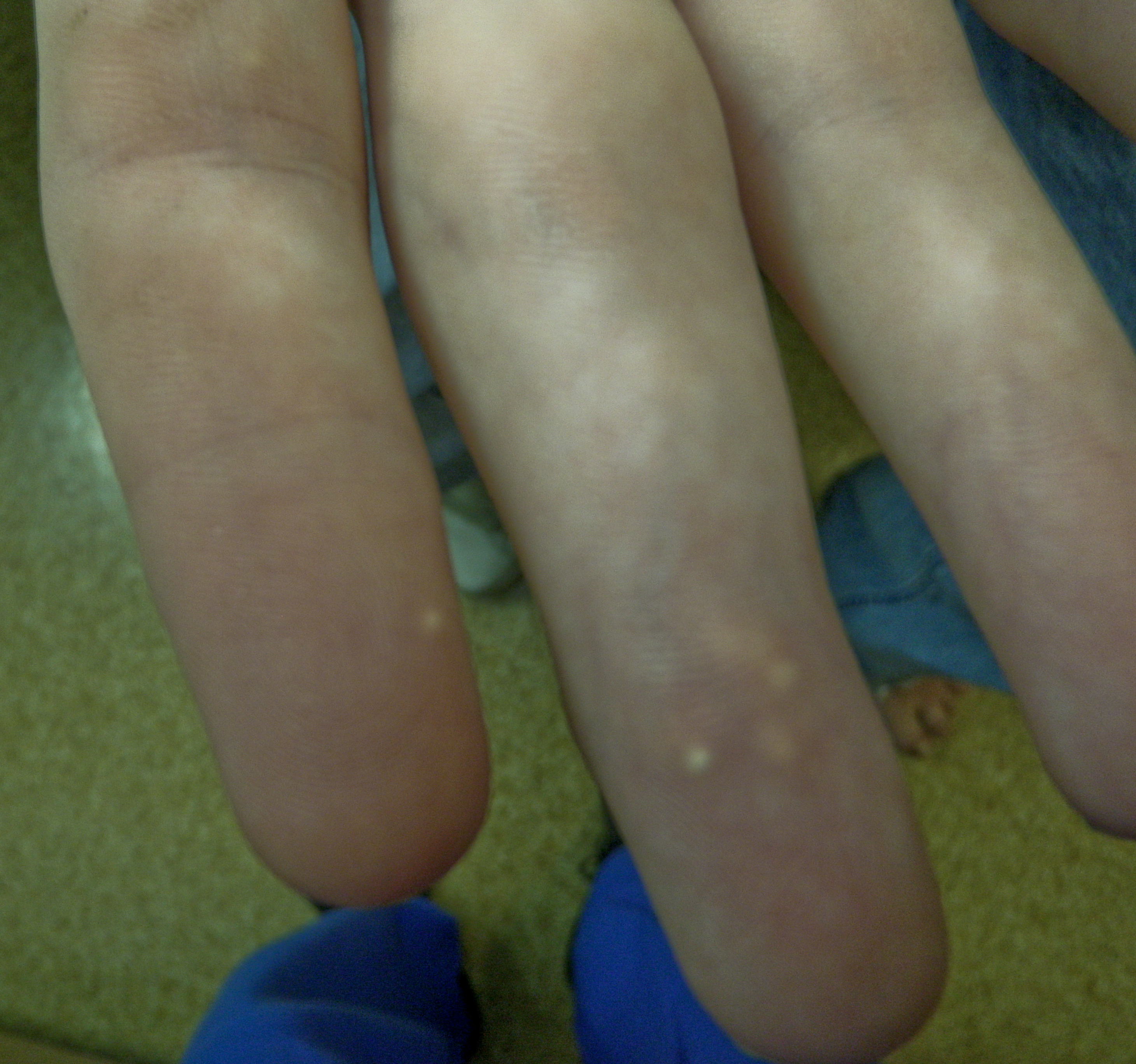
Image Source: Flickr (2010). 5353051607, Licensed under a creative commons licence. Available for free at: https://www.flickr.com/photos/ex_magician/5353051607 (Accessed: 9 January 2024).

Image Source: Raw Pixel (2017). 5964278, Licensed under a creative commons licence. Available for free at: https://www.rawpixel.com/image/5964278/free-public-domain-cc0-photo (Accessed: 4 January 2024).
Gout is excruciating, but acute attacks are self-limiting and usually resolve spontaneously without treatment. In the long term, however, if left untreated, recurrence is common, and several complications may develop. These include:
- Chronic arthritis and subsequent joint damage – this occurs in around 2% of patients with gout and is severe and debilitating. It usually develops around 20 years after the first gout attack.
- Renal stones – hyperuricaemia leads to the formation of urate renal stones in around 14% of patients with gout. These are painful and can have additional complications, including sepsis and kidney damage.
- Tophi are manifestations of monosodium urate crystals deposited around the joint site. They cause significant joint disfigurement and can themselves become inflamed or even infected. They occur in approximately 50% of patients after ten years of poorly controlled gout, rising to 72% of patients after 20 years.
In addition to gout, hyperuricaemia in itself confers an increased risk of organ damage, including:
- Cardiovascular disease, with an increased prevalence of hypertension, myocardial infarction, heart failure and stroke amongst patients with gout.
- Chronic kidney disease, with almost 25% of those with gout having CKD stages 3-5
Appropriate treatment (as discussed later) can suppress gout attacks and their recurrence and prevent these long-term consequences of the disease.

Image Source: Flickr (2019). 158161000@N04/49274750631, Licensed under a creative commons licence. Available for free at: https://flickr.com/photos/158161000@N04/49274750631 (Accessed: 9 January 2024).
Serum uric acid levels may also be monitored; these may be low, normal, or high during an acute attack and are more reliably observed two weeks after resolution of a flare. In the UK, a serum urate level >=360 micromol/L (6mg/dL) confirms a diagnosis of gout.
There is a role for imaging diagnostically. X-rays are of limited use; however, they can differentiate between chronic gout and other joint conditions in the late stages. Ultrasound is more sensitive in detecting erosions, tophi and the gout-specific double contour sign (linear urate deposits over hyaline cartilage). Ultrasound is recommended in cases where joint aspiration is not possible or if gout diagnosis is uncertain.

Image Source: Wikimedia Commons (2012). Gout chondrocalcinosis ultrasound, Licensed under a creative commons licence. Available for free at: https://commons.wikimedia.org/wiki/File:Gout_chondrocalcinosis_ultrasound.svg (Accessed: 4 January 2024).
Management
Early treatment of gout is associated with better clinical outcomes, and therefore, general management is by a two-pronged approach: short-term acute flare resolution to manage pain and preserve function, with long-term prevention of recurrence and protection against joint destruction.
Short-term management:
- NSAIDs
- NSAIDs are the first line in the treatment of acute gout flares. They work by interrupting the inflammatory cascade that results in acute symptoms. Patients at high risk of gastrointestinal complications should be co-prescribed a proton pump inhibitor during treatment.
- Corticosteroids
- If more joints are affected, these may be injected intraarticularly in monoarticular gout flares or parenterally. Patients should be counselled on potentially serious adverse effects.
- Colchicine
- Colchicine acts by blocking the neutrophil-endothelial interaction that drives inflammation in gout. It has a narrow therapeutic-to-risk index, so it should be used at its minimally effective dose. Common adverse effects are diarrhoea, nausea and vomiting.
- Interleukin (IL) – 1 inhibitors
- Anakinra and Canakinumab are IL1 antagonists. They are second-line therapies for acute gout flares that may be effective in patients for whom first-line treatments were ineffective or contraindicated.
Long-term management
- Lifestyle modifications
- Patients should be counselled on the importance of weight loss (if indicated) and dietary modifications, limiting intake of alcohol, purines and high fructose corn syrup.
- Urate lowering drugs
- Xanthine oxidase inhibitors (e.g. allopurinol, febuxostat)
- This class of drugs is the first line in lowering urate in patients with hyperuricaemia. Allopurinol should be reserved for patients without renal impairment, whilst Febuxostat should be used cautiously in those with cardiovascular disease, where allopurinol has failed and the patient has been counselled on its risks.
- Uricosuric agents (e.g. probenecid, pegloticase)
Patients must be counselled that gout relapse is common after discontinuing urate-lowering drug treatment.
Who gets urate-lowering drugs?
Urate lowering drugs are typically recommended in patients with gout with:
- >= 1 subcutaneous tophi
- Evidence of radiographic damage attributable to gout
- Frequent gout flares (>= two annually)
- Patients who are on diuretics have chronic gouty arthritis, or who have moderate-severe CKD (stage >=3), serum urate concentration >540micromol/L (>9mg/dL), or urolithiasis may also benefit from these treatments.
Pharmacological prophylaxis against acute flares (e.g. NSAIDs, low dose colchicine, low dose oral steroid) is advisable for all patients with gout during initiation and titration of urate-lowering drugs (usually first 3-6 months).
Lesch-Nyhan Syndrome
Lesch-Nyhan disease is a rare inborn error of purine metabolism characterised by a deficiency of the enzyme hypoxanthine-guanine phosphoribosyltransferase (HPRT) encoded on the long arm of the X chromosome at Xq26-q27. It is extremely rare, with an estimated prevalence of between 1 case per 235,000 and 1 case per 380,000 live births, and usually affects male patients due to its X-linked mode of inheritance. Most mutations are expected to occur de novo because male patients with classic Lesch-Nyhan disease do not reproduce.
Hypoxanthine-guanine phosphoribosyltransferase (HPRT) is a critical enzyme mediating the recycling of purines (hypoxanthine and guanine) into their respective nucleotide pools. In its absence, this does not happen; instead, they are degraded to uric acid. This combination of reduced purine salvage with accompanying de novo purine synthesis results in marked overproduction of uric acid. Typically, renal clearance limits serum uric acid levels. However, levels remain high despite this, resulting in an increased risk of urate crystal deposition in the joints, kidneys and subcutaneous tissues. Untreated, this can lead to many complications, including the development of gouty arthritis, nephrolithiasis, and subcutaneous tophi.

Image Source: Wikimedia Commons (2018). Purine Salvage Pathway, Licensed under a creative commons licence. Available for free at: https://commons.wikimedia.org/wiki/File:Purine_Salvage_Pathway.svg (Accessed: 4 January 2024).
Clinical features
This includes:
- Self-injurious behaviour
- This is the most striking feature of Lesch-Nyhan disease. It typically starts between the ages of 2 and 5, frequently with finger and lip biting, although progression with subsequent loss of tissue in fingers, lips, tongue and oral mucosa is typical.
- Cognitive impairment
- This is usually seen as a failure to reach motor milestones such as crawling, sitting or walking, with below-average levels of intelligence, mainly involving attention and mental flexibility.
- Hyperkinetic features
- Involuntary movements are common, often presenting later in the disease course. These include generalised action dystonia, plus additional extraneous movements in the face, neck and limbs, giving twisted postures that limit voluntary movement.
- Features of hyperuricaemia
- The presence of orange ‘sand’ crystals in the nappy is not uncommon and may be the earliest manifestation of the disorder, so clinicians should screen it. These form secondary to uric acid crystal formation in the urinary tract and subsequent microhaematuria.
- Hyperuricaemia can also result in nephrolithiasis, gouty arthritis and subcutaneous tophi, particularly in the ears.
- The neuro-behavioural features of LND, including hyperkinetic motor disorder, attentional deficits and abnormal behaviour, are proposed to be secondary to decreased dopamine content in the brain’s basal ganglia and extensive grey and white matter abnormalities. However, the mechanism of this and its relation to HPRT deficiency remains unclear.
Diagnosis
Lesch Nyhan disease is usually diagnosed early in life, typically before the age of 4 years old. It should be suspected in a patient with delayed development paired with a hyperkinetic disorder or, more typically, self-injurious behaviour, particularly in high uric acid levels.
Formal diagnosis involves confirmation of clinical suspicion with hypoxanthine-guanine phosphoribosyltransferase (HPRT) molecular gene analysis, usually also with analysis of HPRT enzyme activity. The absence of HPRT enzyme in cells from any tissue can confirm the diagnosis.
Most patients will have elevated serum and urine uric acid levels secondary to HPRT deficiency; therefore, these investigations typically form part of the work-up to diagnosis. As already described, they lack sufficient sensitivity and specificity for definitive diagnosis, which relies on genetic testing.
Brain imaging, such as CT or MRI scanning, is not essential for diagnosing Lesch-Nyhan disease. However, it can help exclude other conditions. Typically, these scans report no obvious structural malformations or signal changes in these patients.
Differentials include – cerebral palsy, diseases with developmental delay, and diseases with dystonia at a young age.
Management
Currently, there is no curative treatment for Lesch-Nyhan disease; therefore, supportive management and lifelong monitoring to prevent complications remain the mainstay. This often requires the coordinated efforts of many different specialities, including paediatricians and physical therapists.
Allopurinol is vital in all patients to reduce serum uric acid levels and decrease the risk of complications associated with hyperuricaemia. It does this by inhibiting the conversion of xanthine and hypoxanthine to uric acid, and dosage should be tailored to the patient’s serum uric acid concentrations and renal function. It has no impact on the neurological features of the disease.
Treatment for patients who develop nephrolithiasis varies from increasing fluid intake and urine alkalinisation with potassium citrate for smaller stones to lithotripsy or surgery required for larger stones. Regular ultrasound (these stones are radiolucent) is recommended to detect subclinical nephrolithiasis.
Although patients show extrapyramidal features secondary to dopamine deficiency, the role of dopamine replacement therapy is inconsistent and of little value in reducing the burden of these features.
Muscle relaxants, including baclofen and dantrolene, can be helpful in patients who exhibit spasticity.
The most challenging aspect of management remains the difficulty in controlling behavioural abnormalities. No pharmacological treatment has been successful at consistently reducing self-injurious behaviour, and likewise, these behaviours do not respond to psychological treatments either. Furthermore, negative reinforcement has been shown to worsen this behaviour. The most remarkable outcomes have been seen by recognising these behaviours are beyond the patient’s control, positively reinforcing good behaviour, ignoring the bad and providing an environment of active engagement in which the patient feels understood. Despite this, some form of physical restraint remains a mainstay to ensure patient safety.
Patients with Lesch-Nyhan disease require a great deal of support; they do not usually develop the ability to sit unsupported or mobilise and generally need assistance with all activities of daily living. Few with the condition live beyond 40 years, with sudden unexpected death a relatively common phenomenon.
Complete Guide to Clinical Genetics ST3 Application and Recruitment (2027)
Written by Medibuddy. Last updated: October 2026. Clinical Genetics is

Complete Guide to Clinical Radiology ST3 Application and Recruitment (2027)
Recruitment paused: NHS England is not running Clinical Radiology ST3+ recru

Complete Guide to Paediatric Surgery ST3 Application and Recruitment in United Kingdom (2026)
Written by Medibuddy. Last updated: October 2026. If you’re planning


Take your subscriptions with you
Our mobile app allows you to access your interview and exam question banks wherever you are.

