


 Close
Close
 6th February 2024
Primary gout
Gout is a type of inflammatory arthritis characterised by acute flares of severe pain, heat and swelling of joints. It is caused by the formation of monosodium urate crystals in and around the joints. It is usually due to hyperuricaemia, although notably, gout can occur without hyperuricaemia, and hyperuricaemia does not always cause gout.
Uric acid is a metabolite of purines, and at physiological pH (as in the body), it exists in its ionised form, urate. Usually, urate levels are maintained by homeostasis by effective excretion by the kidneys (70%) and the gastrointestinal tract (30%). Excessive production or consumption (high purine foods include offal, game, oily fish and seafood) can overwhelm these clearance mechanisms, and levels rise. Likewise, hyperuricaemia ensues when clearance mechanisms are defective, such as in chronic kidney disease.
Clinical features
Clinical features of gout include:
Joint pain
This is typically rapid onset, severe pain associated with redness, heat and swelling. The most commonly affected joints are metatarsophalangeal joints, followed by tarsometatarsal and knee joints. The condition is typically monoarticular. However, oligoarticular (<4 joints involved) or even polyarticular cases have been described.
Within the joint, urate crystals activate phagocytes, triggering a cytokine-driven inflammatory response that results in neutrophilic synovitis.
Tophi
These are hard nodules of sodium urate crystals around joints, palpable underneath the skin, usually on extensor surfaces. They are representative of longstanding, untreated gout.

Image Source: Flickr (2010). 5353667874, Licensed under a creative commons licence. Available for free at: https://www.flickr.com/photos/ex_magician/5353667874 (Accessed: 9th January 2024).
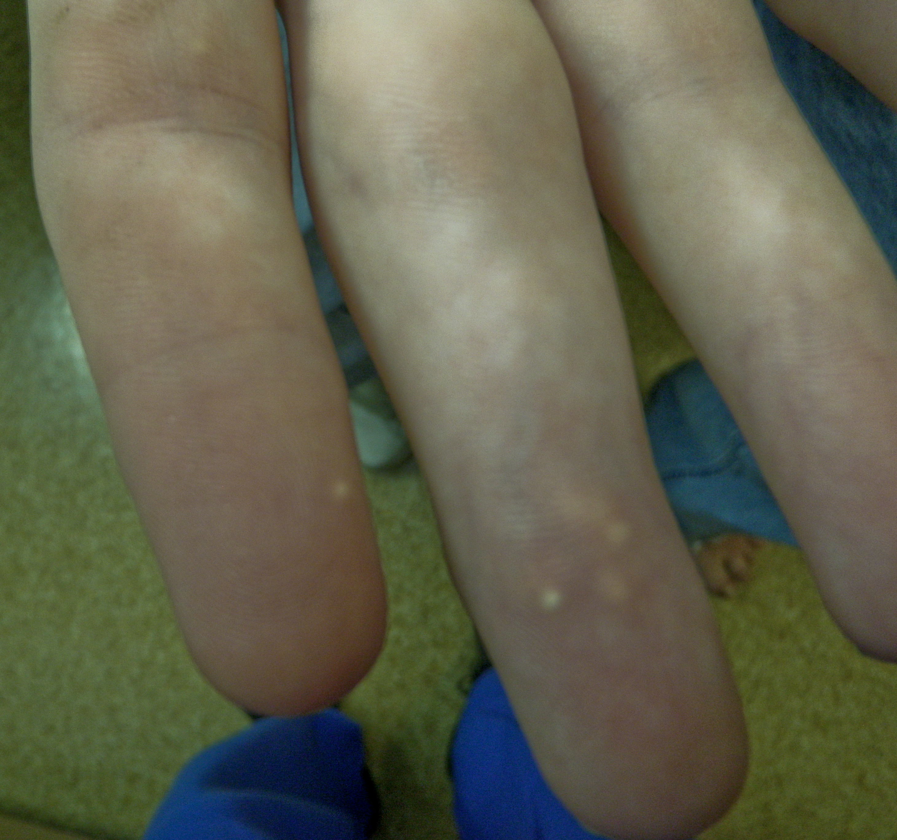
Image Source: Flickr (2010). 5353051607, Licensed under a creative commons licence. Available for free at: https://www.flickr.com/photos/ex_magician/5353051607 (Accessed: 9 January 2024).

Image Source: Raw Pixel (2017). 5964278, Licensed under a creative commons licence. Available for free at: https://www.rawpixel.com/image/5964278/free-public-domain-cc0-photo (Accessed: 4 January 2024).
Gout is excruciating, but acute attacks are self-limiting and usually resolve spontaneously without treatment. In the long term, however, if left untreated, recurrence is common, and several complications may develop. These include:
- Chronic arthritis and subsequent joint damage – this occurs in around 2% of patients with gout and is severe and debilitating. It usually develops around 20 years after the first gout attack.
- Renal stones – hyperuricaemia leads to the formation of urate renal stones in around 14% of patients with gout. These are painful and can have additional complications, including sepsis and kidney damage.
- Tophi are manifestations of monosodium urate crystals deposited around the joint site. They cause significant joint disfigurement and can themselves become inflamed or even infected. They occur in approximately 50% of patients after ten years of poorly controlled gout, rising to 72% of patients after 20 years.
In addition to gout, hyperuricaemia in itself confers an increased risk of organ damage, including:
- Cardiovascular disease, with an increased prevalence of hypertension, myocardial infarction, heart failure and stroke amongst patients with gout.
- Chronic kidney disease, with almost 25% of those with gout having CKD stages 3-5
Appropriate treatment (as discussed later) can suppress gout attacks and their recurrence and prevent these long-term consequences of the disease.
Diagnosis
Gout can be clinically diagnosed in patients with a suggestive history and examination. This may be definitively confirmed by arthrocentesis of the affected joint, revealing the presence of monosodium urate crystals (see image below)

Image Source: Flickr (2019). 158161000@N04/49274750631, Licensed under a creative commons licence. Available for free at: https://flickr.com/photos/158161000@N04/49274750631 (Accessed: 9 January 2024).
Serum uric acid levels may also be monitored; these may be low, normal, or high during an acute attack and are more reliably observed two weeks after resolution of a flare. In the UK, a serum urate level >=360 micromol/L (6mg/dL) confirms a diagnosis of gout.
There is a role for imaging diagnostically. X-rays are of limited use; however, they can differentiate between chronic gout and other joint conditions in the late stages. Ultrasound is more sensitive in detecting erosions, tophi and the gout-specific double contour sign (linear urate deposits over hyaline cartilage). Ultrasound is recommended in cases where joint aspiration is not possible or if gout diagnosis is uncertain.

Image Source: Wikimedia Commons (2012). Gout chondrocalcinosis ultrasound, Licensed under a creative commons licence. Available for free at: https://commons.wikimedia.org/wiki/File:Gout_chondrocalcinosis_ultrasound.svg (Accessed: 4 January 2024).
Management
Early treatment of gout is associated with better clinical outcomes, and therefore, general management is by a two-pronged approach: short-term acute flare resolution to manage pain and preserve function, with long-term prevention of recurrence and protection against joint destruction.
Short-term management:
- NSAIDs
- NSAIDs are the first line in the treatment of acute gout flares. They work by interrupting the inflammatory cascade that results in acute symptoms. Patients at high risk of gastrointestinal complications should be co-prescribed a proton pump inhibitor during treatment.
- Corticosteroids
- If more joints are affected, these may be injected intraarticularly in monoarticular gout flares or parenterally. Patients should be counselled on potentially serious adverse effects.
- Colchicine
- Colchicine acts by blocking the neutrophil-endothelial interaction that drives inflammation in gout. It has a narrow therapeutic-to-risk index, so it should be used at its minimally effective dose. Common adverse effects are diarrhoea, nausea and vomiting.
- Interleukin (IL) – 1 inhibitors
- Anakinra and Canakinumab are IL1 antagonists. They are second-line therapies for acute gout flares that may be effective in patients for whom first-line treatments were ineffective or contraindicated.
Long-term management
- Lifestyle modifications
- Patients should be counselled on the importance of weight loss (if indicated) and dietary modifications, limiting intake of alcohol, purines and high fructose corn syrup.
- Urate lowering drugs
- Xanthine oxidase inhibitors (e.g. allopurinol, febuxostat)
- This class of drugs is the first line in lowering urate in patients with hyperuricaemia. Allopurinol should be reserved for patients without renal impairment, whilst Febuxostat should be used cautiously in those with cardiovascular disease, where allopurinol has failed and the patient has been counselled on its risks.
- Uricosuric agents (e.g. probenecid, pegloticase)
Patients must be counselled that gout relapse is common after discontinuing urate-lowering drug treatment.
Who gets urate-lowering drugs?
Urate lowering drugs are typically recommended in patients with gout with:
- >= 1 subcutaneous tophi
- Evidence of radiographic damage attributable to gout
- Frequent gout flares (>= two annually)
- Patients who are on diuretics have chronic gouty arthritis, or who have moderate-severe CKD (stage >=3), serum urate concentration >540micromol/L (>9mg/dL), or urolithiasis may also benefit from these treatments.
Pharmacological prophylaxis against acute flares (e.g. NSAIDs, low dose colchicine, low dose oral steroid) is advisable for all patients with gout during initiation and titration of urate-lowering drugs (usually first 3-6 months).
Complete Guide to Clinical Genetics ST3 Application and Recruitment (2027)
Written by Medibuddy. Last updated: October 2026. Clinical Genetics is

Complete Guide to Clinical Radiology ST3 Application and Recruitment (2027)
Recruitment paused: NHS England is not running Clinical Radiology ST3+ recru

Complete Guide to Paediatric Surgery ST3 Application and Recruitment in United Kingdom (2026)
Written by Medibuddy. Last updated: October 2026. If you’re planning


Take your subscriptions with you
Our mobile app allows you to access your interview and exam question banks wherever you are.

