


 Close
Close
 12th April 2024
Disorders of the posterior pituitary
Hypothalamus
The hypothalamus is the master control centre for the endocrine system located in the base of the brain below the third ventricle and just above the optic chiasm and pituitary gland. The hypothalamus sends signals directly to the pituitary gland to either stimulate or inhibit the release of hormones.
Posterior pituitary
The pituitary gland consists of the anterior and posterior pituitary glands. It is a direct continuation of the hypothalamus connected by the infundibulum (pituitary stalk) and rests on the sella turcica below the optic chiasm. The posterior pituitary gland, also known as the neurohypophysis, arises from the neural ectoderm at the floor of the forebrain. It is supplied by the inferior hypophyseal artery and drains into the inferior hypophyseal vein. The pituitary gland does not produce hormones; it stores them. The two hormones, vasopressin and oxytocin, are synthesised in the hypothalamus and released from the posterior pituitary gland directly into the systemic circulation. In the hypothalamus, the paraventricular nuclei primarily produces oxytocin and the supraoptic nuclei primarily produces vasopressin. The posterior pituitary gland is made up of axons extending from neuronal cell bodies in the paraventricular and supraoptic nuclei located in the hypothalamus.
| Hypothalamus | ||
|---|---|---|
| Hormones | ADH / vasopressin | Oxytocin |
| Synthesised in the supraoptic nuclei in the hypothalamus | Synthesised in the paraventricular nuclei in the hypothalamus | |
| Posterior Pituitary Gland | ||
| Target organ | ADH acts on kidneys (collecting duct, distal convoluted tubule) to increase permeability of water | Oxytocin acts on the uterus and mammary glands for uterine contraction and lactation |
Table 1: summarising the hormones synthesised in the hypothalamus and released by the posterior pituitary gland to act on their target organs
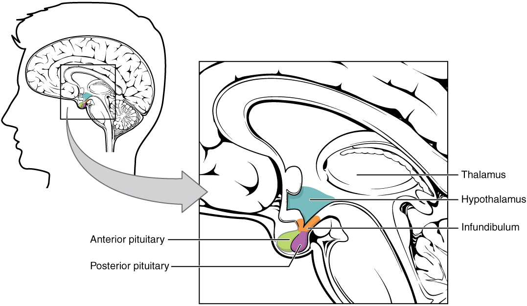
Image Source: Wikimedia Commons (2013). The_Hypothalamus-Pituitary_Complex, Licensed under a creative commons licence. Available for free at: https://en.m.wikipedia.org/wiki/File:1806_The_Hypothalamus-Pituitary_Complex.jpg (Accessed: 11 April 2024).

Image Source: Wikimedia Commons (1918). Anterior_pituitary, Licensed under a creative commons licence.
https://en.wikipedia.org/wiki/Anterior_pituitary#/media/File:Gray1180.png(Accessed: 11 April 2024).
Oxytocin
Oxytocin has two main sites of action: the uterus and the mammary gland.(1) Oxytocin stimulates the myoepithelial layer in the uterus to contract to facilitate delivery. This sends neural signals to the brain to enhance oxytocin secretion, this positive feedback is broken once the baby is delivered. Increased amounts of maternal oxytocin are released in the third stage of labour (after delivery) as it has a role in stopping bleeding after labour, facilitating uterine involution and infant bonding. Positive feedback regulates the release of oxytocin in breastfeeding, suckling the nipple stimulates the paraventricular nucleus in the hypothalamus to produce more oxytocin. This leads to contractions of the myoepithelial cells surrounding the alveoli and ducts in the mammary glands to expel milk, triggering the ‘let down reflex.’.
Antidiuretic hormone (ADH)
Vasopressin, also known as antidiuretic hormone (ADH), regulates water excretion by the kidneys. It is also a potent vasoconstrictor and can be used either directly or in synthetic form to achieve haemostasis, such as in severe gastrointestinal bleeding. ADH acts as an additional stimulus for ACTH release from corticotrophs during foetal development. When the body is dehydrated the extracellular fluid is detected by the osmoreceptors in the hypothalamus (supraoptic nucleus) to stimulate the release of ADH. This acts on the distal convoluted tubule and collecting duct of the kidneys via aquaporin 2 channels to increase the permeability of water, hence leading to water resorption and increased urine concentration. The osmoreceptors also stimulate the hypothalamic thirst centre.
Syndrome of inappropriate antidiuretic hormone secretion (SIADH)
Syndrome of inappropriate ADH secretion (SIADH), also known as syndrome of inappropriate antidiuresis (SIAD), is a disorder characterised by the following:
- Hyponatraemia
- low serum osmolality <270mOsm/kg
- inappropriately high urine osmolality above 100mOsmol/kg.
Normally, when dehydrated, the high serum osmolality causes increased ADH release to increase water reabsorption. Conversely, with low serum osmolality, e.g. in water intoxication, there is a decrease in ADH release to reduce water retention. However, in SIADH increased ingestion of water does not lead to suppression of ADH, therefore causing concentrated urine, reduced urine volume and the increased water retention dilutes the serum sodium levels. The increase in total body water (TBW) causes increased urinary sodium excretion, worsening the hyponatraemia.(²)
SIADH can be secondary to tumours such as small cell lung cancer, brain disorders (trauma or infection), pneumonia, cytotoxic therapy (chemotherapy or radiotherapy), narcotics, analgesics, hypothyroidism, and hypoadrenalism. Rare mutations in the Type 2 vasopressin receptor (AVPR2) can have the same effect as anti-epileptic drugs such as carbamazepine. The ‘gain in function’ mutation to AVPR2 can increase the expression of Type 2 aquaporin water channels, enhancing the sensitivity to ADH.
Signs and symptoms of SIADH include headache, nausea and vomiting, abnormal neurological signs as well as impaired consciousness. In severe cases, there may be coma or convulsions and death. It is one of the commonest causes of hyponatraemia in hospitals, therefore it is important to identify and treat the underlying cause. Treatment consists of restricting fluid intake of up to 1L per day and replacing the sodium lost in the urine. However, it is important to avoid correcting hyponatraemia too rapidly, as increasing sodium levels more than 10 mmol/L per 24 hrs can lead to irreversible brain damage (central pontine demyelination) and death. Vaptans can be used in chronic SIADH, they antagonise the V2 receptors lowering aquaporin water channels in the collecting ducts, thus reducing water re-absorption. Demeclocycline can also be used as it diminishes the action of ADH in the collecting duct, effectively inducing nephrogenic diabetes insipidus.
Diabetes insipidus (DI)
Diabetes insipidus is a metabolic disorder caused by either reduced production or response to ADH causing reduced water re-absorption. This manifests as polydipsia, polyuria (urinary volume greater than 50ml/kg/hr) and the formation of inappropriate hypotonic (dilute) urine, which reduces urine osmolality. This can be divided into two different types: central DI and nephrogenic DI.
Central DI (relating to the hypothalamus and posterior pituitary gland) results when not enough ADH is produced or released. This can be due to pituitary surgery, craniopharyngioma, infiltrative pituitary stalk lesions, traumatic brain injury (TBI), subarachnoid haemorrhage, congenital hypothalamic-pituitary defects, alcohol, phenytoin and autoimmune disorders. (¹, ³)As central DI results from reduced ADH, it can be treated with a synthetic ADH analogue such as desmopressin.
Nephrogenic DI is the result of renal insensitivity to ADH. This can be due to lithium treatment, chronic kidney disease (CKD), chronic hypercalcaemia or hypokalaemia. Nephrogenic DI can be treated with adequate fluid intake, salt restriction and diuretics to reduce polyuria. Both types of DI can be caused by genetic mutations, they may also be associated with hypernatraemia and present as a medical emergency.
Diabetes insipidus can be confirmed by a serum osmolality above 293mOsm/kg, whereas the urine osmolality would be below 300mOsm/kg. A water deprivation test can be used to differentiate the type of DI. Water deprivation is carried out over 8 hrs with repeated measurements of urine and serum osmolality. A synthetic vasopressin analogue is given, if it is caused by central DI, the urine osmolality increases ≥ 750mOsm/kg. However, if there is no change in the urine osmolality after desmopressin is given or urine osmolality remains ≤ 750mOsm/kg, this indicates that the kidney is unable to use this synthetic vasopressin and points towards nephrogenic DI.
Cerebral Salt Wasting
Cerebral salt wasting is characterised by the following:
- Hyponatraemia < 135mEq/L with low plasma osmolality
- Inappropriately elevated urine osmolality > 100mosmol/kg (usually above 300mosmol/kg)
- Urine sodium concentration above 40mEq/l
- Low serum uric acid concentration due to urate wasting in urine
It is believed that the impaired sodium resorption may be due to increased Brain Natriuretic peptide (BNP) typically released in patients with brain injuries or due to diminished central sympathetic activity. Both mechanisms cause inhibition of the renin-angiotensin system. Normally, sympathetic stimulation triggers sodium, urate and water resorption in the proximal tubule. However, diminished sympathetic input results in reduced sodium and urate absorption as well as ADH release due to the volume depletion. The reduced renin and aldosterone released can explain the absence of hypokalaemia in CSW, as there is a failure of aldosterone to be released in response to volume depletion.
CSW can be caused by subarachnoid haemorrhage, head trauma, intracranial neoplasm, metastatic neoplasm, infections such as meningitis, encephalitis, CNS surgery. In patients with neurosurgical procedures CSW would typically manifest within the first 10 days. However, CSW only accounts for a small percentage of hyponatraemia in patients with brain injury.
It can be difficult to differentiate between SIADH and CSW, however one key feature is clinical evidence of volume depletion e.g. hypotension, decreased skin turgor, elevated haematocrit and possibly increased BUN/ serum creatinine ratio. Response to isotonic saline could also help distinguish, as restoration of euvolaemia in CSW should remove the stimulus to release ADH resulting in dilute urine and correction of hyponatraemia. Whereas isotonic saline worsens the hyponatraemia in SIADH. However, this is not carried out in practice, as treating hyponatraemia with isotonic saline in patients with cerebral disease can cause fatal levels of hyponatraemia.
In contrast to SIADH, which is managed with fluid restriction, in patients with subarachnoid haemorrhage fluid restriction could worsen the volume depletion, hypotension and lead to cerebral infarction. So, instead, CSW is managed with 3% hypertonic saline to raise serum sodium levels. Salt tablets can also be given once they can take oral medications, as well as mineralocorticoids.
| DI | SIADH | CSW | |
|---|---|---|---|
| Urine output | ↑ reduced ADH, urinate more | ↓ more ADH, urinate less | ↑ elevated urinary sodium, urinate more |
| Serum sodium | ↑ urinate more, concentrated serum | ↓ retain fluid, dilute serum | ↓ urinary loss of sodium |
| Urine sodium | ↓ concentrate serum, less sodium in urine | ↑ urinate less water, urine more concentrated | ↑ elevated urinary sodium |
| Serum osmolality | ↑ concentrate serum | ↓ retain fluid, dilute serum | ↓ less serum sodium due to urinary losses |
| Urine osmolality | ↓ urinate more water | ↑ urinate less, concentrate urine | ↑ elevated urinary sodium |
| Volume status | Euvolaemic or Hypovolaemic | Hypervolaemic or euvolaemic | Hypovolaemic |
Table 2 detailing the differences between DI, SIADH and CSW
References
- Welt CK. Hypothalamic-pituitary axis. UpToDate2023.
- Holt RIG, Hanley NA. Essential Endocrinology and Diabetes: Wiley; 2021.
- Cuzzo B PS, Lappin SL. Physiology, Vasopressin. StatPearls. 2023.
- Palmer BF. Cerebral Salt Wasting. Up to Date. 2024.
- Norwitz ER. Physiology of parturition at term. UpToDate2024.
- Sterns RH. Pathophysiology and etiology of the syndrome of inappropriate antidiuretic hormone secretion (SIADH). UpToDate2021.
- Gross P. Clinical management of SIADH. Ther Adv Endocrinol Metab. 2012;3(2):61-73.
- Ball S. Diabetes insipidus. BMJ Best Practice 2021.
- Ganapathy MK TP. Anatomy, Head and Neck, Pituitary Gland. StatPearls. 2023.
- Patel H JR, Tiwari V. Physiology, Posterior Pituitary. StatPearls. 2024.
- Saper CB, Lowell BB. The hypothalamus. Current Biology. 2014;24(23):R1111-R6.
- Nicolaides NC WH, Bornstein SR, et al. Adrenal Cortex: Embryonic Development, Anatomy, Histology and Physiology. NIH. 2023.
- Yasir M MO. Syndrome of Inappropriate Antidiuretic Hormone Secretion. StatPearls. 2023.
- Tandukar S, Sterns RH, Rondon-Berrios H. Osmotic Demyelination Syndrome following Correction of Hyponatremia by ≤10 mEq/L per Day. Kidney360. 2021;2(9):1415-23.
- Makita N, Manaka K, Sato J, Iiri T. Chapter Four – V2 vasopressin receptor mutations. In: Litwack G, editor. Vitamins and Hormones. 113: Academic Press; 2020. p. 79-99.
Complete Guide to Clinical Genetics ST3 Application and Recruitment (2027)
Written by Medibuddy. Last updated: October 2026. Clinical Genetics is

Complete Guide to Clinical Radiology ST3 Application and Recruitment (2027)
Recruitment paused: NHS England is not running Clinical Radiology ST3+ recru

Complete Guide to Paediatric Surgery ST3 Application and Recruitment in United Kingdom (2026)
Written by Medibuddy. Last updated: October 2026. If you’re planning


Take your subscriptions with you
Our mobile app allows you to access your interview and exam question banks wherever you are.

