


 Close
Close
 26th June 2023
Disorders of the Spinal Cord
Background Neuroanatomy
Spinal Cord Neuroanatomy
This will have been covered in greater detail in the neuroanatomy section; please refer to this accordingly. The following points are a quick recap to aid understanding of spinal cord disorders.
The intervertebral disc is made up of:
- Annulus fibrosus
- Nucleus pulposus
Compression on the spinal cord caused by intervertebral disc herniation is a result of the nucleus pulposus pushing through a defect in the annulus fibrosus. Reversible risk factors include high body weight, poor posture, occupations involving bending, sitting and lifting, lack of physical activity, smoking, and intrinsic risk factors such as the presence of diabetes mellitus and chondrodystrophies.
The layers of the spinal cord are continuous with the meninges:
- The dura mater has a periosteal and meningeal layer (on top of the spinal arachnoid mater) which separate caudal to the foramen magnum. It is this separation that forms the epidural space (a bit of a misnomer considering it is technically the intradural space.)
- Within the epidural space lie connective tissue, lymphatics, fat, nerve roots and vertebral venous plexus. Of note, this space does not contain any cerebrospinal fluid (CSF). It is this space that epidural anaesthesia targets.
- Beneath the spinal arachnoid mater is the subarachnoid space which contains CSF. It is this space that a lumbar puncture, intrathecal medication, or spinal anaesthesia targets.
- The pia mater forms denticulate ligaments which anchor the spinal cord.
The spinal cord ends at L3 at birth and L1-L2 in adulthood. Below this level is the cauda equina.
The C1-C7 spinal nerves exit above the atlas and above their respective vertebrae. C8 and below exit below their respective vertebrae.
The spinal cord is made of:
- Grey matter
- Dorsal horn – sensory dorsal horn neurons
- Lateral horn and lateral grey column – preganglionic visceral motor neurons that project to the sympathetic ganglia
- Ventral horn – motor neurons supplying striated muscle
- White matter
- Posterior columns – fasciculus cuneatus and gracilis – ascending sensory information from somatic mechanoreceptors
- Lateral columns – lateral corticospinal tracts, posterior spinocerebellar tract, rubrospinal tract
- Anterior columns – spinothalamic tracts, anterior spinocerebellar tract, vestibulospinal tract, olivospinal tract
For more information on the ascending and descending tracts, please refer to the ‘Basic neuroanatomy’ section.
The vascular supply of the spinal cord is composed of:
- Multiple radicular vessels
- At each intervertebral foramen supplying the corresponding nerve root
- C1 to T12 supplied by vertebral and thyrocervical trunk
- T3 to T7 supplied by intercostal arteries
- T8 downwards by the Great artery of Adamkiewicz
- Three longitudinal vessels:
- One anterior spinal artery
- Two posterior spinal arteries in the posterior lateral sulcus
Disruption of this vascular supply, e.g. by aortic dissection, vasculitis, or thrombus, can lead to spinal cord infarctions, which differ in presentation depending on the affected vessel and can be syndromic, e.g. anterior spinal artery syndrome, covered later in this section.
Cauda Equina syndrome and Conus Medullaris syndromes
Both syndromes are relatively uncommon and approached very similarly in the practical day-to-day work on a medical unit, i.e. if suspected, a digital rectal examination, sensory body map of the lower limbs to both touch and pinprick, pre-void and post-void bladder scan should be performed and MRI imaging with neurosurgical referral made, as both are neurosurgical emergencies that warrant emergent surgical intervention.
Conus Medullaris syndrome is the result of a lesion to, or of, the spinal cord itself at the conus medullaris (termination of spinal cord at L1-L2), and hence presents with bilateral symmetrical signs and is usually due to spinal cord intrinsic lesions, e.g. intrinsic tumours such as ependymomas or astrocytomas, spinal cord metastases, sarcoidosis, and demyelinating lesions.
Cauda Equina syndrome results from a lesion to the nerve roots that make up the cauda equina and is usually caused by compression from intervertebral disc herniation or trauma, with other causes being epidural lesions, e.g. epidural metastases and abscesses, leptomeningeal carcinomatosis, schwannoma of the nerve roots, and rarely inflammatory causes, e.g. arachnoiditis, cytomegalovirus polyradiculitis.
Clinical features
| Signs and Symptoms | Cauda Equina Syndrome | Conus Medullaris syndrome |
|---|---|---|
| Pain distribution | Severe, radicular
Unilateral, asymmetric Mainly in perineum, thighs, legs, back |
Bilateral, symmetric over perineum and thighs |
| Motor loss | Gradual, unilateral
Asymmetric, more marked Atrophy may occur Usually no fasciculations |
Sudden, bilateral
Symmetric Fasciculations may be present |
| Sensory loss pattern | Saddle distribution
May be unilateral and asymmetric All modalities affected |
Saddle distribution
Bilateral, usually symmetric Dissociation of sensation |
| Reflex loss | Patellar and ankle reflexes may be lost | Only ankle reflex lost |
| Bladder symptoms | Late and less marked, can be both incontinence or urgency | Early and marked |
| Rectal symptoms | Late and less marked | Early and marked |
| Sexual function | Less marked impairment | Erection and ejaculation impaired |
Spinal Cord Compression
A spinal cord compression can be due to a large variety of traumatic and atraumatic causes similar to Cauda Equina and Conus Medullaris syndromes. Traumatic causes include intervertebral disc herniation and compression from vertebral fractures, while atraumatic causes include degenerative spondylosis with resulting myelopathy, spinal epidural abscesses or haematomas, and primary and metastatic spinal cord tumours. A spinal cord compression is a medical emergency, and suspicion should prompt an urgent MRI scan and treatment with urgent glucocorticoids, typically dexamethasone which downregulates vascular endothelial growth factor and prostaglandin E2, decreasing spinal cord oedema. Prompt referral to neurosurgery, and if indicated, oncology teams for rescue radiotherapy should be considered.
Spinal cord syndromes
Complete Cord Transection

Distribution of motor and sensory loss in complete lower thoracic spinal cord transection.
Image Source: Dr D Chang – own work
In a complete cord transection, there is a loss of motor function (corticospinal), pain and temperature sense (spinothalamic), and touch and proprioception sense (dorsal column) below the lesion, which can result in paraplegia or tetraplegia with loss of all sensory input depending on the level of the lesion. The affected limbs will initially be flaccid and areflexic due to spinal shock before progressing to hypertonic and hyper-reflexive clinical findings with bilateral upgoing plantar reflexes (Babinski’s sign).
Autonomic dysfunction is also a feature, with disruption to urinary and rectal sphincter functions mediated by sympathetic/parasympathetic pathways, which can manifest as anhidrosis, impaired temperature control and loss of the cremasteric reflex (stroking of the medial thigh causing the cremaster muscle to contract and pull up the ipsilateral testicle toward the inguinal canal.)
Its aetiology is primarily due to traumatic and inflammatory causes. Inflammatory causes include multiple sclerosis and postinfectious inflammation from viral, bacterial, and fungal causes, and it can be a very rare complication of vaccination.
It can also result from trauma, such as a traumatic spinal injury or herniated intervertebral disc, and other mechanical causes, such as tumour and metastatic disease.
Prognosis of recovery in traumatic cases is poor with a less than 5 percent chance of recovery of function; recovery in nontraumatic cases is dependent on aetiology.
Brown-Séquard Syndrome
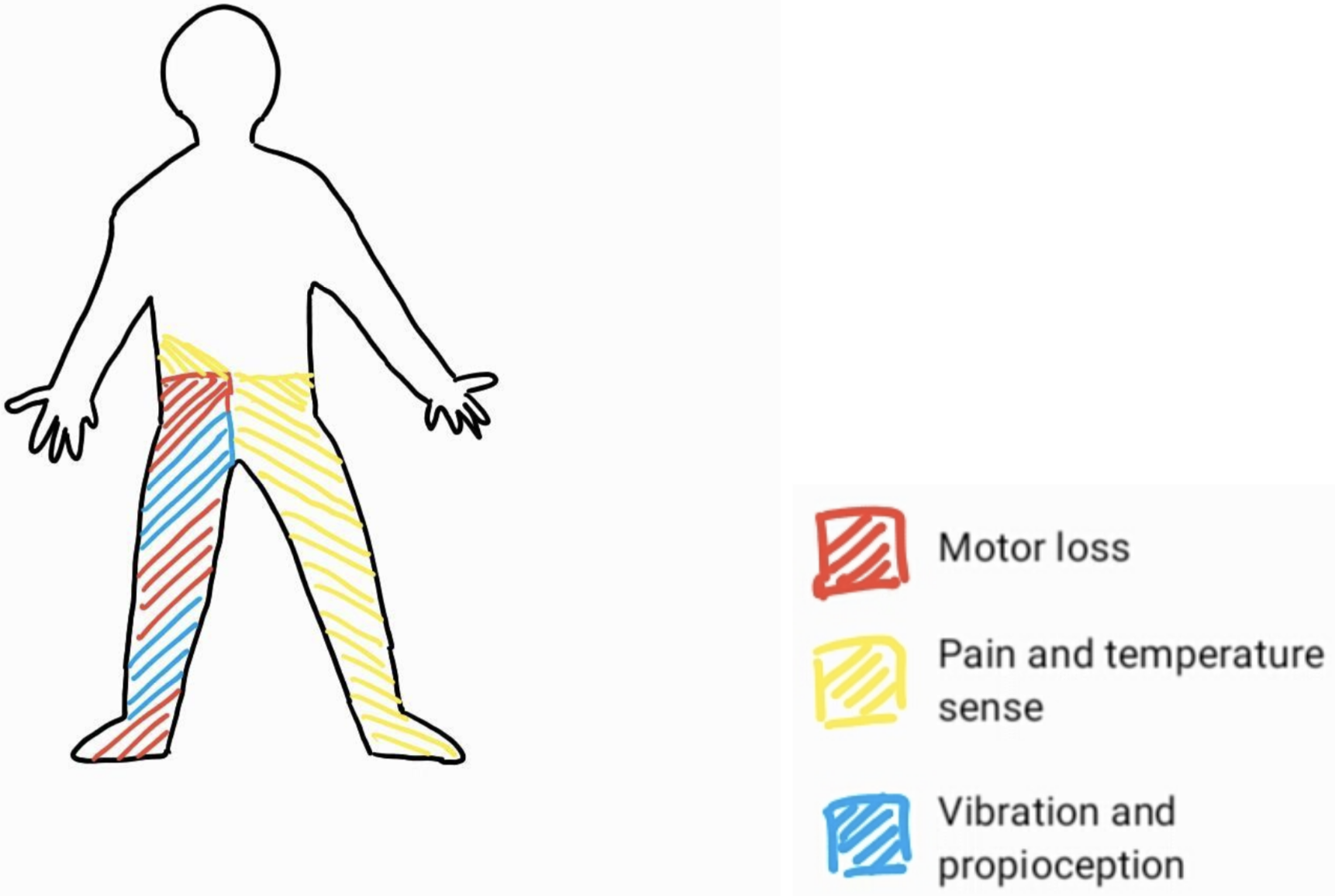
Distribution of motor and sensory loss in right hemisection of the lower thoracic spinal cord.
Image Source: Dr D Chang – own work
Brown-Séquard syndrome describes a pattern of sensorimotor loss resulting from damage to one half of the spinal cord (i.e. hemisection of the cord) causing:
- Ipsilateral loss of vibration sense and proprioception, with hemiparesis.
- Contralateral loss of pain and temperature sense, with a clinical sensory level usually 1 to 2 levels below the actual lesion.
- Ipsilateral loss of all sensory modalities at the level of the lesion, with radicular pain.
- Ipsilateral flaccid paralysis at the level of the lesion.
Brown-Séquard syndrome is usually caused by a traumatic spinal injury or intramedullary tumours confined to one side of the spinal cord, but an important cause of Brown-Séquard syndrome is transverse myelitis (see below).
This syndrome has the most favourable prognosis among the incomplete spinal cord syndromes, with 75% of patients able to ambulate independently at discharge from rehabilitation (75%) and nearly 70% perform functional skills and activities of daily living independently and favourable bladder and bowel function profile with continent bladder and bowel function achieved in 89% and 82%, respectively.
Central Cord Syndrome

Distribution of motor and sensory loss in central cord syndrome from a large lesion affecting the corticospinal tracts, spinothalamic tracts, and dorsal columns at the level of the cervical spine.
Image Source: Dr D Chang – own work
Central cord syndrome is the most common incomplete spinal cord syndrome and has a varied presentation depending on how large the lesion is, which determines how much cord is involved. It is insidious in onset, affecting first the anterior and lateral spinothalamic tracts, causing loss of pain and temperature with preservation of position, vibration, and touch sensation. Lesions causing central pattern spinal cord syndrome typically arise at the level of the cervical spinal cord. Sensory loss here presents in a vest-like pattern over the shoulders with a spectrum of range and severity with size of lesion. Sacral sensation is usually spared.
Larger lesions can progress posteriorly to include the dorsal columns, affecting vibration and proprioception below the lesion, anteriorly to the anterior horns, and laterally involving the corticospinal tracts, causing a flaccid paralysis with fasciculations, with eventual atrophy at the level of the lesion and spastic paralysis of muscles below the lesion.
The aetiology of a central pattern syndrome is most commonly from a traumatic hyperextension injury involving the cervical cord, which specifically injures the central part of the cord, typically the central corticospinal tracts (as opposed to fractures/trauma causing other complete or incomplete spinal cord syndromes). This can result in quadriplegia with some recovery of lower limb function and a disproportionately greater upper limb weakness, worse distally than proximally. This is more common in people with cervical osteoarthritis and cervical spondylosis, where an existing stenosis, e.g. from osteophytes, increases the vulnerability of the spinal cord to injury. Bladder dysfunction, e.g. urinary retention, priapism and autonomic dysreflexia may occur.
In traumatic central cord syndrome, the prognosis is generally favourable, with patients recovering substantial neurological function after the injury with 90 percent return of motor function by three years, progressing in an ascending order with return of lower limb function, then bladder control and eventually proximal upper limbs and hands. Patients undergoing surgical intervention are more likely to attain pre-injury mobility status.
Atraumatic causes are much rarer and include neoplastic causes such as intramedullary spinal cord tumours and syringomyelia.
Syringomyelia

Distribution of pain and temperature loss in syringomyelia.
Image Source: Dt D Chang – own work
Syringomyelia is the development of a CSF filled syrinx within the spinal cord parenchyma or central canal that can enlarge and compress surrounding tracts, typically at the C8/T1 level. It is typically associated with type 1 Arnold-Chiari malformations in which there is downward displacement of the cerebellar tonsils beneath the foramen magnum into the cervical spinal canal, blocking the normal CSF flow between the spinal canal and intracranial space. Syringomyelia can also be associated with a tethered cord (in which the caudal spinal cord can become fixed to surrounding inelastic tissue) and with spina bifida, tumours, meningitis and arachnoiditis.
Syringomyelia typically presents with bilateral pain and temperature insensitivity in a cape-like distribution over the upper limbs due to the compression of the spinothalamic tract decussation in the anterior white commissure, with preserved touch and vibration sense below the level of the syrinx. Symptoms can be static for years but may worsen in association with Valsalva manoeuvres, e.g. coughing or sneezing/heavy lifting/straining, which can cause enlargement and extension of the syrinx.
Enlargement of the syrinx causes the progression of the central cord syndrome, compressing anterior horn cells and corticospinal tracts, leading to a variable presentation of syringomyelia depending on the size of the syrinx and which structures are being compressed. This can include spasticity or paralysis of the legs, ataxia, or fasciculations.
If the syrinx is higher in the cord, extending into the medulla of the brainstem, this is termed syringobulbia, which can result in cerebellar and lower motor cranial nerve nuclei signs.
Investigation is with MRI and dynamic CSF flow MRI studies, with CT myelograms as an alternative in the event of MRI contraindications.
Treatment is typically with surgical intervention to improve CSF flow and is dependent on the cause, e.g. a Chiari malformation indicating a craniocervical decompression if associated with type 1 Arnold-Chiari malformation, or with decompressive laminectomy, syringosubarachnoid or syringoperitoneal shunts.
Posterior Column Syndrome

Distribution of vibration and proprioception loss in posterior column syndrome of the cervical spinal cord.
Image Source: Dr D Chang – own work
Posterior column syndrome is the most uncommon incomplete spinal cord syndrome, manifesting as a loss of proprioception and vibratory sense, and two-point discrimination distal to the lesion, deep tendon areflexia and a sensory ataxia (due to loss of joint position sense). Romberg sign is positive, and a stomping gait is present which can be exacerbated by removing visual cues, e.g. by closing eyes. There is hypotonia in limbs distal to the lesion, but power is usually preserved. If the lesion is at the level of the cervical cord, it can be associated with Lhermitte’s sign, in which neck flexion elicits neuropathic pain and paraesthesia down the arms.
The aetiology or posterior cord syndrome is classically due to neurosyphilis with tabes dorsalis, but can happen due to thrombosis or occlusion of the posterior spinal arteries, mechanical causes such as localised compression from epidural tumours, haematomas or abscesses, and inflammatory causes such as multiple sclerosis. Friedreich’s ataxia (see the section below) can also manifest primarily as a posterior cord syndrome before progressing to involve corticospinal tracts.
In the case of tabes dorsalis as a late complication of neurosyphilis, posterior column degeneration is also accompanied by dorsal root ganglion demyelination. Tabes dorsalis progressively develops over years, manifesting with the posterior cord syndrome as above, accompanied with lancinating pain and dysesthesias and can progress to cause bladder dysfunction, Argyll Robertson pupils (see Cranial Nerve Disorders section) and can cause a Charcot arthropathy. Treatment is with intramuscular benzylpenicillin, noting that patients undergoing treatment may experience a Jarisch Herxheimer reaction where the death of the Treponema pallidum spirochetes release endotoxins that provoke pyrexia, tachycardia, rigors, and rashes 6 to 48 hours post initiation of therapy.
Posterolateral Column Syndrome
Posterolateral column syndrome, also known as ‘Subacute Combined Degeneration of the spinal cord’ (SACD), is similar to a posterior column syndrome, but also involves the lateral corticospinal tracts. This is a slow and insidious spinal cord syndrome characterised by ascending bilateral limb paraesthesia, manifesting as loss of position and vibratory sense, progressing to weakness with ataxia, and eventually a bilateral spastic paresis. Pain and temperature sense is unaffected. Clinical findings may show a sensory ataxia with positive Romberg’s sign and bilateral spastic paresis with a positive Babinski sign.
It is usually caused by metabolic aetiology, most commonly a prolonged vitamin B12 deficiency. This is why a patient with folate deficiency should always have B12 levels checked, as symptoms of B12 deficiency can be exacerbated and masked if only the folate deficiency is treated. Subacute Combined Degeneration of the spinal cord can also be caused by vitamin E deficiency and copper deficiency, causing loss of myelin initially at the dorsal columns (governing vibration, coarse touch, and proprioception) and then at the lateral columns, including spinocerebellar and corticospinal tracts, but sparing the spinothalamic tracts.
Diseases which exacerbate these nutritional deficiencies, e.g. pernicious anaemia causing vitamin B12 deficiency, cystic fibrosis or abetalipoproteinemia causing loss of absorption of vitamin E, or Menkes disease, an X-linked recessive mutation in copper transport proteins, can all cause SACD. Of note, nitrous oxide administration (e.g. as anaesthesia, analgesia, or as a recreational drug) can induce a functional B12 deficiency and precipitate SACD in already B12 deficient individuals, by inactivating vitamin B12. This is assessed by checking methylmalonic acid (MMA) and homocysteine levels.
Treatment of SACD is with intramuscular hydroxocobalamin injections to replace vitamin B12 levels.
Anterior Cord Syndrome

Distribution of motor and sensory loss in anterior cord syndrome of the cervical spinal cord.
Image Source: Dr D Chang – own work
Anterior cord syndrome is an incomplete spinal cord syndrome affecting the anterior two-thirds of the spinal cord. In general, an anterior cord syndrome manifests as bilateral motor paralysis below the level of the lesion, with associated bilateral loss of pain and temperature sense. Lateral horn involvement can cause an autonomic dysfunction, including anhidrosis, hypotension, neurogenic bowel, bladder, and sexual dysfunction.
There are two primary ‘sub-syndromes’ depending on whether the anterior horn cells are affected (anterior horn cell syndrome) or whether the vascular supply to the anterior cord is the aetiology of the syndrome (anterior spinal artery syndrome).
In anterior horn cell syndrome, there is involvement of the anterior grey matter horns of the spinal cord, causing ipsilateral or bilateral flaccid motor paralysis with atrophy and fasciculations. The aetiology is varied and includes:
- Infections: Poliomyelitis, West Nile virus
- Iatrogenic: Post-radiation myelitis
- Metabolic: Hexosaminidase A deficiency (aka lysosomal storage disorder), GM2 gangliosidosis (aka Tay Sachs disease)
- Hereditary: Spinal muscular atrophies
Anterior spinal artery syndrome is typically vascular in aetiology, with disruption of the anterior spinal artery or the great artery of Adamkiewicz from a thrombus, ruptured aortic aneurysm, aortic dissection, or vasculitis. Disruption of vascular supply, e.g. in severe hypotension or a cocaine associated vasospasm, can cause a similar presentation. Onset is abrupt with deficits occurring minutes to hours post insult. Clinical features may include radicular back or neck pain and acute flaccid paralysis with loss of pain and temperature and autonomic dysfunction. Symptoms may progress to bilateral spastic para/quadriparesis over days to weeks. Touch, proprioception, and vibration sense are preserved due to the sparing of the dorsal column medial lemniscus tracts.
Treatment involves management of the underlying cause, e.g. fluid/vasopressors for hypotension, as well as supportive therapies, including physiotherapy and occupational therapy, social and psychological support along with pressure area care, bladder catheterisation, venous thromboembolism prophylaxis and stress ulcer protection. Prognosis is poor and many patients do not recover motor, sensory or autonomic function.
Specific spinal cord pathologies
Motor Neurone Disease
Motor neurone disease is a group of chronic sporadic and hereditary neurodegenerative diseases characterised by axonal degeneration of neurons responsible for somatic motor function in the motor cortex, anterior horn cells, corticospinal tracts, and cranial nerve nuclei. It can affect the upper motor, lower neurons or both simultaneously. There is usually no sensory loss or sphincter disturbance.
The incidence of motor neuron disease is 2-3 in 100,000, with a prevalence of 5-7 in 100,000. The median age of onset is 60 years. Prognosis is poor, with an average life expectancy of 2 to 4 years after diagnosis. Poor prognostic markers include female gender, older age of onset, bulbar involvement, and respiratory involvement.
Aetiology
There is a genetic basis to MND which can be sporadic or hereditary. Approximately 10 percent of individuals with amyotrophic lateral sclerosis (ALS), a common form of MND, have familial disease. The estimated lifetime risk of sporadic ALS is 1 in 400 with a male to female preponderance of 3:2. Four genes, the C9ORF72 (chromosome 9 open reading frame), TARDBP (TAR DNA binding protein), SOD1 (superoxide dismutase), and FUS (fused in sarcoma) RNA binding protein) genes are associated with up to 70 percent cases of familial ALS. C9ORF72 has been associated with 40 percent cases of familial and 10 percent cases of sporadic MND. C9ORF72 mutations, which lead to a hexanucleotide repeat expansion, have also been found in cases of fronto-temporal dementia, suggesting a link or possibly spectrum between the diseases. The neuropathological hallmark of MND is the presence of ubiquitinated proteinaceous inclusions in motor neurons containing TDP-43 (transactive response DNA binding protein 43), together with skeletal muscle and cortical motor atrophy.
Clinical features
Phenotypes are based upon the site of origin (spinal/limb versus bulbar/respiratory) and the severity of neurological involvement, which can be characterised by:
- asymmetrical versus symmetrical
- proximal versus distal
- upper versus lower limb predominance
- degree of upper motor to lower motor neuron signs
- presence or absence of bulbar symptoms
- presence or absence of respiratory involvement
The four large subtypes of MND are:
- Amyotrophic lateral sclerosis (ALS)
- Typically limb onset, progressing asymmetrically from distal to proximal muscles
- Loss of motor neurons in cortex and anterior horn results in mixed upper motor neuron (hyperreflexia and spastic weakness) and lower motor neuron signs (flaccid paralysis, muscle atrophy, hyporeflexia/areflexia, and fasciculations).
- Bulbar involvement is documented in one fifth to one third of presentations, along with respiratory and psychiatric symptoms (frontotemporal dementia).
- Typical clinical presentation is with distal asymmetrical manifestations such as difficulty turning keys/door knobs, writing, unsteady gait, or frequent falls.
- A hypercatabolic state can develop disproportionately to physical activity, the cause of which is not yet known.
- Progressive bulbar palsy
- This is typically a bulbar-onset ALS which has rapid progression and worse prognosis; there are reports of an isolated bulbar palsy which is more slowly progressive and has better prognosis.
- Affects CN 9 to 12, causing a bulbar palsy with lower motor neuron signs, e.g. flaccid fasciculating tongue, quiet nasal speech, absent jaw jerk, and loss of gag reflex.
- Mimics include Guillain Barré syndrome, myasthenia gravis and central pontine myelinolysis.
- Classic progressive bulbar palsy may then develop into ALS phenotype as above.
- Progressive muscular atrophy
- Purely anterior horn cell lesion resulting in lower motor neuron phenotype.
- Asymmetrical distal to proximal progression, typically asymmetric weakness with distal limb atrophy
- Rare cause of MND (5%).
- Better prognosis with a slower rate of progression.
- Primary lateral sclerosis
- Loss of Betz cells in motor cortex – primarily upper motor neuron signs
- Spinal motor neurons not affected, therefore no muscle atrophy
- Marked spastic paraparesis with pseudobulbar palsy, with stiffness
- Rarest form of MND (1-3%)
- Slowly progressive
Investigations
The variability of clinical findings in MND and the lack of a clear biological diagnostic marker can make absolute diagnosis a challenge. The El Escorial and Airlie House diagnostic criteria for ALS include:
- Presence of lower motor neuron degeneration by clinical, electrophysiological, or neuropathological evidence.
- Presence of upper motor neuron degeneration by clinical evidence.
- Progressive spread of signs and symptoms within a region to another region determined by history and examination.
- The absence of electrophysiological, pathological, and neuroimaging evidence of other disease processes that might explain the observed clinical signs.
Thus, investigations are focused on both excluding other pathology and identifying criteria that would fulfil an MND diagnosis. Tests to exclude other pathologies may include thyroid function tests, B12/folate, CK, immunoglobulins, serum electrophoresis, virology, receptor antibodies and lumbar puncture. Imaging may include MRI head and spine, and functional tests such as nerve conduction studies and electromyography. A muscle biopsy can also be considered. Neurofilament light peptide and phosphorylated neurofilament heavy neuropeptides in cerebrospinal fluid (CSF) have been associated with ALS, but these are not specific and can also be found in Alzheimer’s dementia and multiple sclerosis.
Management
Management of MND is with a multi-disciplinary approach, a combination of the neurologist, physiotherapy, occupational therapy, dietitian, specialist nurse, family, and community team.
Disease modifying modalities include Riluzole, an NMDA (N-methyl-D-aspartate) receptor antagonist with antiglutamatergic action, which has been found to improve survival by 3 months. Edaravone is a newer drug; it is a member of the substituted 2-pyrazolin-5-one class and is thought to act as an antioxidant with a neuroprotective effect, impeding disease progression. Where there is respiratory involvement, non-invasive ventilation overnight has been shown to extend survival and improve quality of life.
Symptomatic treatment includes managing oral secretions with hyoscine, amitriptyline, propantheline, botulinum into parotid glands, and preventing thick bronchial secretions with carbocisteine and nebulised saline. Spasticity can be managed with tizanidine (α2 receptor agonist), baclofen, physiotherapy and botulinum injections. Pain management is with analgesia, such as NSAIDs, opioids, and cannabinoids, gabapentinoids, and tricyclic antidepressants.
Due to the hypercatabolic state and dysphagia associated with MND, nutrition is of high importance and feeding via a gastrostomy tube can be considered. Pressure sores and damage should be monitored closely, and VTE prophylaxis considered. Constipation treatment with laxatives, and bladder control with oxybutynin, tolterodine or a long-term catheter can be considered. The need for psychological support, social support and help with communication cannot be overstated.
Friedreich’s Ataxia
Friedreich’s ataxia is an autosomal recessive neurodegenerative disease caused by a mutation (typically a GAA trinucleotide repeat expansion in 98% of cases) in the FXN gene on chromosome 9, resulting in reduced production of frataxin. It is the most common of the inherited ataxias with an incidence of 1:150,000 and results in both spinal cord axonopathy and demyelination, with widespread alteration in cellular phospholipid metabolism, affecting mainly the dorsal root ganglia, posterior columns of the spinal cord, corticospinal tracts, dorsal spinocerebellar tracts and also the heart. Progressive destruction of the dentate nucleus with atrophy of Betz cells also occurs intracranially.
Clinical features are loss of vibration sense and proprioception with progressive gait and limb ataxia, absent lower limb reflexes and extensor plantar responses with limb weakness and atrophy. Dysarthria and dysphagia can also develop, and many patients will suffer from cardiomyopathy, kyphoscoliosis, and foot deformities such as pes cavus.
Investigations include MRI imaging, EMG, nerve conduction studies, echocardiogram, and genetic testing. Nerve conduction studies will characteristically show absent sensory nerve action potentials and absent spinal somatosensory evoked potentials, with motor nerve conduction velocities reduced to a lesser extent than sensory nerve action potentials.
Management is supportive, with a focus on rehabilitation and orthoses, with management of any complications, e.g. heart failure. Prognosis is variable and dependent on age and the specific genetic mutation in the FXN gene, with patients surviving into their 30s and some living into their 60s.
Poliomyelitis
Poliomyelitis is a consequence of poliovirus infection leading to damage of lower motor neurons and resulting in an acute flaccid paralysis. It is primarily a lower motor neuron lesion and results in the destruction of the anterior horns and a subsequent pure motor deficit. Depending on the level of the damage, this can lead to bulbar involvement and acute respiratory failure.
The causative agent is the Poliovirus which is transmitted faecal orally, and replicates in the oropharynx and small intestine before spreading to the bloodstream and CNS.
Treatment is primarily supportive with no specific treatment, thus prevention of the infection with vaccination is vastly preferable.
Due to widespread vaccination since the 1950s, the incidence of polio has dramatically declined and has been virtually eradicated from most countries.
Complete Guide to Clinical Genetics ST3 Application and Recruitment (2027)
Written by Medibuddy. Last updated: October 2026. Clinical Genetics is

Complete Guide to Clinical Radiology ST3 Application and Recruitment (2027)
Recruitment paused: NHS England is not running Clinical Radiology ST3+ recru

Complete Guide to Paediatric Surgery ST3 Application and Recruitment in United Kingdom (2026)
Written by Medibuddy. Last updated: October 2026. If you’re planning


Take your subscriptions with you
Our mobile app allows you to access your interview and exam question banks wherever you are.

