


 Close
Close
 5th April 2024
Disorders of Sodium
Sodium ions are the major cation in extracellular fluid and contribute a large part to its osmotic pressure. Normal serum sodium concentration is between 135-145 mmol/L. Loss of water from the extracellular space results in increased sodium concentration, a cause of hypernatraemia, defined by serum sodium exceeding 145 mmol/L. Hyponatraemia is defined by serum sodium concentration less than 135 mmol/L.
Hyponatraemia
Causes of hyponatraemia can be divided based on fluid status:
- Hypovolaemic hyponatraemia due to renal and extra renal losses
- Euvolaemic hyponatraemia includes SIADH; hypothyroidism, glucocorticoid deficiency, primary polydipsia and drugs including antipsychotics and thiazide like diuretics.
- Hypervolaemic hypernatraemia occurs with congestive cardiac failure; cirrhosis, nephrotic syndrome, acute and chronic kidney disease.
Psuedohyponatraemia may occur in hypertriglyceridaemia or hyperproteinemia, giving a falsely low sodium when the actual serum sodium concentration is normal.
Clinical manifestations
Signs and symptoms of sodium imbalance will depend on the severity and speed of onset. Mild hyponatremia can be asymptomatic but nausea, confusion and headaches may manifest with moderate sodium increases. Severe hyponatremia causes vomiting, seizures, sleep abnormalities and coma.
Investigations
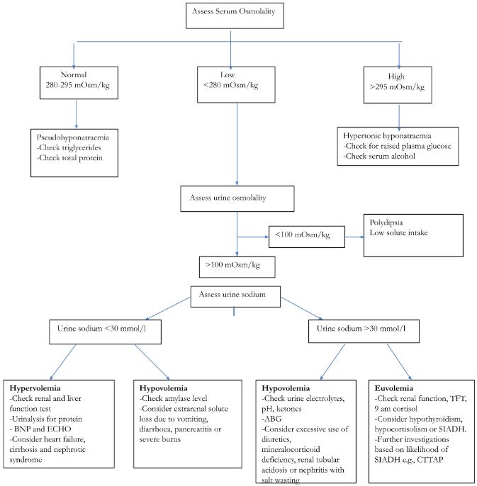
Initial investigations include renal function tests; serum and urine osmolality, urine sodium and thyroid function tests (TFT). The investigation algorithm and different causes of hyponatraemia are shown in the image below.
Syndrome of inappropriate antidiuretic hormone
Syndrome of inappropriate antidiuretic hormone (SIADH) secretion causes hyponatraemia, characterised by low plasma osmolality with inappropriate urinary response. Diagnosis requires elevated urine sodium, euvolemic fluid status and for other causes of euvolemic hyponatremia to be excluded. Causes of SIADH can be divided into:
- Neurological: stroke, trauma, epilepsy and haemorrhage
- Respiratory: COPD, sarcoidosis, asthma and acute respiratory failure
- Medications: carbamazepine, phenytoin, risperidone, quetiapine, duloxetine, gabapentin
- Malignancy: lung cancer, genitourinary and gastrointestinal cancers
Algorithm for investigations in hyponatraemia
Management
Management depends on the cause, and where hyponatremia is due to congestive cardiac failure with a hypervolemic fluid status, fluid restriction and diuretics are used.
Hyponatraemia secondary to gastrointestinal losses will require fluid replacement with 0.9% sodium chloride.
In SIADH related hyponatremia, treating the cause; fluid restriction and medications such as tolvaptan and demeclocycline are used.
In severe symptomatic hyponatremia, emergency correction using 150 ml of 1.8% or 3% sodium chloride over 20 minutes is used, with frequent sodium monitoring. The aim is to increase sodium concentration by 4-6 mmol in 24 hours and once stable, correction is slowed, aiming to increase sodium by 10 mmol in 24 hours.
Osmotic demyelination syndrome
Correcting hyponatremia too quickly can cause rapid changes in osmolality and disrupt the blood brain barrier. This is toxic to and causes damage to the myelin sheath. Presentation includes progressive neurological deficits and weakness; behavioural changes, encephalopathy, quadriplegia and locked in syndrome.
Hypernatraemia
High plasma sodium > 145 mmol/L is usually the result of water depletion but can be caused by excess salt intake, diabetes insipidus and administration of hypertonic sodium. Low serum protein may result in falsely raised sodium levels. The cause can be determined by the timeline of events with accompanying serum and urine sodium testing. Hypernatraemia can be classified according to the patient’s fluid status:
- Hypovolemic hypernatraemia from excessive sweating and dehydration; vomiting, diarrhoea, diuretic use and kidney disease.
- Euvolemic hypernatraemia due to fever, diabetes insipidus and drugs including lithium.
- Hypervoleamic hypernatraemia causes include hyperaldosteronism, excess administration of hypertonic saline or sodium bicarbonate and rarely, from excess salt intake
- Low serum protein may result in falsely raised sodium
Clinical manifestations
Early symptoms include polydipsia, anorexia, nausea and weakness. Severe symptoms include confusion, myoclonic jerks and seizures.
Management
Acute hypernatraemia with onset over hours can be corrected rapidly using dextrose 5% and normal saline. The usual rate is 3-6ml/kg/hour with regular monitoring of serum sodium concentrations. Once serum sodium is below 145 mmol/L the infusion rate should be reduced to 1ml/kg/hour to restore sodium concentrations around 140 mmol/L.
In chronic hypernatraemia, present longer than 48 hours, 5% dextrose fluids are used with the aim to lower sodium concentrations by 10 mmol/L over a 24-hour period.
Diabetes insipidus
Diabetes insipidus (DI) is characterised by hypernatraemia, polydipsia, polyuria and inappropriately dilute urine. Cranial DI is due to a lack of production of arginine vasopressin (AVP), also known as antidiuretic hormone (ADH). Nephrogenic DI occurs from a lack of response to AVP. The hormone is secreted from the posterior pituitary and acts at the distal convoluted tubule and collecting ducts of the nephron to increase water reabsorption.
Causes of cranial DI include:
- Pituitary surgery
- Craniopharyngioma
- Traumatic brain injury
- Subarachnoid haemorrhage
- Wolfram syndrome
Risk factors for nephrogenic DI include:
- Lithium therapy
- Chronic kidney disease
- Chronic hypercalcaemia or hypokalemia
Initial investigations include serum and urine sodium testing, water deprivation test and MRI brain where cranial DI is suspected.
Water deprivation test
This is also called the desmopressin stimulation test (see table below) and can distinguish between psychogenic polydipsia, cranial and nephrogenic DI. Patients are advised to avoid fluids up to eight hours before the test, following which urine osmolality is measured.
In primary polydipsia, water deprivation alone causes urine osmolality to be high and desmopressin need not be given as the high urine osmolality rules out diabetes insipidus. Administration of desmopressin in primary polydipsia has little effect on urine osmolality.
Where the urine osmolality is low, synthetic desmopressin (DDAVP) is administered either 20 mcg intranasally or 2 ug intramuscularly and urine osmolality remeasured.
In cranial DI, where the patient lacks AVP, administration of desmopressin causes water reabsorption by the kidneys and an increase in urine osmolality. In nephrogenic DI, the urine osmolality will remain low before and after desmopressin administration as the kidneys are unable to respond to desmopressin.
| Post dehydration osmolality (mOsm/kg) | Post DDAVP osmolality (mOsm/kg) | ||
| Diagnosis | Serum | Urine | Urine |
| Cranial DI | >300 | < 300 | >400-600 |
| Nephrogenic DI | >300 | < 300 | <300 |
| Primary polydipsia | <295 | > 600 | > 600 |
The water deprivation test
Management for cranial DI includes administration of desmopressin. Nephrogenic DI is treated with dietary salt restriction, diuretics and adequate fluid intake.
References
Water deprivation test https://mft.nhs.uk/app/uploads/2023/03/Water-deprivation-test-Adults.pdf
Complete Guide to Clinical Genetics ST3 Application and Recruitment (2027)
Written by Medibuddy. Last updated: October 2026. Clinical Genetics is

Complete Guide to Clinical Radiology ST3 Application and Recruitment (2027)
Recruitment paused: NHS England is not running Clinical Radiology ST3+ recru

Complete Guide to Paediatric Surgery ST3 Application and Recruitment in United Kingdom (2026)
Written by Medibuddy. Last updated: October 2026. If you’re planning


Take your subscriptions with you
Our mobile app allows you to access your interview and exam question banks wherever you are.

