


 Close
Close
 8th February 2024
Endocrine Physiology
Overview of hormones
Classification of hormones
Hormones are chemical messengers produced by cells, glands or organs into the circulation, exerting their effects elsewhere in the body.
Classification of hormones is according to their chemical composition:
- Polypeptides – E.g. the hypothalamic and pituitary hormones, growth hormones
- Monoamines (i.e., single amino acid) – E.g. catecholamines (adrenaline and noradrenaline), thyroxine, serotonin
- Steroids – E.g. aldosterone, sex hormones
Comparing and contrasting the three classifications:
| Polypeptides | Monoamines | Steroids |
|---|---|---|
| Stored prior to release | Catecholamines and serotonin are stored prior to release
Thyroxine is bound to thyroglobulin |
Released immediately |
| Generally polar and water-soluble | Some are polar (e.g., adrenaline), whilst others are carried by proteins. | Lipophilic and hydrophobic, so it requires a carrier protein for transport in circulation. |
| Bind to transmembrane protein, causing transduction and amplification of signals via cAMP (second messenger) | Adrenaline binds to membrane receptors.
Thyroid binds to nuclear receptors. |
Bind to intracellular receptor, resulting in gene transcription. |
| Fast-acting | Catecholamines are fast-acting
Thyroid hormones are slow-acting (due to its action in gene transcription) |
Slow-acting |
Regulation of hormones
Regulation of hormone secretion can be achieved by the negative feedback mechanism or the positive feedback mechanism.
Negative feedback mechanism
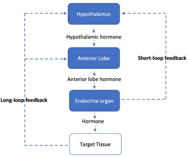
The action of one hormone either directly or indirectly inhibits the secretion of another hormone. It is self-limiting. The majority of homeostasis within our body relies on negative feedback loops.
Long-loop feedback
Hormone feeds back to the hypothalamic-pituitary axis
E.g. High levels of the T3 hormone suppress the secretion of TSH
Short-loop feedback
Anterior pituitary hormone feeds back on the hypothalamus to inhibit the secretion of hypothalamic-secreting hormone
E.g. Growth hormone suppresses the release of Growth-hormone releasing hormone
Positive feedback mechanism
The action of one hormone either directly or indirectly stimulates the secretion of another hormone. It is self-reinforcing. It is more uncommon than a negative feedback mechanism. For example, luteinising hormone (LH) levels rise just prior to ovulation due to the positive effect of oestrogen on the anterior pituitary gland.
References
Tortora, G. J., & Derrickson, B. (2017). Tortora’s principles of anatomy and physiology. John Wiley and Sons.
Kumar, P. and Clark, M., (2012). Kumar & Clark’s clinical medicine. 8th ed.
The hypothalamus and the pituitary gland
Hypothalamus
The hypothalamus acts as a link between the brain, autonomic nervous system and the endocrine system. Some of its functions include:
- Regulating temperature
- Regulating body water
- Regulating sleep-wake cycle
- Regulating sexual function
- Moderating behaviour
- Controlling pituitary function
The hypothalamic regulatory hormones which control pituitary function can be classified into:
Releasing hormones
| Hypothalamic hormone | Pituitary hormone |
| Corticotropin-releasing hormone
(CRH) |
Adrenocorticotropic hormone
(ACTH) |
| Gonadotropin-releasing hormone
(GnRH) |
Luteinising hormone and Follicle-stimulating hormone
(LH and FSH) |
| Thyrotropin-releasing hormone
(TRH) |
Thyroid-stimulating hormone
(TSH) |
| Growth hormone-releasing hormone
(GHRH) |
Growth hormone
(GH) |
| Prolactin-releasing hormone
(PRH) |
Prolactin (PRL) |
Inhibitory hormones
| Hypothalamic hormone | Pituitary hormone |
| Dopamine | Prolactin |
| Somatostatin | TSH, FSH, GH |
Pituitary gland
The pituitary gland lies directly beneath the hypothalamus within the sella turcica and is connected to the hypothalamus by the pituitary stalk. The pituitary gland is divided anatomically (from its embryological origin) and functionally into:
| Anterior Pituitary
“adenohypophysis” |
Posterior pituitary
“neurohypophysis” |
| Hypothalamic hormones either stimulate or inhibit anterior pituitary hormones. | Hormones are synthesised in the paraventricular and supraoptic nuclei of the hypothalamus. |
| Vascular connection via the long portal vein | Transported via the neural connection between the hypothalamus and posterior pituitary |
| Hormones secreted:
Adrenocorticotropic hormone (ACTH) Follicle-stimulating hormone (FSH) Luteinising hormone (LH) Thyroid-stimulating hormone (TSH) Growth hormone (GH) Prolactin (PRL) |
Hormones secreted:
Oxytocin Antidiuretic hormone (ADH) |
Anterior pituitary hormones
The anterior pituitary gland secretes multiple hormones. We summarise the action of each hormone below.
Adrenocorticotropic hormone (ACTH)
ACTH and melanocyte-stimulating hormone (MSH) are derived from a common precursor molecule, pro-opiomelanocortin (POMC), synthesised in the hypothalamus and cleaved in the anterior pituitary. Its secretion is mediated by the corticotropin-releasing hormone released by the hypothalamus (via negative feedback control from cortisol).
The hormone levels follow a circadian rhythm due to diurnal variation of cortisol levels (lowest at midnight and highest at 0800). It serves to increase the secretion of cortisol and cholesterol concentration, which will, in turn, increase the cortisol level.
High levels of ACTH are typically seen in Cushing’s and Addison’s disease. Low ACTH levels are typically seen in secondary and tertiary adrenal insufficiency.
Luteinising hormone (LH) and Follicle-stimulating hormone (FSH)
LH, FSH and TSH originate from the same glycoprotein family. All these hormones have two subunits; an α subunit is identical in LH, FSH and TSH, whilst the β subunit is unique and specific to each hormone.
The secretion of FSH and LH is mediated by hypothalamic gonadotropin-releasing hormone (GnRH). The secretion of GnRH is mediated by circulating androgen levels ( oestrogen and testosterone). FSH levels are high in the follicular phase (to develop follicles) and fall just preceding ovulation. Both LH and FSH peak at mid-ovulatory cycle (LH more so than FSH), triggering ovulation
LH acts on the Leydig cell receptor, controls the development of corpus luteum and secretes progesterone in females and testosterone production in males. FSH acts on the Sertoli cell receptor, and it controls the development and maturation of Graafian follicles in females, the growth of seminiferous tubules, and the development of sperm in males
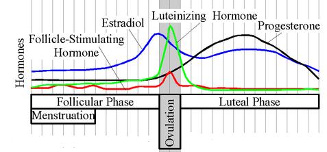
Image Source: Wikimedia Commons (2006). Estradiol Cycle, Licensed under a creative commons licence. Available for free at: https://upload.wikimedia.org/wikipedia/commons/c/c7/Estradiol.Cycle.jpg (Accessed: 4 January 2024).
High levels of LH and FSH can be seen in post-menopause and primary gonadal failure. Low levels of FSH and LH can be seen in hypopituitarism.
Thyroid-stimulating hormone (TSH)
TSH is a glycoprotein and is made up of α subunits and β subunits. The thyrotropin-releasing hormone controls its release (via negative feedback control of free T4 and T3). Its secretion will increase the release of T4 and T3 hormones and encourage the growth of the thyroid gland.
Growth hormone (GH)
GH is a single-chain polypeptide with a structure similar to prolactin. It is secreted in a pulsatile manner and is controlled by growth hormone-releasing hormone (GHRH). GHRH stimulates the synthesis and secretion of growth hormone and somatostatin, inhibiting the secretion of GH. Somatomedins are produced when GH acts on the target tissue. It inhibits the secretion of GH via its direct action on the anterior pituitary and by stimulating somatostatin secretion.
Stimuli for the secretion of GH include sleep, stress, vigorous exercise, starvation, hypoglycaemia and bromocriptine. Its secretion is inhibited by somatostatin, hyperglycaemia, obesity and increased cortisol.
GH stimulates lipolysis, protein synthesis and growth of bone, cartilage and connective tissue. It also stimulates hyperglycaemia by reducing glucose uptake into tissues. Growth hormone deficiency results in short stature and delayed puberty. Growth hormone excess can result in gigantism (childhood) or acromegaly (after puberty), increasing organ and periosteal bone growth.
Prolactin
Prolactin is a single-chain polypeptide structurally similar to growth hormone. It displays a pulsatile secretion with its peak serum concentration at night and nadir during the morning. Its secretion is stimulated by pregnancy (oestrogen), breast-feeding, thyrotropin-releasing hormone, dopamine antagonists and stress. Its secretion is inhibited by dopamine, dopamine agonist (bromocriptine), and prolactin.
Prolactin has a vital role in lactogenesis and breast development. It also inhibits ovulation and spermatogenesis. Prolactin deficiency can result from disruption to the anterior pituitary gland, reducing lactation. Prolactin hypersecretion occurs due to prolactinomas and hypothalamic disorders, and this presents with galactorrhoea, ovulation failure and amenorrhoea.
Posterior pituitary gland
Antidiuretic hormone (ADH)
ADH is a nonapeptide that acts on the distal convoluted tubules and collecting ducts, resulting in increased water permeability and vasoconstriction.
Its secretion is stimulated by increased serum osmolality, nausea, pain, hypoglycaemia, opiates and certain drugs (e.g., nicotine). Secretion is inhibited by reduced serum osmolality, alcohol and ANP.
Oxytocin
Oxytocin is a nonapeptide that acts on the uterus and breast tissue, resulting in uterine contraction and the milk ejection reflex.
Labour stimulates its secretion (predominantly during the 2nd stage, especially in response to cervical dilation) and breast suckling.
References
Tortora, G. J., & Derrickson, B. (2017). Tortora’s principles of anatomy and physiology. John Wiley and Sons.
Kumar, P. and Clark, M., (2012). Kumar & Clark’s clinical medicine. 8th ed.
Adrenal gland
Overview
The adrenal gland produces several types of steroids, including:
18-carbon steroids: – Oestrogen
19-carbon steroids: – Androgen
21-carbon steroids: – Progesterone (precursor of all the other 21-carbon steroids)
– Glucocorticoid (hydroxylation at C-17)
– Mineralocorticoid (hydroxylation at C-21)

Image Source: Wikimedia Commons (2014). Adrenal Steroids Pathways, Licensed under a creative commons licence. Available for free at: https://upload.wikimedia.org/wikipedia/commons/f/f8/Adrenal_Steroids_Pathways.svg(Accessed: 4 January 2024).
The adrenal cortex is divided into three distinct zones, each producing a different hormone.
The zona glomerulosa secretes aldosterone, and ACTH controls its secretion, increasing potassium levels and the renin-angiotensin system ( reduced perfusion pressure stimulates renin secretion that induces aldosterone secretion via angiotensin II). At the distal convoluted tubules, aldosterone stimulates sodium reabsorption and potassium excretion. This leads to a net increase in circulating plasma volume by osmosis.
Glucocorticoids like cortisol and cortisone are produced within the zona fasciculata. These hormones circulate within the body bound to corticosteroid-binding globulin. Its circulation follows a circadian rhythm with the highest levels during the day. ACTH mediates its secretion but exerts a negative feedback effect on the pituitary gland to reduce the level of ACTH secreted. Glucocorticoids act to increase serum glucose concentration via increased glycolysis, lipolysis, gluconeogenesis and protein catabolism. It is also an anti-inflammatory mediator and can result in immunosuppression.
The sex hormones are produced mainly within the zona reticularis, the deepest portion of the adrenal cortex. Androstenedione and testosterone are produced within the adrenal cortex, while the final production of dihydrotestosterone and oestradiol occurs in the peripheral tissues.
Below are the pathophysiology of disorders affecting the adrenal cortex:
| Adrenal disorder | Features | Hormone levels |
| Addison’s disease
(Primary adrenal insufficiency) |
Hyperpigmentation, decreased pubic and axillary hair in women, weakness, weight loss, N&V, hypoglycaemia, hyperkalaemia, metabolic acidosis, | Reduced cortisol → Increased ACTH
∴Treat by glucocorticoids and mineralocorticoid replacement |
| Cushing’s syndrome
(e.g., exogenous steroids, primary adrenal hyperplasia) |
Central obesity, moon face, buffalo hump, striae, hypertension, hyperglycaemia, muscle wasting | Increased cortisol → Reduced ACTH |
| Cushing’s disease
(Overproduction of ACTH) |
Similar features to Cushing’s syndrome | Increased ACTH
∴ Treat by removal of ACTH-secreting tumour |
| Conn’s syndrome
(Aldosterone-secreting tumour) |
Hypertension, hypokalaemia, metabolic alkalosis | Increased aldosterone → Reduced Renin |
| 21β-Hydroxylase deficiency | Symptoms of glucocorticoids & and mineralocorticoids deficiency
Virilisation of women, early growth of pubic and axillary hair |
Reduced glucocorticoids & mineralocorticoids → Increased ACTH
∴ Treat by glucocorticoids and mineralocorticoid replacement Increased androgens |
| 17α-Hydroxylase deficiency | Symptoms of glucocorticoid deficiency and mineralocorticoid excess
Lack of pubic and axillary hair |
Reduced glucocorticoids (and adrenal androgens) → Increased ACTH
Increased mineralocorticoids ∴ Treat by glucocorticoid replacement and aldosterone antagonist |
The adrenal medulla is the deepest region of the adrenal gland and is generally made of chromaffin cells. This region of the adrenal gland secretes catecholamines. The preganglionic sympathetic nerves stimulate the secretion of catecholamines. The catecholamines will result in increased glycogenolysis, gluconeogenesis and lipolysis.
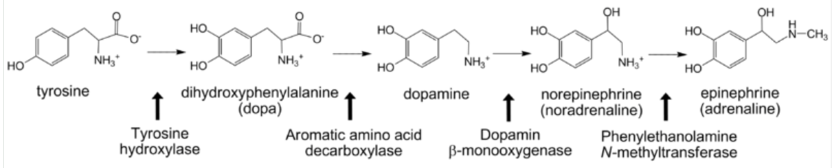
Image Source: Wikimedia Commons (2009). Catecholamine biosynthesis, Licensed under a creative commons licence. Available for free at: https://upload.wikimedia.org/wikipedia/commons/8/8d/Catecholamine_biosynthesis.png (Accessed: 4 January 2024).
References
Tortora, G. J., & Derrickson, B. (2017). Tortora’s principles of anatomy and physiology. John Wiley and Sons.
Kumar, P. and Clark, M., (2012). Kumar & Clark’s clinical medicine. 8th ed.
Thyroid and parathyroid gland
Thyroid gland
The thyroid gland is a highly vascular structure consisting of 2 lobes joined by the isthmus. It contains colloid-containing follicles filled with thyroglobulin, precursors to thyroid hormone. A layer of cuboidal epithelial cells surrounds these follicles.
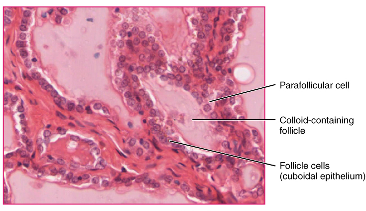
Image Source: Wikimedia Commons (2013). Thyroid follicle cells, Licensed under a creative commons licence. Available for free at: https://upload.wikimedia.org/wikipedia/commons/f/f1/Thyroid_follicle_cells.jpg (Accessed: 4 January 2024).
There are several types of thyroid hormones. The predominant form of circulating thyroid hormone is T4 ( thyroxine). Most T4 is usually protein-bound, with only 0.05% of the hormones existing free within the serum. T4 is peripherally converted into T3 (triiodothyronine) within the peripheral tissues. While both T4 and T3 can bind to the thyroid hormone receptor to exert its action, T3 is far more biologically active when compared to T4. It is responsible for the majority of the thyroid hormone’s biological activity.
TSH regulates thyroid hormone synthesis, and its precursor is thyroglobulin. Follicular cells are the site of synthesis for thyroglobulin, synthesised from tyrosine. Thyroglobulin is packaged within secretory vesicles and is secreted into the follicular lumen. At the same time, iodide ions are actively transported into the follicular lumen. Within the lumen, peroxidase will oxidise iodide into iodine. Iodine will react with tyrosine attached to thyroglobulin to form mono-iodotyrosine and di-iodotyrosine. These molecules will then conjugate to form T3 and T4. This will then be repackaged into the follicular cell by pinocytosis, and TSH stimulates this. Lysosomes will react with thyroglobulin to release the T3 and T4 from thyroglobulin. T3 and T4 are released into the bloodstream, where most will bind to plasma proteins.
T3 has a half-life of approximately one day, while T4 has a half-life of 1 week. These hormones are typically broken down within the liver.
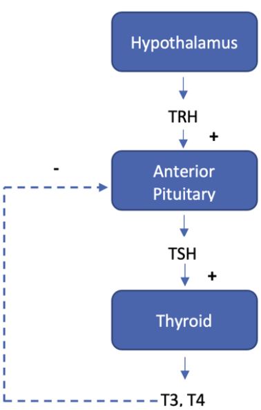
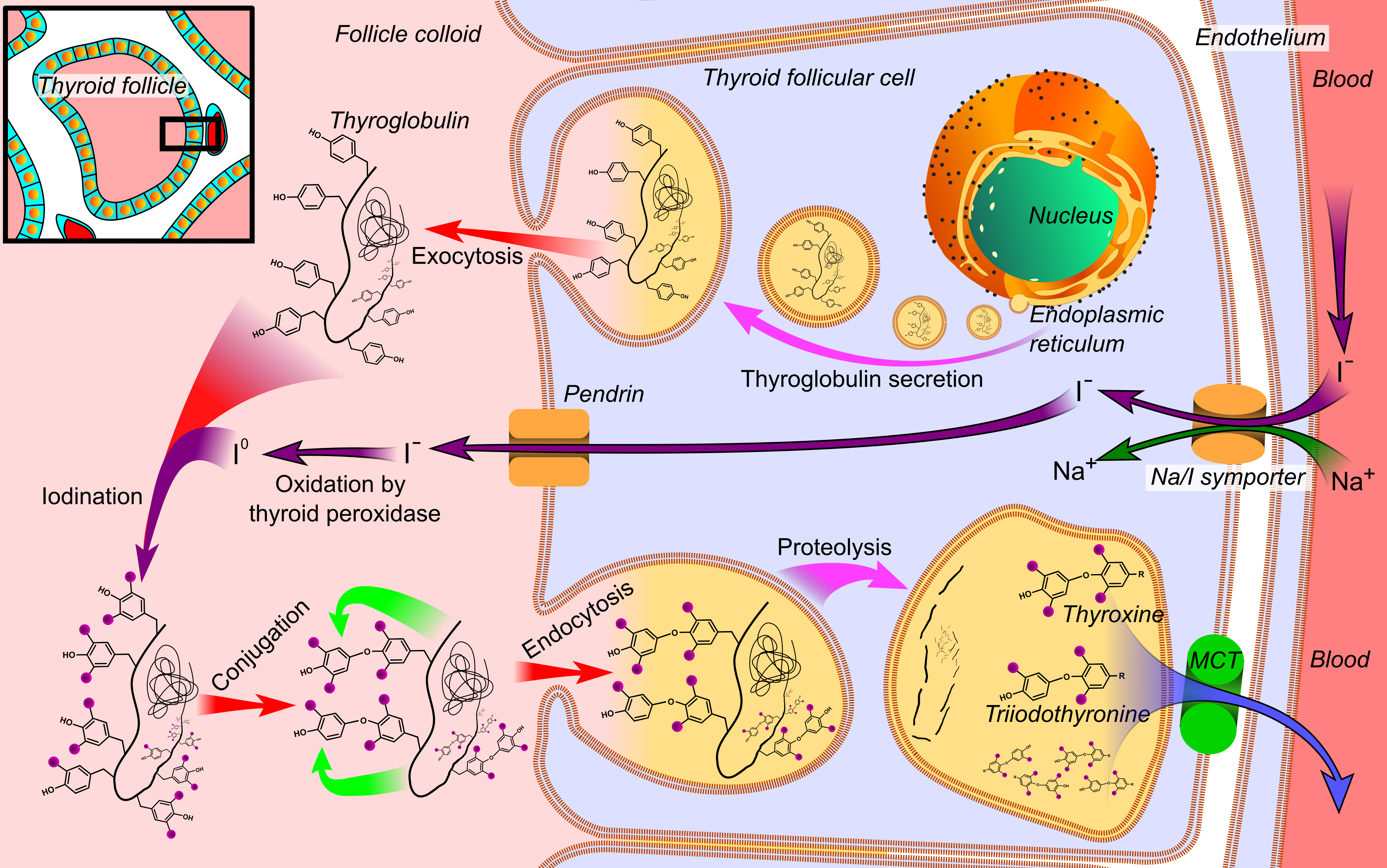
Image Source: Wikimedia Commons (2014). Thyroid hormone synthesis, Licensed under a creative commons licence. Available for free at: https://upload.wikimedia.org/wikipedia/commons/8/82/Thyroid_hormone_synthesis.png (Accessed: 4 January 2024).
The thyroid hormone is responsible for several actions, including:
- Growth: needed for normal growth and stature, acts synergistically with growth hormone to promote bone formation
- Metabolic: Increases metabolic rate by increasing oxygen consumption, increasing sensitivity to catecholamines, increases protein breakdown, increases calcium turnover from bone
- Nervous system: needed for development and maturation of the nervous system for normal myelination (Thyroid hormone deficiency causes irreversible mental retardation)
- Genitourinary: needed for normal gonadal function
We will highlight the impact of increased and reduced levels of thyroid hormones on the normal human physiology:
| Hyperthyroidism | Hypothyroidism | |
| Tachycardia, High output cardiac failure, AF, warm peripheries | Cardiorespiratory system | Bradycardia, Ischaemic heart disease, Low-output cardiac failure, cold peripheries, peripheral oedema |
| Irritable, psychosis, goitre, hyperreflexia, eye changes (exophthalmos, lid lag, ophthalmoplegia) | Central nervous system | Fatigued, flat affect, hyporeflexia |
| Weight loss, diarrhoea | Gastrointestinal system | Weight gain, constipation |
| Oligo/amenorrhoea | Genitourinary system | Menorrhagia, infertility |
| Tremor, proximal muscle weakness | Musculoskeletal system | Proximal myopathy, muscular hypertrophy, hair thinning, dry skin |
| Grave’s disease (antibodies to TSH receptor)
Thyroid neoplasm |
Causes | Thyroiditis (e.g., Hashimoto’s)
Iodine deficiency Congenital cretinism Thyroidectomy |
| Propylthiouracil
Thyroidectomy Iodine-131 β-blockers (treating hypermetabolic symptoms) |
Treatment | Thyroid hormone replacement |
Table 2: Comparison of the symptoms, causes and treatment of hyperthyroidism and hypothyroidism. Own table.
Calcium and the parathyroid gland
Calcium metabolism is tightly regulated by parathyroid hormone and vitamin D. They act on the kidneys, intestines and bones to maintain calcium homeostasis.
Parathyroid gland
The parathyroid gland is four separate glands found on the posterior aspect of the thyroid gland. The embryological origins of the gland are from the third and fourth pharyngeal pouches.
It aids in calcium homeostasis by secreting parathyroid hormone (PTH). The chief cells of the parathyroid gland are responsible for the synthesis and secretion of PTH. Specific G-protein receptors are found on the surface of the chief cells, the calcium-sensing receptor. This allows the cell to identify low circulating calcium levels and respond by secreting PTH.
Mutations affecting the calcium-sensing receptor results can result in pathology. Gain of function mutation results in autosomal dominant hypocalcaemia with hypercalciuria, which presents similarly to hypoparathyroidism. Loss of function mutation results in familial hypocalciuric hypercalcemia.
Parathyroid hormone
The parathyroid hormone acts to regulate calcium levels on a short time scale. It acts on two main sites:
- Bones:
- PTH binds onto osteoblasts and increases the expression of RANKL. This stimulates the production of nuclear factor kappa-B ligand. This increases the differentiation of osteoblasts into osteoclasts. This shifts the balance of bone homeostasis towards bone resorption, increasing serum calcium levels.
- Kidneys
- It acts to increase calcium reabsorption. Phosphate absorption is also inhibited. Reduced phosphate ions in the circulation promote increased serum-ionised calcium ions as phosphate ions will form insoluble salts with calcium.
- PTH also stimulates the conversion of 25-hydroxycholecalciferol into 1,25-dihydroxycholecalciferol by upregulating the production of 1-alpha hydroxylase in the proximal convoluted tubules.
Vitamin D
Further information on vitamin D can be found in the Vitamins section.
Its primary site of action is in the intestinal tract, which stimulates calcium and phosphate absorption into the circulation. It also acts on the distal convoluted tubules by increasing calcium and phosphate absorption. Increased vitamin D levels also encourage bone mineralisation.
Calcitonin
This hormone is secreted by the parafollicular cells of the thyroid gland. Its primary function is to oppose the actions of PTH and vitamin D3. Its secretion is stimulated by raised calcium levels and gastrin.
Its main action is to reduce serum calcium levels. It achieves this by reducing bone osteoclast activity and inhibiting calcium and phosphate reabsorption in the kidneys.
References
Tortora, G. J., & Derrickson, B. (2017). Tortora’s principles of anatomy and physiology. John Wiley and Sons.
Kumar, P. and Clark, M., (2012). Kumar & Clark’s clinical medicine. 8th ed.
Pancreas
Overview
The pancreas is responsible for both endocrine and exocrine functions. The islet of Langerhans contributes towards the endocrine function of the pancreas and consists of 3 main cell types:
| Cell type | Location | Function |
| Alpha | The outer rim of islet cells | Secrete glucagon |
| Beta | Central islet | Secrete insulin |
| Delta | Intermixed | Secrete somatostatin & gastrin |
Insulin
Insulin consists of an α chain and β chain linked by two disulphide bridges. It is synthesised from proinsulin within the β-cells. Hydrolysis of the C-peptide molecule from proinsulin produces the active insulin molecule.
Its secretion is stimulated by glucose, fatty acids, parasympathetic response, gut hormones (e.g., gastrin, secretin, CCK, glucagon) and certain drugs (e.g., β agonists, sulphonylureas). Sympathetic response, β-blockers, dopamine and somatostatin inhibit its secretion.
It aids in reducing the serum concentration of glucose by stimulating glucose uptake by cells. This increases glycogen and lipid production within the liver while reducing gluconeogenesis and ketogenesis. Adipose tissues will increase their fatty acid synthesis and lipid storage. Muscles will increase their synthesis of glycogen and proteins and increase amino acid uptake. It is worth noting that insulin has no effect on glucose transport in the brain, kidneys, intestine and red blood cells.
The primary condition that involves insulin is diabetes mellitus ( both type 1 and type 2). Type 2 diabetes mellitus is caused by insulin resistance, which can be associated with several conditions, including obesity, pregnancy, polycystic ovarian syndrome, cystic fibrosis and acromegaly.
Glucagon
Glucagon is a polypeptide that increases blood glucose concentrations by increasing glycogenolysis, gluconeogenesis and lipolysis. It also stimulates urea production. Its secretion is stimulated by decreasing blood glucose levels.
Somatostatin
Somatostatin is a polypeptide hormone produced by the pancreas and the neurons within the hypothalamus. It suppresses the endocrine and exocrine functions of the pancreas. Along the rest of the digestive tract, it inhibits the secretion of other GI hormones, reducing gastric emptying and smooth muscle contractions. In the CNS, it inhibits the release of prolactin, GNRH and TSH.
References
Tortora, G. J., & Derrickson, B. (2017). Tortora’s principles of anatomy and physiology. John Wiley and Sons.
Kumar, P. and Clark, M., (2012). Kumar & Clark’s clinical medicine. 8th ed.
Complete Guide to Clinical Genetics ST3 Application and Recruitment (2027)
Written by Medibuddy. Last updated: October 2026. Clinical Genetics is

Complete Guide to Clinical Radiology ST3 Application and Recruitment (2027)
Recruitment paused: NHS England is not running Clinical Radiology ST3+ recru

Complete Guide to Paediatric Surgery ST3 Application and Recruitment in United Kingdom (2026)
Written by Medibuddy. Last updated: October 2026. If you’re planning


Take your subscriptions with you
Our mobile app allows you to access your interview and exam question banks wherever you are.

