


 Close
Close
 9th February 2024
Thyroid gland
The thyroid gland is a highly vascular structure consisting of 2 lobes joined by the isthmus. It contains colloid-containing follicles filled with thyroglobulin, precursors to thyroid hormone. A layer of cuboidal epithelial cells surrounds these follicles.
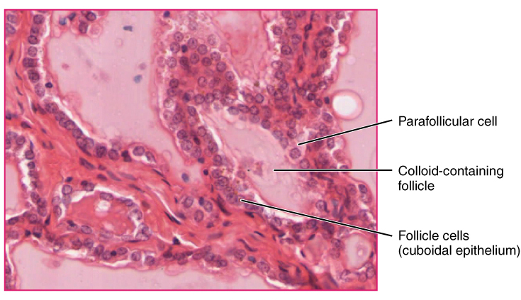
Image Source: Wikimedia Commons (2013). Thyroid follicle cells, Licensed under a creative commons licence. Available for free at: https://upload.wikimedia.org/wikipedia/commons/f/f1/Thyroid_follicle_cells.jpg (Accessed: 4 January 2024).
There are several types of thyroid hormones. The predominant form of circulating thyroid hormone is T4 ( thyroxine). Most T4 is usually protein-bound, with only 0.05% of the hormones existing free within the serum. T4 is peripherally converted into T3 (triiodothyronine) within the peripheral tissues. While both T4 and T3 can bind to the thyroid hormone receptor to exert its action, T3 is far more biologically active when compared to T4. It is responsible for the majority of the thyroid hormone’s biological activity.
TSH regulates thyroid hormone synthesis, and its precursor is thyroglobulin. Follicular cells are the site of synthesis for thyroglobulin, synthesised from tyrosine. Thyroglobulin is packaged within secretory vesicles and is secreted into the follicular lumen. At the same time, iodide ions are actively transported into the follicular lumen. Within the lumen, peroxidase will oxidise iodide into iodine. Iodine will react with tyrosine attached to thyroglobulin to form mono-iodotyrosine and di-iodotyrosine. These molecules will then conjugate to form T3 and T4. This will then be repackaged into the follicular cell by pinocytosis, and TSH stimulates this. Lysosomes will react with thyroglobulin to release the T3 and T4 from thyroglobulin. T3 and T4 are released into the bloodstream, where most will bind to plasma proteins.
T3 has a half-life of approximately one day, while T4 has a half-life of 1 week. These hormones are typically broken down within the liver.
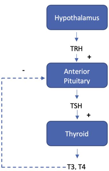
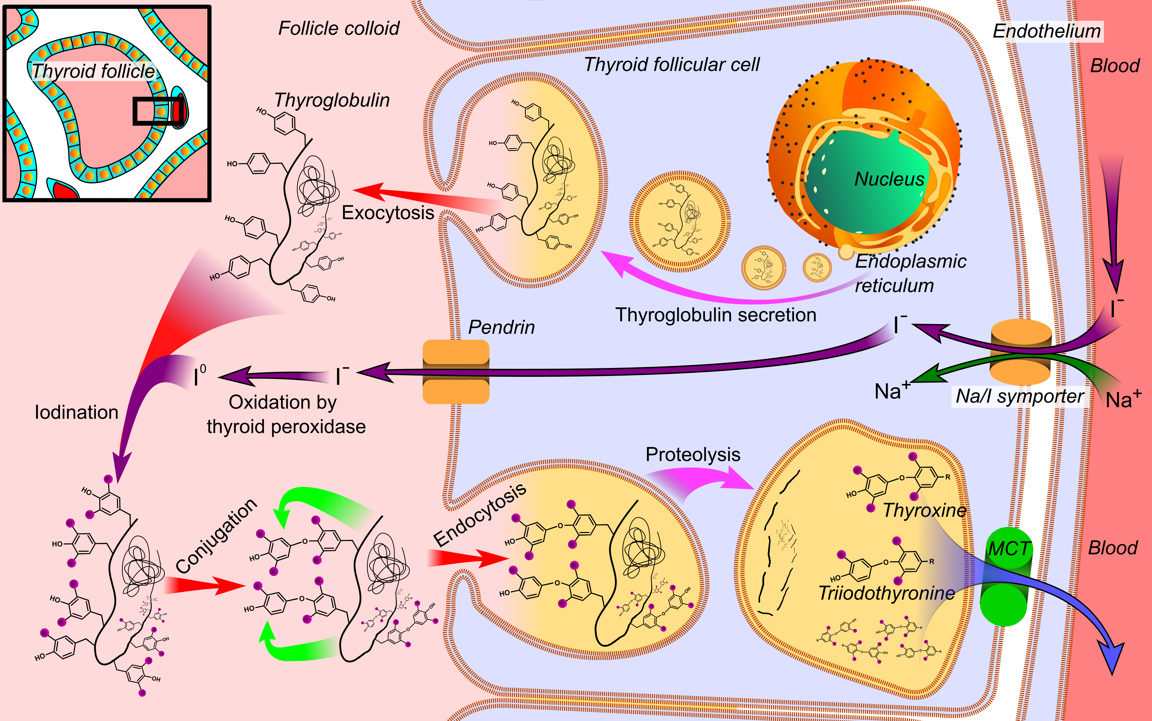
Image Source: Wikimedia Commons (2014). Thyroid hormone synthesis, Licensed under a creative commons licence. Available for free at: https://upload.wikimedia.org/wikipedia/commons/8/82/Thyroid_hormone_synthesis.png (Accessed: 4 January 2024).
The thyroid hormone is responsible for several actions, including:
- Growth: needed for normal growth and stature, acts synergistically with growth hormone to promote bone formation
- Metabolic: Increases metabolic rate by increasing oxygen consumption, increasing sensitivity to catecholamines, increases protein breakdown, increases calcium turnover from bone
- Nervous system: needed for development and maturation of the nervous system for normal myelination (Thyroid hormone deficiency causes irreversible mental retardation)
- Genitourinary: needed for normal gonadal function
We will highlight the impact of increased and reduced levels of thyroid hormones on the normal human physiology:
| Hyperthyroidism | Hypothyroidism | |
| Tachycardia, High output cardiac failure, AF, warm peripheries | Cardiorespiratory system | Bradycardia, Ischaemic heart disease, Low-output cardiac failure, cold peripheries, peripheral oedema |
| Irritable, psychosis, goitre, hyperreflexia, eye changes (exophthalmos, lid lag, ophthalmoplegia) | Central nervous system | Fatigued, flat affect, hyporeflexia |
| Weight loss, diarrhoea | Gastrointestinal system | Weight gain, constipation |
| Oligo/amenorrhoea | Genitourinary system | Menorrhagia, infertility |
| Tremor, proximal muscle weakness | Musculoskeletal system | Proximal myopathy, muscular hypertrophy, hair thinning, dry skin |
| Grave’s disease (antibodies to TSH receptor)
Thyroid neoplasm |
Causes | Thyroiditis (e.g., Hashimoto’s)
Iodine deficiency Congenital cretinism Thyroidectomy |
| Propylthiouracil
Thyroidectomy Iodine-131 β-blockers (treating hypermetabolic symptoms) |
Treatment | Thyroid hormone replacement |
Table 2: Comparison of the symptoms, causes and treatment of hyperthyroidism and hypothyroidism. Own table.
Calcium and the parathyroid gland
Calcium metabolism is tightly regulated by parathyroid hormone and vitamin D. They act on the kidneys, intestines and bones to maintain calcium homeostasis.
Parathyroid gland
The parathyroid gland is four separate glands found on the posterior aspect of the thyroid gland. The embryological origins of the gland are from the third and fourth pharyngeal pouches.
It aids in calcium homeostasis by secreting parathyroid hormone (PTH). The chief cells of the parathyroid gland are responsible for the synthesis and secretion of PTH. Specific G-protein receptors are found on the surface of the chief cells, the calcium-sensing receptor. This allows the cell to identify low circulating calcium levels and respond by secreting PTH.
Mutations affecting the calcium-sensing receptor results can result in pathology. Gain of function mutation results in autosomal dominant hypocalcaemia with hypercalciuria, which presents similarly to hypoparathyroidism. Loss of function mutation results in familial hypocalciuric hypercalcemia.
Parathyroid hormone
The parathyroid hormone acts to regulate calcium levels on a short time scale. It acts on two main sites:
- Bones:
- PTH binds onto osteoblasts and increases the expression of RANKL. This stimulates the production of nuclear factor kappa-B ligand. This increases the differentiation of osteoblasts into osteoclasts. This shifts the balance of bone homeostasis towards bone resorption, increasing serum calcium levels.
- Kidneys
- It acts to increase calcium reabsorption. Phosphate absorption is also inhibited. Reduced phosphate ions in the circulation promote increased serum-ionised calcium ions as phosphate ions will form insoluble salts with calcium.
- PTH also stimulates the conversion of 25-hydroxycholecalciferol into 1,25-dihydroxycholecalciferol by upregulating the production of 1-alpha hydroxylase in the proximal convoluted tubules.
Vitamin D
Further information on vitamin D can be found in the Vitamins section.
Its primary site of action is in the intestinal tract, which stimulates calcium and phosphate absorption into the circulation. It also acts on the distal convoluted tubules by increasing calcium and phosphate absorption. Increased vitamin D levels also encourage bone mineralisation.
Calcitonin
This hormone is secreted by the parafollicular cells of the thyroid gland. Its primary function is to oppose the actions of PTH and vitamin D3. Its secretion is stimulated by raised calcium levels and gastrin.
Its main action is to reduce serum calcium levels. It achieves this by reducing bone osteoclast activity and inhibiting calcium and phosphate reabsorption in the kidneys.
Complete Guide to Clinical Genetics ST3 Application and Recruitment (2027)
Written by Medibuddy. Last updated: October 2026. Clinical Genetics is

Complete Guide to Clinical Radiology ST3 Application and Recruitment (2027)
Recruitment paused: NHS England is not running Clinical Radiology ST3+ recru

Complete Guide to Paediatric Surgery ST3 Application and Recruitment in United Kingdom (2026)
Written by Medibuddy. Last updated: October 2026. If you’re planning


Take your subscriptions with you
Our mobile app allows you to access your interview and exam question banks wherever you are.

