


 Close
Close
 25th June 2023
Intracranial haemorrhages
Intracranial haemorrhage is roughly subdivided into intra-axial and extra-axial haemorrhage. Intra-axial haemorrhage involves bleeding within the brain itself, typically seen as intraparenchymal or intraventricular haemorrhage in the context of stroke. Extra-axial haemorrhage occurs within the skull but outside of the brain tissue and includes subdural, extradural, and subarachnoid haemorrhage. These are more relevant in the context of head injury.
Extra-axial haemorrhages
Extradural haemorrhage
Caused by laceration of arteries or veins running along the inner aspect of the skull, often accompanied by associated skull fracture. The middle meningeal artery is typically implicated. Characterised clinically by a lucid interval where the patient is asymptomatic and not critically ill. However, once the haematoma reaches a large enough size, it can lead to significant mass effect with midline shift and trans-tentorial herniation and rapid progression to coma and death. These patients typically require close neuro-monitoring and emergency haematoma evacuation in the setting of neurological deterioration.
CT findings: Hyperdense collection in extradural space, extent is limited by suture lines. Forms a crescenteric or lens appearance.

Extradural haemorrhage with associated skull fracture.
Image Source: Wikimedia Commons (2018). EpiduralHematoma, Licensed under a creative commons licence. Available for free at: https://commons.wikimedia.org/wiki/File:EpiduralHematoma.jpg (Accessed on: 11 January 2023).

Subdural haemorrhage
Subdural haemorrhage occurs due to damage to the bridging veins that cross the subdural space and leads to blood collecting between the dura mater and arachnoid mater of the meninges. Signs and symptoms of subdural haemorrhages typically develop more slowly than extradural haemorrhage. Typically, patients with acute subdural haemorrhage will present with headache, confusion, loss of consciousness and focal neurological deficits in the context of a recent head injury. However, chronic subdural haemorrhage may present with behavioural change, disorientation, focal neurological deficit, or amnesia weeks after a traumatic injury, or without a history of witnessed head injury.
Patients with atrophic brain volume, such as the elderly, or with a history of alcohol excess are particularly vulnerable to subdural haemorrhage.
CT findings:
Acute subdural: Hyperdense crescent crescent-shaped homogeneously hyperdense extra-axial collection that spreads diffusely over the affected hemisphere. May see associated midline shift secondary to mass effect.
Chronic subdural: Similar in appearance to acute subdural haematoma, however, over time the collection becomes hypodense with respect to the cerebral cortex and may become isodense with the CSF.
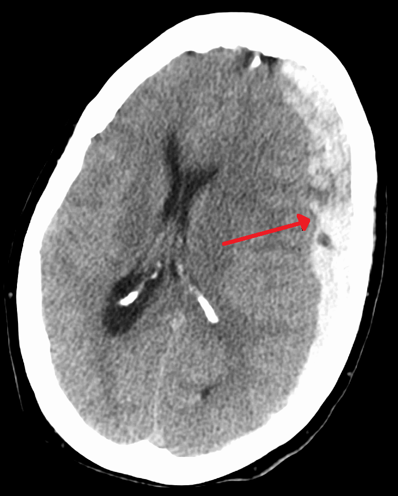
Acute subdural haematoma with midline shift.
Image Source: Wikimedia Commons (2012). Subduralandherniation, Licensed under a creative commons licence. Available for free at: https://commons.wikimedia.org/wiki/File:Subduralandherniation.PNG (Accessed on: 11 January 2023).

Subarachnoid haemorrhage
Subarachnoid haemorrhage (SAH) occurs when there is an arterial or venous injury to the vessels coursing through the subarachnoid space overlying the brain parenchyma.
CT findings: Abnormal hyperdensity in the subarachnoid space.

Subarachnoid haemorrhage with extension of blood into the sulci.
Image Source: Wikimedia Commons (2010). SubarachnoidP, Licensed under a creative commons licence. Available for free at: https://en.wikipedia.org/wiki/File:SubarachnoidP.png (Accessed on: 11 January 2023).

Complete Guide to Clinical Genetics ST3 Application and Recruitment (2027)
Written by Medibuddy. Last updated: October 2026. Clinical Genetics is

Complete Guide to Clinical Radiology ST3 Application and Recruitment (2027)
Recruitment paused: NHS England is not running Clinical Radiology ST3+ recru

Complete Guide to Paediatric Surgery ST3 Application and Recruitment in United Kingdom (2026)
Written by Medibuddy. Last updated: October 2026. If you’re planning


Take your subscriptions with you
Our mobile app allows you to access your interview and exam question banks wherever you are.

