


 Close
Close
 7th March 2024
Heart Valve Disorders
Aortic Valve
Background
An appreciation of the four chambers of the heart (see image below) and their orientation in the body will facilitate understanding of the location of murmurs and their radiation. Cardiac murmurs are heard loudest at the area above the respective valve and radiate in the direction of the turbulent flow. Systolic and diastolic murmurs are summarised in Tables 1 and 2.
To understand heart valve disorders, the following points are to be kept in mind for the MRCP:
- Blood normally moves towards ventricles in diastole
- Blood normally moves away from ventricles in systole
- The left sided circulation is a high-pressure system and the right side is a low-pressure system
- Stenosis produces pressure overload on the place from where blood is going
- Regurgitation produces volume overload on the place where it is coming back to
- Ventricles hypertrophy in response to pressure overload and dilate in response to volume overload
Table 1. Diastolic murmurs
| Diastolic murmurs
(Blood flows towards ventricle) |
Stenosis: | Mitral stenosis (MS) |
| Tricuspid stenosis (TS) | ||
| Regurgitation: | Aortic Regurgitation (AR) | |
| Pulmonary Regurgitation (PR) | ||
Table 2. Systolic murmurs
| Systolic Murmurs
(Blood flows away from ventricle) |
Stenosis: | Aortic stenosis (AS) |
| Pulmonary stenosis (PS) | ||
| Regurgitation: | Mitral Regurgitation (MR) | |
| Tricuspid Regurgitation (TR) | ||
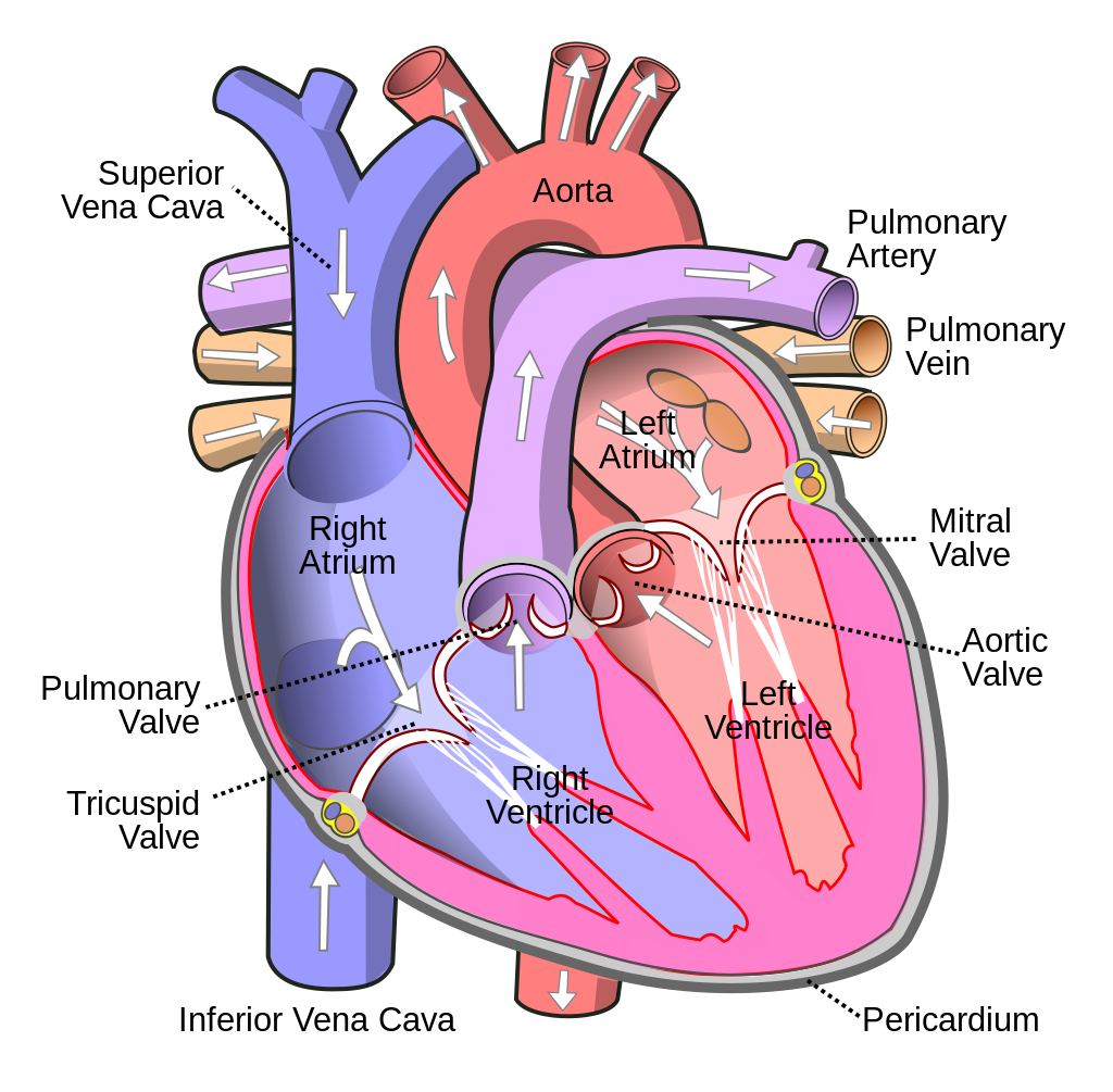 Image Source: Wikimedia Commons (2006). Heart, Licensed under a creative commons licence. Available for free at: https://simple.wikipedia.org/wiki/Heart (Accessed: 25 January 2024).
Aortic stenosis
Any disorder involving the aortic valve will generate pressure/volume overload on the left ventricle. Over time, this will affect the left atrium. An aortic murmur will be heard loudest over the aortic area.
Aortic stenosis (AS) is characterised by an ejection systolic murmur radiating to the carotids. The pathology is due to a stenosed aortic valve causing blood to be squeezed past the narrowed valve opening. Causes of AS include:
- Degenerative calcification. The most common cause in patients > 65 years
- Bicuspid aortic valve. This is the most common cause in patients < 65 years
- Congenital e.g. William’s syndrome where there is supra valvular stenosis
- Post rheumatic fever
- Sub valvular e.g. HOCM
Clinical manifestations
AS can vary from asymptomatic to low cardiac output symptoms which includes chest pain, dyspnoea, syncope and pulmonary oedema. The severity of the stenosis can manifest in symptoms ranging from reduced exercise tolerance to sudden cardiac death (SCD). The classical symptoms are only in 30-40% of individuals with AS. When symptoms appear, survival without surgery is, on average, 2-3 years. SCD occurs in 1-2% of asymptomatic AS patients per year. Symptoms of severe AS include:
- Narrow pulse pressure and small volume pulse due to little blood escaping the ventricle
- Slow rising pulse due to a narrow outflow
- Soft or absent S2 due to a narrow pulse pressure and restricted leaflet motion.
- Displaced apex beat due to left ventricular hypertrophy.
- Delayed ejection systolic murmur
- Paradoxical split S2 and palpable thrill
Diagnosis
While AS may be clinically silent in many cases, a broad range of symptoms, as outlined above, can lead to a suspicion of disease.
- Chest x-ray may show cardiomegaly due to left ventricular hypertrophy and detect abnormalities of aortic contour or size.
- Echocardiography is key to confirming the diagnosis and severity of aortic stenosis, assessing valve calcification, LV function and wall thickness. It can also detect other valve pathologies and provide prognostic information.
- Transoesophageal echocardiography is used when images on a transthoracic echo prove insufficient. A pressure gradient of over 40mmHg is diagnostic of severe aortic stenosis and is an indication for surgery. Aortography done as part of a coronary angiogram may also support a finding of AS.
- Exercise testing may unmask symptoms and is recommended for risk stratification of asymptomatic patients with severe aortic stenosis. Exercise echocardiography provides additional prognostic information by assessing the increase in mean pressure gradient and change in LV function. Although the severity of valvular calcification is prognostically important in an asymptomatic patient, the most important predictor is LV function.
- Coronary angiography is essential prior to transarterial valve implantation (TAVI) and surgical AV valve replacement (SAVR) to determine the potential need for concomitant revascularisation.
- Cardiac CT (CCT) provides information on calcium score, anatomy of the aortic root and ascending aorta, and feasibility of vascular access.
- Myocardial fibrosis (due to amyloidosis, for example) is a major driver of LV decompensation in aortic stenosis. This can be detected and quantified using cardiac magnetic resonance (CMR).
- Natriuretic peptides predict symptom-free survival and outcome in normal and low-flow severe aortic stenosis. They can be used to arbitrate the source of symptoms in patients with multiple potential causes and identify those with high-risk asymptomatic aortic stenosis who may benefit from early intervention.
Management
This depends on the symptoms and severity of AS.
- Early intervention is recommended for all patients with symptomatic severe aortic stenosis. The only exception is for those in whom intervention is unlikely to improve quality of life or survival. Symptomatic patients without severe AS can be managed medically with conventional heart failure therapy. It is important to note that angiotensin-converting enzyme inhibitors (ACEI) are safe in AS, provided that BP is monitored carefully. They provide beneficial myocardial effects before the onset of symptoms and after TAVI and SAVR but are not generally recommended.
- Coexisting hypertension should be treated to avoid additional afterload, although medication (particularly vasodilators) should be titrated to avoid symptomatic hypotension.
- Replacement should also be considered in asymptomatic patients with a valvular gradient > 40 mmHg with features of LVSD.
- Asymptomatic patients without severe aortic stenosis or evidence of LV dysfunction are managed with watchful waiting and prompt intervention at symptoms onset. Intervention such as TAVI or SAVR is recommended in symptomatic patients with high-gradient AS, regardless of LVEF.
- Balloon valvuloplasty is reserved for patients with critical AS who are not fit for valve replacement and has a high rate of recurrence.
Aortic Regurgitation
Aortic regurgitation (AR) is characterised by an early decrescendo diastolic murmur. There is no correlation between the presence of murmur and severity of AR or aetiology. A wide pulse pressure may be found and is caused by blood returning to the left ventricle from the aorta during diastole, causing a drop in diastolic pressure. To compensate, there is peripheral vasoconstriction. A water hammer pulse, also called a collapsing pulse, is characteristic of AR. Other signs in AR include:
- Austin Flint murmur. In severe aortic regurgitation, the regurgitant jet of blood can exert pressure on the anterior mitral valve cusps during diastole, resulting in a mid/late diastolic murmur like that of and virtually indistinguishable from mitral stenosis.
- The first heart sound may be absent in severe AR
AR can be caused by primary disease of the aortic valve cusps and/or abnormalities of the aortic root and ascending aortic geometry.
Degenerative tricuspid and bicuspid aortic regurgitation are the most common aetiologies in high-income countries.
Valvular causes include:
- Rheumatic fever
- Bicuspid aortic valve
- Infective endocarditis
- Connective tissue diseases e.g., RA/SLE
AR due to aortic root disease can occur in:
- Hypertension
- Syphilis
- Marfan’s, Ehler-Danlos syndrome
- Aortic dissection
- Spondyloarthropathies (e.g., ankylosing spondylitis)
Diagnosis
Echocardiography is key to confirm valve anatomy, quantify aortic regurgitation, define the morphology of the aorta and determine the feasibility of valve-sparing aortic surgery or valve repair.
CMR should be used to quantify AR when echocardiographic measurements are equivocal or discordant with clinical findings. In patients with aortic dilatation, a cardiac CCT is recommended. CMR can be used for follow-up, but indication for surgery should preferably be based on CCT measurements.
Management
Acute aortic regurgitation may require urgent surgery. It is mainly caused by infective endocarditis and aortic dissection but may also occur after blunt chest trauma and iatrogenic complications during catheter-based cardiac interventions.
In symptomatic patients who are fit for surgery with severe AR, replacement is recommended irrespective of the LVEF. Replacement is also recommended in symptomatic and asymptomatic patients with severe aortic regurgitation undergoing coronary artery bypass grafting (CABG) or surgery of the ascending aorta or another valve.
In asymptomatic patients with severe aortic regurgitation, impairment of LV function [LVEF ≤50% or left ventricular end-systolic diameter (LVESD) >50 mm] is associated with worse outcomes and surgery should therefore be pursued when these cut-offs are reached.
TAVI may be considered in experienced centres for selected patients with aortic regurgitation and ineligible for SAVR. Medical therapy, especially ACEI or dihydropyridines, may provide symptomatic improvement in individuals with chronic severe aortic regurgitation in whom surgery is not feasible. In patients who undergo surgery but continue to suffer from heart failure or hypertension, ACEI, angiotensin receptor blockers (ARBs), and beta-blockers are useful.
All asymptomatic patients with severe aortic regurgitation and normal LV function should be followed up at least every year.
References
- Alec Vahanian et al. ESC/EACTS Scientific Document Group, 2021 Guidelines for the management of valvular heart disease: Developed by the Task Force for the management of valvular heart disease of the European Society of Cardiology (ESC). European Heart Journal, Volume 43, Issue 7, 14 February 2022. https://doi.org/10.1093/eurheartj/ehab395
- Braunwald’s Heart Disease: A Textbook of Cardiovascular Medicine, Single Volume, 11e
- Aortic Stenosis BMJ Best Practice. [Online] https://bestpractice.bmj.com/topics/en-gb/325
- Aortic Regurgitation BMJ Best Practice. [Online] https://bestpractice.bmj.com/topics/en-gb/324
Pulmonary Valve
Pulmonary Stenosis
Pulmonary stenosis (PS) is mostly a congenital condition and is characterised by an ejection systolic murmur heard best in the pulmonary area. It causes a widely split S2. In 80% to 90% of all cases, PS is found at the level of the valve, but it can also occur below the level of the valve or distally in the pulmonary arteries. It can be asymptomatic. Symptoms range from those primarily due to right heart failure, such as dyspnoea, fatigue and peripheral edema to profound cyanosis and sudden death. PS is also associated with an increased risk of arrhythmias. Causes of PS include:
- Carcinoid syndrome
- Pellagra
- Tetralogy of Fallot
- Congenital
- LEOPARD syndrome
- Noonan syndrome
Diagnosis
Like other valvular diseases, echocardiography with Doppler study remains the mainstay of investigation, showing an abnormal morphology of the pulmonary valve and an increased transvalvular gradient across the pulmonary valve during systole (>10 mmHg). ECG is normal in 30-65% of patients but in moderate to severe PS can show right axis deviation and RV conduction delay. There may also be right atrial enlargement with tall and peaked P waves in lead II and in precordial leads V1 to V3.
Arterial blood gas or pulse oximetry may show a low arterial oxygen saturation.
Diagnostic cardiac catheterisation is indicated prior to therapeutic relief of moderate to critical stenosis. It is also indicated when peripheral pulmonary stenosis is suspected to determine its severity and anatomy. It shows an increased transvalvular gradient (>10 mmHg).
Management
Mild disease is usually managed with watchful waiting. It requires only sequential cardiology follow-up into adulthood. Moderate to severe disease can be treated with percutaneous balloon pulmonary valvuloplasty (PBPV).
Surgical valvuloplasty is used in patients not amenable to PBPV or those with multiple levels of fixed obstruction.
Valvectomy is also an option with or without a transannular patch. In patients with respiratory distress or cyanosis, supplemental oxygen with or without alprostadil is used in addition to the above.
Pulmonary Regurgitation
Any disease of the left side of the heart or the lungs that results in significant pulmonary hypertension and dilated pulmonary arteries may lead to acquired pulmonary regurgitation (PR). PR may also be due to a congenital defect. Acquired PR typically results from surgical repair of Tetralogy of Fallot; pulmonary stenosis, atresia, carcinoid syndrome, rheumatic fever or as a result of infective endocarditis.
Isolated PR is rarely symptomatic; however, large regurgitant volume in the presence of dilated right ventricle may be associated with exertional dyspnoea, easy fatigability, and intermittent chest pain.
PR is characterised by an early diastolic murmur along the left sternal border in the second intercostal space with the patient in full inspiration, called a Graham Steell Murmur. This murmur is often heard in patients with chronic cor pulmonale as a result of chronic obstructive pulmonary disease.
Since most PR is secondary to another cause, treatment revolves around treating the underlying cause. Surgical management is rarely required, with the mainstay of treatment being medical management of heart failure.
References
- Pulmonary Stenosis BMJ Best Practice. [Online] https://bestpractice.bmj.com/topics/en-gb/475
- Pulmonary Regurgitation BMJ Best Practice. [Online] https://bestpractice.bmj.com/topics/en-us/474
Mitral Valve
Mitral Stenosis
The aetiology of mitral stenosis (MS) is mostly rheumatic or degenerative.
Rheumatic fever is the most common cause of mitral stenosis worldwide. Its prevalence has greatly decreased in industrialised countries, but it remains a significant healthcare problem in developing countries and affects young patients.
Rarer causes of mitral stenosis include congenital deformity of the valve, carcinoid syndrome, use of ergot and/or serotonergic drugs such as fenfluramine, SLE, mitral annular calcification due to ageing, and amyloidosis.
Clinical manifestations
MS usually manifests with dyspnoea or orthopnoea. Patients can have a malar flush, distended neck veins and a low volume pulse which is irregular due to atrial fibrillation.
It is characterised by a mid-late diastolic rumbling murmur heard best in expiration with presystolic accentuation because at the end of diastole the atria contract, squirting the last of the blood into the ventricle. A loud S1 with an opening snap (due to thickened valve leaflets and high LA pressures) and a loud P2 may be heard.
In severe MS, the murmur lasts longer and the opening snap comes closer to S2. A severely calcified valve will result in no opening snap.
Diagnosis
The ECG may show atrial fibrillation and chest x-ray may show a double right heart border indicating an enlarged left atrium, prominent pulmonary artery and Kerley B lines.
Echocardiography is the preferred method for diagnosis, assessment of severity, and haemodynamic consequences of mitral stenosis. A TOE should be performed to exclude LA thrombus before percutaneous mitral commissurotomy or after an embolic episode, or when TTE is suboptimal.
Stress testing is indicated in patients with no symptoms or symptoms equivocal or discordant with the severity of mitral stenosis. Exercise echocardiography is also used.
Management
Diuretics, beta-blockers, digoxin, non-dihydropyridine calcium channel blockers and ivabradine can improve symptoms.
Anticoagulation with vitamin K antagonist (warfarin) with a target INR between 2 and 3 is indicated in patients with AF. Patients with moderate-to-severe mitral stenosis and AF should be kept on warfarin and not receive NOACs.
Definitive therapy requires a mechanical solution by percutaneous balloon commissurotomy, surgical mitral valve repair, or valve replacement. Definitive therapy should be offered to patients with severe symptomatic disease who are unlikely to benefit from medical therapy alone, and may be considered in some patients with severe asymptomatic disease.
A systematic review found a lack of evidence concerning the effectiveness of the various treatments for valvular heart disease in pregnancy. Diuretics are given to control mild symptoms. If symptoms worsen and heart failure threatens the life of the mother and/or foetus, balloon valvotomy can be performed safely.
The contraindications for percutaneous mitral balloon valvotomy are as follows:
- Moderate to severe MR.
- Heavily calcified mitral valve.
- LA thrombus.
- Concomitant coronary artery disease or other valve disease requiring surgery.
Mitral Regurgitation
Mitral regurgitation (MR) may present with dyspnoea, usually on exertion, palpitations, and/or decreased exercise tolerance. It typically presents as a holosystolic blowing murmur at the apex, radiating to the axilla.
A transthoracic echocardiogram is the diagnostic test of choice in identifying the presence, severity, and mechanism of MR. Colour Doppler flow and continuous-wave Doppler studies can assess the severity of regurgitation, left ventricular dimensions, size and function of the right ventricle, and pulmonary artery systolic pressure.
Surgical treatment is the best available treatment option, although percutaneous approaches have gained significant potential. Asymptomatic chronic MR with LVEF, more than 60% are treated with ACEi and beta blockers. All patients with MR may be offered antibiotic prophylaxis prior to dental and/or urological procedures, depending on their risk profiles. The most common complications of MR include AF, pulmonary hypertension, LV dysfunction, CCF and prosthesis stenosis.
References
- Mitral Stenosis BMJ Best Practice. [Online] https://bestpractice.bmj.com/topics/en-gb/323
- Mitral Regurgitation BMJ Best Practice. [Online] https://bestpractice.bmj.com/topics/en-gb/322
- Alec Vahanian et al. ESC/EACTS Scientific Document Group, 2021 Guidelines for the management of valvular heart disease: Developed by the Task Force for the management of valvular heart disease of the European Society of Cardiology (ESC). European Heart Journal, Volume 43, Issue 7, 14 February 2022. https://doi.org/10.1093/eurheartj/ehab395
Tricuspid Valve
Any disorder involving the tricuspid valve tends to generate pressure/volume overload on the right ventricle and can, over time, affect the right atrium. This can manifest as right heart failure with systemic edema. The murmur is generally heard over the tricuspid area.
Tricuspid Stenosis
Tricuspid stenosis (TS) is usually a late complication of acute rheumatic fever. It is also seen in Group A streptococcal pharyngitis, metastatic carcinoid tumours and IVDU as a complication of infective endocarditis. It rarely occurs in the absence of associated mitral/aortic disease.
Clinical manifestations
It usually presents with dyspnoea, exercise intolerance, abdominal swelling/ascites, peripheral oedema, atrial fibrillation and hepatomegaly.
Associated symptoms of carcinoid syndrome, such as episodic facial flushing, watery diarrhoea or bronchoconstriction, may be present.
Clinical examination shows a murmur similar to that heard with mitral stenosis. There should be a high index of suspicion for TS if other signs of right heart involvement are present. The JVP waveform shows prominent a waves. The murmur increases in intensity with inspiration and squatting.
Diagnosis
Doppler echocardiography is crucial as the tricuspid valve can appear normal in 2D imaging. The presence of an abnormal appearing tricuspid valve with normal appearing mitral and aortic valves should prompt consideration for carcinoid heart disease and dissuade the clinician from rheumatic heart disease as the aetiology.
Right atrial enlargement with dilated systemic and hepatic veins is consistent with more severe TS. Biochemically, there may be evidence of hepatic involvement, endocarditis or carcinoid syndrome (increased 24h urinary 5 HIAA).
Management
Congenital disease is usually treated with surgery and preoperative alprostadil. TS, as a result of carcinoid heart disease, is treated with fluid and sodium restriction, loop diuretics and somatostatin analogues along with surgical treatment or balloon dilatation. Endocarditis is treated with antibiotics with or without surgery.
Tricuspid Regurgitation
Moderate or severe tricuspid regurgitation (TR) is observed in 0.55% of the general population and its prevalence increases with age, affecting about 4% of the patients aged 75 years or more.
Aetiology
Aetiology is secondary in over 90% of cases, due to pressure and/or volume overload mediated RV dilatation or enlarged right atrium and tricuspid annulus due to chronic AF.
Cardiac implantable electronic device-lead implantation leads to progressive tricuspid regurgitation in 20-30% of patients.
Other causes include pulmonary hypertension (e.g. Secondary to COPD), rheumatic heart disease, infective endocarditis, Ebstein’s anomaly and carcinoid syndrome.
Mild or moderate tricuspid regurgitation without abnormal valve anatomy, ventricular function, or pulmonary artery pressure is not necessarily abnormal but is estimated to be present in over 50% of asymptomatic young adults.
Clinical manifestations
Tricuspid regurgitation usually presents with fatigue, dyspnoea, and lower extremity oedema. Additional complaints may include abdominal distension and early satiety.
Clinical examination reveals an abnormal and prominent v wave and a left lower parasternal systolic murmur (holosystolic or less than holosystolic, depending on the severity of haemodynamic derangement) which is increased on inspiration (Carvallo’s sign).
Diagnosis
ECG may show AF, evidence of RV strain or evidence of previous myocardial infarction. An echocardiogram is needed to confirm the diagnosis. In secondary TR, annular dilatation is seen with a raised pulmonary artery pressure.
Management
Treatment is focused on managing the underlying cause and regular follow up. Medical management of heart failure and risk factor modification is also advised. Severe disease can be treated with annuloplasty or tricuspid valve replacement. Surgery should be considered for medically refractory right heart failure, imminent or existing atrial fibrillation, or progressive right ventricular dilation and dysfunction.
References
- Tricuspid Stenosis BMJ Best Practice. [Online] https://bestpractice.bmj.com/topics/en-gb/473
- Tricuspid Regurgitation BMJ Best Practice. [Online] https://bestpractice.bmj.com/topics/en-gb/472
- Alec Vahanian et al., ESC/EACTS Scientific Document Group, 2021 ESC/EACTS Guidelines for the management of valvular heart disease: Developed by the Task Force for the management of valvular heart disease of the European Society of Cardiology (ESC) and the European Association for Cardio-Thoracic Surgery (EACTS), European Heart Journal, Volume 43, Issue 7, 14 February 2022, Pages 561–632, https://doi.org/10.1093/eurheartj/ehab395
Complete Guide to Clinical Genetics ST3 Application and Recruitment (2027)
Written by Medibuddy. Last updated: October 2026. Clinical Genetics is

Complete Guide to Clinical Radiology ST3 Application and Recruitment (2027)
Recruitment paused: NHS England is not running Clinical Radiology ST3+ recru

Complete Guide to Paediatric Surgery ST3 Application and Recruitment in United Kingdom (2026)
Written by Medibuddy. Last updated: October 2026. If you’re planning


Take your subscriptions with you
Our mobile app allows you to access your interview and exam question banks wherever you are.

