


 Close
Close
 7th March 2024
Aortic Valve
Background
An appreciation of the four chambers of the heart (see image below) and their orientation in the body will facilitate understanding of the location of murmurs and their radiation. Cardiac murmurs are heard loudest at the area above the respective valve and radiate in the direction of the turbulent flow. Systolic and diastolic murmurs are summarised in Tables 1 and 2.
To understand heart valve disorders, the following points are to be kept in mind for the MRCP:
- Blood normally moves towards ventricles in diastole
- Blood normally moves away from ventricles in systole
- The left sided circulation is a high-pressure system and the right side is a low-pressure system
- Stenosis produces pressure overload on the place from where blood is going
- Regurgitation produces volume overload on the place where it is coming back to
- Ventricles hypertrophy in response to pressure overload and dilate in response to volume overload
Table 1. Diastolic murmurs
| Diastolic murmurs
(Blood flows towards ventricle) |
Stenosis: | Mitral stenosis (MS) |
| Tricuspid stenosis (TS) | ||
| Regurgitation: | Aortic Regurgitation (AR) | |
| Pulmonary Regurgitation (PR) | ||
Table 2. Systolic murmurs
| Systolic Murmurs
(Blood flows away from ventricle) |
Stenosis: | Aortic stenosis (AS) |
| Pulmonary stenosis (PS) | ||
| Regurgitation: | Mitral Regurgitation (MR) | |
| Tricuspid Regurgitation (TR) | ||
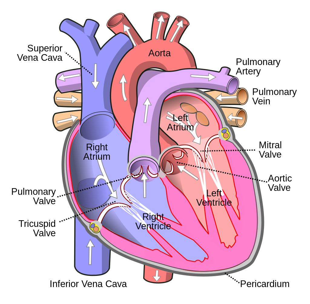 Image Source: Wikimedia Commons (2006). Heart, Licensed under a creative commons licence. Available for free at: https://simple.wikipedia.org/wiki/Heart (Accessed: 25 January 2024).
Aortic stenosis
Any disorder involving the aortic valve will generate pressure/volume overload on the left ventricle. Over time, this will affect the left atrium. An aortic murmur will be heard loudest over the aortic area.
Aortic stenosis (AS) is characterised by an ejection systolic murmur radiating to the carotids. The pathology is due to a stenosed aortic valve causing blood to be squeezed past the narrowed valve opening. Causes of AS include:
- Degenerative calcification. The most common cause in patients > 65 years
- Bicuspid aortic valve. This is the most common cause in patients < 65 years
- Congenital e.g. William’s syndrome where there is supra valvular stenosis
- Post rheumatic fever
- Sub valvular e.g. HOCM
Clinical manifestations
AS can vary from asymptomatic to low cardiac output symptoms which includes chest pain, dyspnoea, syncope and pulmonary oedema. The severity of the stenosis can manifest in symptoms ranging from reduced exercise tolerance to sudden cardiac death (SCD). The classical symptoms are only in 30-40% of individuals with AS. When symptoms appear, survival without surgery is, on average, 2-3 years. SCD occurs in 1-2% of asymptomatic AS patients per year. Symptoms of severe AS include:
- Narrow pulse pressure and small volume pulse due to little blood escaping the ventricle
- Slow rising pulse due to a narrow outflow
- Soft or absent S2 due to a narrow pulse pressure and restricted leaflet motion.
- Displaced apex beat due to left ventricular hypertrophy.
- Delayed ejection systolic murmur
- Paradoxical split S2 and palpable thrill
Diagnosis
While AS may be clinically silent in many cases, a broad range of symptoms, as outlined above, can lead to a suspicion of disease.
- Chest x-ray may show cardiomegaly due to left ventricular hypertrophy and detect abnormalities of aortic contour or size.
- Echocardiography is key to confirming the diagnosis and severity of aortic stenosis, assessing valve calcification, LV function and wall thickness. It can also detect other valve pathologies and provide prognostic information.
- Transoesophageal echocardiography is used when images on a transthoracic echo prove insufficient. A pressure gradient of over 40mmHg is diagnostic of severe aortic stenosis and is an indication for surgery. Aortography done as part of a coronary angiogram may also support a finding of AS.
- Exercise testing may unmask symptoms and is recommended for risk stratification of asymptomatic patients with severe aortic stenosis. Exercise echocardiography provides additional prognostic information by assessing the increase in mean pressure gradient and change in LV function. Although the severity of valvular calcification is prognostically important in an asymptomatic patient, the most important predictor is LV function.
- Coronary angiography is essential prior to transarterial valve implantation (TAVI) and surgical AV valve replacement (SAVR) to determine the potential need for concomitant revascularisation.
- Cardiac CT (CCT) provides information on calcium score, anatomy of the aortic root and ascending aorta, and feasibility of vascular access.
- Myocardial fibrosis (due to amyloidosis, for example) is a major driver of LV decompensation in aortic stenosis. This can be detected and quantified using cardiac magnetic resonance (CMR).
- Natriuretic peptides predict symptom-free survival and outcome in normal and low-flow severe aortic stenosis. They can be used to arbitrate the source of symptoms in patients with multiple potential causes and identify those with high-risk asymptomatic aortic stenosis who may benefit from early intervention.
Management
This depends on the symptoms and severity of AS.
- Early intervention is recommended for all patients with symptomatic severe aortic stenosis. The only exception is for those in whom intervention is unlikely to improve quality of life or survival. Symptomatic patients without severe AS can be managed medically with conventional heart failure therapy. It is important to note that angiotensin-converting enzyme inhibitors (ACEI) are safe in AS, provided that BP is monitored carefully. They provide beneficial myocardial effects before the onset of symptoms and after TAVI and SAVR but are not generally recommended.
- Coexisting hypertension should be treated to avoid additional afterload, although medication (particularly vasodilators) should be titrated to avoid symptomatic hypotension.
- Replacement should also be considered in asymptomatic patients with a valvular gradient > 40 mmHg with features of LVSD.
- Asymptomatic patients without severe aortic stenosis or evidence of LV dysfunction are managed with watchful waiting and prompt intervention at symptoms onset. Intervention such as TAVI or SAVR is recommended in symptomatic patients with high-gradient AS, regardless of LVEF.
- Balloon valvuloplasty is reserved for patients with critical AS who are not fit for valve replacement and has a high rate of recurrence.
Aortic Regurgitation
Aortic regurgitation (AR) is characterised by an early decrescendo diastolic murmur. There is no correlation between the presence of murmur and severity of AR or aetiology. A wide pulse pressure may be found and is caused by blood returning to the left ventricle from the aorta during diastole, causing a drop in diastolic pressure. To compensate, there is peripheral vasoconstriction. A water hammer pulse, also called a collapsing pulse, is characteristic of AR. Other signs in AR include:
- Austin Flint murmur. In severe aortic regurgitation, the regurgitant jet of blood can exert pressure on the anterior mitral valve cusps during diastole, resulting in a mid/late diastolic murmur like that of and virtually indistinguishable from mitral stenosis.
- The first heart sound may be absent in severe AR
AR can be caused by primary disease of the aortic valve cusps and/or abnormalities of the aortic root and ascending aortic geometry.
Degenerative tricuspid and bicuspid aortic regurgitation are the most common aetiologies in high-income countries.
Valvular causes include:
- Rheumatic fever
- Bicuspid aortic valve
- Infective endocarditis
- Connective tissue diseases e.g., RA/SLE
AR due to aortic root disease can occur in:
- Hypertension
- Syphilis
- Marfan’s, Ehler-Danlos syndrome
- Aortic dissection
- Spondyloarthropathies (e.g., ankylosing spondylitis)
Diagnosis
Echocardiography is key to confirm valve anatomy, quantify aortic regurgitation, define the morphology of the aorta and determine the feasibility of valve-sparing aortic surgery or valve repair.
CMR should be used to quantify AR when echocardiographic measurements are equivocal or discordant with clinical findings. In patients with aortic dilatation, a cardiac CCT is recommended. CMR can be used for follow-up, but indication for surgery should preferably be based on CCT measurements.
Management
Acute aortic regurgitation may require urgent surgery. It is mainly caused by infective endocarditis and aortic dissection but may also occur after blunt chest trauma and iatrogenic complications during catheter-based cardiac interventions.
In symptomatic patients who are fit for surgery with severe AR, replacement is recommended irrespective of the LVEF. Replacement is also recommended in symptomatic and asymptomatic patients with severe aortic regurgitation undergoing coronary artery bypass grafting (CABG) or surgery of the ascending aorta or another valve.
In asymptomatic patients with severe aortic regurgitation, impairment of LV function [LVEF ≤50% or left ventricular end-systolic diameter (LVESD) >50 mm] is associated with worse outcomes and surgery should therefore be pursued when these cut-offs are reached.
TAVI may be considered in experienced centres for selected patients with aortic regurgitation and ineligible for SAVR. Medical therapy, especially ACEI or dihydropyridines, may provide symptomatic improvement in individuals with chronic severe aortic regurgitation in whom surgery is not feasible. In patients who undergo surgery but continue to suffer from heart failure or hypertension, ACEI, angiotensin receptor blockers (ARBs), and beta-blockers are useful.
All asymptomatic patients with severe aortic regurgitation and normal LV function should be followed up at least every year.
References
- Alec Vahanian et al. ESC/EACTS Scientific Document Group, 2021 Guidelines for the management of valvular heart disease: Developed by the Task Force for the management of valvular heart disease of the European Society of Cardiology (ESC). European Heart Journal, Volume 43, Issue 7, 14 February 2022. https://doi.org/10.1093/eurheartj/ehab395
- Braunwald’s Heart Disease: A Textbook of Cardiovascular Medicine, Single Volume, 11e
- Aortic Stenosis BMJ Best Practice. [Online] https://bestpractice.bmj.com/topics/en-gb/325
- Aortic Regurgitation BMJ Best Practice. [Online] https://bestpractice.bmj.com/topics/en-gb/324
Complete Guide to Clinical Genetics ST3 Application and Recruitment (2027)
Written by Medibuddy. Last updated: October 2026. Clinical Genetics is

Complete Guide to Clinical Radiology ST3 Application and Recruitment (2027)
Recruitment paused: NHS England is not running Clinical Radiology ST3+ recru

Complete Guide to Paediatric Surgery ST3 Application and Recruitment in United Kingdom (2026)
Written by Medibuddy. Last updated: October 2026. If you’re planning


Take your subscriptions with you
Our mobile app allows you to access your interview and exam question banks wherever you are.

