


 Close
Close
 22nd July 2023
Hepatic Pathology
Alcohol-related Liver Disease (ALD)
Background
In contrast to many other major diseases, the mortality associated with liver disease has been increasing in recent decades, as demonstrated in the image below. Alcohol is the leading cause of liver disease in the UK. It can cause either acute or chronic liver disease, and the latter may eventually result in cirrhosis.
ALD is associated with chronic and excessive alcohol intake. However, not all those with a significant alcohol history will develop ALD. Other risk factors associated with ALD are smoking and being overweight.
Pathophysiology
There are multiple pathways through which alcohol can damage the liver, including the production of free radicals and oxidative stress.
The histological stages of ALD include: steatosis, alcohol-related steatohepatitis, fibrosis and cirrhosis, and hepatocellular carcinoma (HCC) as a further complication. Similar stages are seen in non-alcohol related fatty liver disease. However, there are a few key histological findings which are more typical of ALD, such as hyaline sclerosing necrosis and fibro-obliterative damage in the hepatic veins, which can be used to distinguish the two.
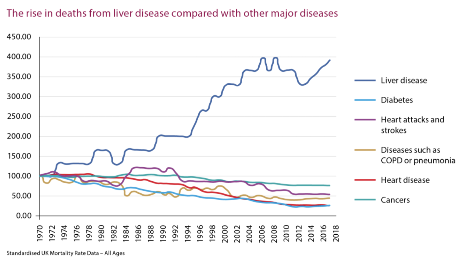
British Liver Trust – Standardised UK Mortality Rate Data.
Image Source: British Liver Trust (no date). Standardised UK Mortality Rate Data – All Ages. Available at: https://britishlivertrust.org.uk/about-us/media-centre/statistics/ (Accessed: 28 June 2023).
Assessment and diagnosis
Taking a thorough alcohol history is key when there is a suspicion of ALD. There are a number of validated tools to screen for alcohol misuse, such as AUDIT, which consists of 10 questions. A link to the AUDIT tool can be found in the References section.
Clinical features
ALD may be asymptomatic or may present with a variety of symptoms, including fatigue, weight loss, anorexia, abdominal discomfort or distension, fever, nausea, vomiting and jaundice.
Findings on examination may be in keeping with chronic liver disease or excess alcohol intake, such as palmar erythema, Dupuytren’s contracture and parotid gland enlargement.
Investigations
Initial investigations should include a full set of liver biochemistry tests (AST, ALT, ALP, GGT, bilirubin and albumin) – i.e. LFTs. AST is often the most raised, and more so than ALT. However, the transaminases can be normal in some people with severe ALD or even cirrhosis, so normal LFTs do not rule out the diagnosis.
Further basic blood tests include an FBC to assess for anaemia and thrombocytopenia, U&Es with magnesium and phosphate levels, and a coagulation screen.
Additional tests may be appropriate to exclude alternative causes of liver dysfunction; for example, a viral hepatitis screen, iron studies and auto-antibodies. This is also known as a ‘non-invasive liver screen’ (NILS).
Ultrasound is the first line imaging investigation, as a non-invasive and cheap way of assessing for pathological markers such as steatosis, and to exclude other pathology, e.g., gallstone disease.
Transient elastography is better for imaging the liver if there is a suspicion of severe fibrosis and cirrhosis, and should be offered to those with a history of chronic and heavy alcohol use. CT and MRI scans may show evidence of liver disease, but are not able to determine aetiology, and offer little additional extra information compared to ultrasound and elastography, so are generally not indicated.
Liver biopsy can be used to help determine the cause of the liver disease but is not necessary in every patient.
Further screening
Hepatocellular carcinoma (HCC) is an important potential complication of ALD. If suspected, α-fetoprotein (AFP) should be measured, as a serum marker strongly associated with HCC. First line imaging remains ultrasound, but CT or MRI may be needed as further investigations if HCC is suspected.
In addition to assessment of the liver, it is important to screen for other alcohol-related organ damage, for example, cardiomyopathy and chronic pancreatitis.
It is also important to include assessment of disease severity.
NICE recommends using the Model for End-Stage Liver Disease (MELD) score, the components of which consist of:
- Dialysis at least twice in the past week or continuous veno-venous haemodialysis for more than 24 hours
- Creatinine level
- Bilirubin level
- INR
- Sodium level
Another commonly used tool is the Child-Pugh score – which uses bilirubin, albumin, INR, the presence of ascites and encephalopathy as its assessment metrics.
Management
The first line treatment – and most important intervention – in the management of ALD is complete abstinence from alcohol.
In the earlier stages of the disease, abstaining from alcohol has the potential to reverse some of the damage that has occurred. For those with more advanced disease, including cirrhosis, there are survival benefits associated with abstinence. It is important to support people in their plans to stop drinking, as some may be at risk of alcohol withdrawal syndrome – see Alcohol withdrawal syndrome section.
Other recommended lifestyle changes include smoking cessation and weight loss.
A multidisciplinary team approach to management is recommended, ideally with input from mental health services and a nutrition expert. People with ALD are highly likely to be malnourished and may require supplementation. This cohort of patients is at high risk of refeeding syndrome and should be monitored closely for this.
Patients should also be monitored and treated for other known complications of alcohol-related cirrhosis. This may include endoscopic surveillance for oesophageal varices and 6-monthly ultrasound scans – with or without AFP serum measurement – to screen for HCC.
Oesophageal varices may be managed with non-selective beta-blockers or endoscopic banding.
Ultimately, those with advanced disease should be considered for liver transplantation. There are criteria when considering if someone may be suitable for a transplant; this often includes a requirement for a period of abstinence and are generally specific to units.
Decompensated liver disease
Cirrhosis secondary to ALD, or any other cause of liver disease, can either be compensated or decompensated. In compensated liver disease, the liver is still able to function, and the patient is relatively well. Decompensation refers to a sudden deterioration in liver function and presents with new or worsened symptoms. Decompensated liver disease is associated with a high mortality rate.
The classic features of decompensation include:
- Jaundice
- Ascites
- Hepatic encephalopathy
- Variceal bleeding
Other indicators of decompensation include hepatorenal syndrome, recurrent infections, and cachexia.
The British Society of Gastroenterology care bundle for decompensated cirrhosis highlights the key areas of management.
The key steps include assessment of liver biochemistry and synthetic function, screening for and treating infection (including an ascitic tap in all patients with ascites to assess for spontaneous bacterial peritonitis), establishing alcohol intake, monitoring and managing alcohol withdrawal, supporting nutrition, and identifying and managing acute kidney injury.
It is important to identify any potential underlying cause for decompensation and treat appropriately.
Additionally, certain features of decompensation have specific management, such as gastrointestinal bleeding, hepatic encephalopathy, and ascites.
Alcohol withdrawal syndrome
Clinical features
People with alcohol dependence are at risk of developing alcohol withdrawal syndrome when they stop or suddenly reduce their alcohol intake.
Alcohol interacts with both GABA and NMDA receptors; chronic use can lead to dysregulation of these receptors. If alcohol consumption is drastically reduced or ceased, this leads to an excess of glutamatergic activity, with reduced GABA activity, which results in overstimulation of the sympathetic nervous system.
Common clinical features of withdrawal include sweating, anxiety, nausea, vomiting, tachycardia and palpitations.
Severe alcohol withdrawal can manifest as ‘delirium tremens’. This is an acute confusional state associated with visual hallucinations, delusions and a coarse tremor. It typically develops 48 hours after the patient’s last drink. This is a medical emergency and can be life-threatening.
Additionally, severe alcohol withdrawal can also cause seizures, which typically manifest 72 hours after the last drink.
Assessment and diagnosis
It is crucial to identify patients with alcohol dependence so that alcohol withdrawal can be monitored for and treatment preempted.
When alcohol withdrawal syndrome is suspected, the severity of symptoms should be assessed in order to guide treatment. The Clinical Institute Withdrawal Assessment for Alcohol Revised (CIWA-Ar) is a validated scoring system to assess severity and is based on key clinical features associated with alcohol withdrawal. (See the References section for more information.
Although alcohol withdrawal is a clinical diagnosis, other investigations should be carried out to support the diagnosis, but also exclude other causes of the presentation.
Management
The mainstay of management of alcohol withdrawal is benzodiazepines. Emergency treatment for seizures is intravenous lorazepam, and delirium tremens can be treated with oral lorazepam or diazepam. Antipsychotics may be used if there is a poor response to benzodiazepines.
For those without seizures or delirium tremens, a benzodiazepine regimen should be initiated. In the majority of people, this would be with chlordiazepoxide, which is a longer-acting benzodiazepine than diazepam and lorazepam.
In addition, all patients should be given thiamine to prevent or treat Wernicke’s encephalopathy. Initially, a high dose parental form is recommended, commonly known as the brand name Pabrinex. Local protocols differ in their recommendations on how long intravenous thiamine should be continued; the patient should then subsequently be established on an oral formulation long term.
Chronic liver disease (CLD)
The following section covers other causes and conditions associated with chronic liver disease (CLD), outside of ALD.
Progression of liver damage over time, due to any cause, may result in cirrhosis. For those with liver cirrhosis, there is a risk of decompensation with features such as jaundice and ascites. The assessment and management of decompensated liver disease has been covered in the Alcohol-related Liver Disease (ALD) section, so this will not be repeated in detail here. However, it is important to consider that it is a potential complication for each individual cause of chronic liver disease.
Non-alcohol related fatty liver disease (NAFLD)
Background
NAFLD is an increasingly common cause of liver disease in the UK and the Western world, in both adults and children. Prevalence increases with age and NAFLD currently affects ~20% population in developed countries. NAFLD is defined as the accumulation of fat (steatosis) in >5% of hepatocytes, in the absence of significant alcohol consumption or any other secondary cause of hepatic steatosis.
Risk factors include central obesity, impaired glucose tolerance, type 2 diabetes mellitus, hypertension, and hyperlipidaemia. NAFLD itself is also a risk factor for type 2 diabetes, hypertension and chronic kidney disease.
Complications may be liver related, e.g. liver failure, portal hypertension and hepatocellular carcinoma, or metabolic related, with cardiovascular disease being the most common cause of death in people with NAFLD.
Pathophysiology
NAFLD encompasses a spectrum of disease with the following stages: hepatic steatosis, non-alcohol related steatohepatitis (NASH), NASH with fibrosis, and hepatic cirrhosis. The disease often forms part of a wider metabolic syndrome, and it is thought that insulin resistance is one of the key mechanisms in the development of NAFLD.
Histological features are similar to those seen in alcohol-related liver disease and include steatosis, hepatocyte ballooning, inflammation and apoptosis and Mallory-Denk bodies.
Assessment and diagnosis
Many people with NAFLD are asymptomatic, which can make identifying cases challenging. Early recognition and intervention are crucial as, in the early stages, NAFLD is potentially reversible. NAFLD should be suspected in those with features of metabolic syndrome.
NAFLD is one of the most common causes for deranged liver blood tests and so may be picked up incidentally. However, not all those with NAFLD will have abnormal blood tests, so normal results cannot be used to exclude the disease. Imaging carried out for other purposes may also find incidental evidence of NAFLD.
A thorough assessment should be carried out, which covers alcohol intake, risk factors and exclusion of other causes of liver diseases (some of which are detailed in the following sections).
Ultrasound imaging can be used to assess for increased hepatic echogenicity – which is indicative of fatty liver changes. However, ultrasound may be normal in those with NAFLD.
The gold standard for diagnosis is liver biopsy and histology; however, this is invasive and costly. Diagnosis is, therefore, usually based on a combination of elements from the history, examination and investigation.
Non-invasive tests can be used to assess the severity of inflammation and fibrosis. NICE suggests using the enhanced liver fibrosis (ELF) test, which generates a score using a combination of three blood markers of fibrosis: tissue inhibitor of metalloproteinases 1, amino-terminal propeptide of type III procollagen and hyaluronic acid.
Transient elastography is a non-invasive test that is increasingly being used to detect fibrosis and cirrhosis.
If a non-invasive test cannot exclude fibrosis, the diagnosis is uncertain or another coexistent liver disease is suspected, then liver biopsy should be considered.
Management
The mainstay of management centres on lifestyle interventions: including physical activity, diet, preventing excess weight gain and avoiding excess alcohol intake. Other cardiovascular risk factors should be identified and treated appropriately, e.g. hypertension.
Those with advanced liver fibrosis should be referred for specialist assessment. NICE recommends consideration of pioglitazone or vitamin E, however, this an off-label use of both medications. Emerging treatment options include GLP-1 agonists and SGLT2 inhibitors.
Bariatric surgery may be considered in some patients and has the potential to reverse steatosis and fibrosis.
For those with end-stage liver disease, transplantation is the only curative treatment option. However, NAFLD may recur in the transplanted liver, especially if contributory lifestyle factors are not addressed.
Hereditary haemochromatosis
Pathophysiology
Haemochromatosis is a genetic condition characterised by dysfunctional iron metabolism. The hereditary form is the most common genetic disease in people of European descent.
It is inherited in an autosomal recessive pattern. The most common causes are C282Y or H63D mutations in the HFE gene on chromosome 6. Clinical penetrance is low, meaning not all those with genetic mutations will develop the disease.
The HFE gene encodes a protein which is involved in the synthesis of hepcidin, the main regulator in iron metabolism. In hereditary haemochromatosis (HH), there is reduced expression of hepcidin, which results in continued absorption of iron despite normal or increased levels of total body iron. In addition, there is excess iron release from macrophages. This leads to excessive iron accumulation in body tissues.
Assessment and diagnosis
The characteristic iron accumulation in body tissues of HH leads to multi-system manifestations, with the liver, heart, brain and pancreas being the organs most affected. Typically, symptoms develop in later life and include fatigue, arthralgia, hyperpigmentation, loss of libido, neurological or psychiatric symptoms, as well as being an important cause of chronic liver disease. Examination findings reveal hepatomegaly, hyperpigmentation which may range from bronzing of the skin to a slate-grey appearance and/or features of porphyria cutanea tarda. The classical triad of bronzed skin, diabetes mellitus and cirrhosis may not be present in all patients, especially in the early stages of the disease.
If HH is suspected, the recommended initial investigations are a full blood count, liver function tests, ferritin and transferrin saturation. A raised ferritin on incidental testing should be interpreted with caution, as ferritin is an acute phase protein, and therefore there are a number of reasons it may be elevated.
If transferrin saturation is raised, HFE genetic testing should be carried out to confirm the diagnosis. Ferritin level guides the severity of iron overload.
Further testing, once the diagnosis is confirmed, should be carried out to assess the effect of iron deposition on tissues, especially the liver. MRI is used to assess the degree of iron content in the liver. Transient elastography is used as a non-invasive method of assessing fibrosis. The gold standard to assess iron deposition is liver biopsy, however, the British Society for Haematology guidelines advise it is no longer required for diagnosis.
Impact on other organs should be assessed with investigations screening for diabetes, cardiomyopathy, conduction abnormalities and hypogonadism.
Screening
First degree family members of those diagnosed with HH should be screened for the disease, with genetic testing in addition to a full blood count, liver function tests, transferrin saturation and ferritin. Unselected population screening is not recommended.
Genetic counselling should be offered to affected families.
Management
Prognosis is variable depending on the degree of organ involvement. Initial measures are lifestyle advice, with patients advised to avoid iron-containing supplements and to avoid excess alcohol intake in order to protect their liver long term.
Further management is guided by the stage of disease, with earlier stages requiring active monitoring only, and later stages being managed with venesection to maintain serum ferritin <50ng/ml. For those in whom venesection is contraindicated (e.g. anaemia, ischaemic heart disease), iron chelation treatment can be used. Deferasirox is an oral iron chelator.
In many cases, treatment can reverse organ dysfunction and improve overall survival. However, arthropathy does not tend to respond to treatment, and hypogonadism is irreversible.
Liver cirrhosis cannot be reversed, and the increased risk of hepatocellular carcinoma (HCC) persists despite treatment. Those with cirrhosis should undergo screening for HCC with AFP and liver ultrasound every 6 months.
Autoimmune hepatic disease
There are three major autoimmune diseases which affect the liver and can lead to chronic liver disease: autoimmune hepatitis, primary biliary cholangitis and primary sclerosing cholangitis. It is thought there may be some degree of overlap between these conditions.
Autoimmune hepatitis (AIH)
Background
The precise underlying aetiology of AIH is unknown, but it is thought to involve altered T-cell function. It is often associated with other autoimmune disorders.
Historically there are three types of AIH, classified by autoantibodies. However, more recently, it has been proposed that AIH type III is not a distinct subgroup, but rather should be included within AIH type I.
Assessment and diagnosis
Clinical presentation varies from asymptomatic to acute hepatitis, or most commonly, the insidious onset of chronic liver disease.
Symptoms include fatigue, anorexia, abdominal discomfort, nausea, pruritus, polyarthralgia of the small joints, amenorrhoea and fluctuating jaundice. One third of adults will have cirrhosis at the time of presentation. On examination, there may be stigmata of chronic liver disease.
Liver function tests will be abnormal at presentation. Often there is a significant transaminitis (elevated AST and ALT) alongside a more modest rise in bilirubin and ALP. However, AIH can also present with a cholestatic picture. Raised IgG is characteristic of AIH. A reduced albumin level and prolonged prothrombin time may also be seen.
The next stage of investigation is to test for the presence of autoantibodies (described in the table below). A liver biopsy should also be performed. Characteristic histological findings are of a periportal infiltrate with inflammation that extends beyond the limiting plate and interface hepatitis (previously known as piecemeal necrosis). In an acute hepatitis presentation, the histology may be atypical.
Other tests may be necessary to exclude other causes of liver disease. The diagnosis cannot be made from one test alone but is a clinical diagnosis based on a combination of features.
| Type | Associated autoantibodies | Notes |
|---|---|---|
| I | Antinuclear antibody (ANA)
Smooth muscle antibody (SMA) Perinuclear anti-neutrophil cytoplasmic antibody (p-ANCA) Anti-soluble liver antigen/liver pancreas antigen (anti-SLA/LP). |
Most common subtype, and more common in women
Associated with HLA-DR3, HLA-DR4 and HLA-DR13 Good treatment response – may not require long term maintenance |
| II | Anti-liver kidney microsomal 1 antibody (LKM1)
Anti-liver cytosol type I antigen (anti-LC1) |
Typically affects children
Associated with HLA-DRB and HLA-DQB1 High rates of relapse on withdrawal of drug therapy – long term maintenance generally required |
| III | Anti-soluble liver antigen/liver pancreas antigen (anti-SLA/LP) | Very similar to type I, increasingly recognised as a subcategory rather than a category in of itself |
Table 1: Different types of AIH
Management
The mainstay of treatment is immunosuppression. In some cases, e.g. older patients with asymptomatic disease and mild hepatitis, it may be appropriate not to treat, but to actively monitor.
The recommended initial treatment is combination therapy of a corticosteroid plus azathioprine. Prior to treatment with azathioprine, patients should have thiopurine methyltransferase (TPMT) activity measured. TPMT deficiency can lead to bone marrow toxicity if an affected patient is given immunosuppressants.
For patients with decompensated liver disease or those with acute severe AIH, combination therapy is not the recommended first line, but instead corticosteroid monotherapy is preferred.
Long-term maintenance therapy may be required. Patients should be monitored for side effects relating to the use of corticosteroids and azathioprine.
It may also be appropriate to screen for HCC with regular AFP and liver ultrasound – as with any form of chronic liver disease.
Primary biliary cholangitis (PBC)
Background
Previously known as primary biliary cirrhosis, PBC is an autoimmune condition which affects the bile ducts. The exact aetiology is not fully understood.
The disease is characterised by destruction of the epithelial cells in the smaller intrahepatic bile ducts, alongside portal tract inflammation. PBC is a progressive disease which generally leads to chronic – and then end-stage – liver disease. However, there is a subgroup of PBC patients with a lower risk of disease progression.
The condition is considerably more common in women with a female to male ratio of 10:1. Typically, people present in middle age; 55-65 years is the peak age range for diagnosis.
PBC is associated with other autoimmune conditions such as Hashimoto’s thyroiditis, Sjogren’s disease and coeliac disease. There may also be a positive family history for PBC.
Assessment and diagnosis
The most common presenting features are fatigue and pruritus. However, people can be asymptomatic and, therefore, may present as incidental abnormal liver function tests. In advanced disease, there may be symptoms and signs in keeping with chronic liver disease and cirrhosis.
The diagnosis of PBC is made on the presence of the following findings:
- Cholestatic LFTs: elevated ALP and/or GGT
- Serum autoantibodies: antimitochondrial antibodies (AMA) or PBC-specific antinuclear antibodies (ANA)
- Liver histology: demonstrating bile duct lesions with portal tract inflammation and granulomas
Liver biopsy is now used less often to diagnose PBC, due to the accuracy of serological markers, and the risks of biopsy. Ultrasound scans do not demonstrate PBC but are indicated as part of the assessment in order to exclude other causes of cholestasis, such as gallstones.
Management
The goals of management are symptom control and prevention of disease progression. The first line treatment targeting disease progression is ursodeoxycholic acid. For the proportion of patients who do not respond, obeticholic acid is the second line. Cholestyramine is used for symptomatic relief of pruritus. Rifampicin or naltrexone can be used as second line agents.
Primary sclerosing cholangitis (PSC)
Background
PSC is a condition which is likely to be immune-mediated; however, like AIH and PBC, the exact aetiology is not fully understood.
The disease is characterised by inflammation of the medium and large-sized bile ducts, leading to strictures and progressive fibrosis.
Unlike most other autoimmune related conditions, PSC is much more common in men. There is a strong association with inflammatory bowel disease (IBD), particularly ulcerative colitis. It is important to note that IBD may develop before or after PSC.
Assessment and diagnosis
Many people with PSC are asymptomatic. When present, common presenting symptoms include fatigue, pruritus and upper abdominal pain. In more advanced disease, patients may present with features in keeping with chronic liver disease, for example, jaundice or abdominal distension.
Initial investigations include LFT, which typically shows a cholestatic pattern (raised ALP and GGT). In more advanced disease, bilirubin will also be elevated. There are no diagnostic autoantibodies associated with PSC, however, it may be appropriate to test for these to exclude related conditions like PBC and AIH.
Imaging of the biliary tree is crucial to diagnose PSC. An abdominal ultrasound would be the usual first step, as a non-invasive test to exclude other causes of bile duct disease. CT imaging may show bile duct thickening and/or duct dilatation. However, MRCP is the gold-standard diagnostic modality, and can also provide information about the distribution of disease. ERCP can also be diagnostic, however, is more invasive and carries more risk; therefore, it should only be done if MRCP has not been diagnostic or if there is potential for therapeutic intervention.
The typical features on MRCP are multiple strictures, giving a ‘beaded’ appearance of the intra and extrahepatic ducts.
Liver biopsy is generally not required, if there is enough evidence to support a diagnosis of PSC. Histology itself is not diagnostic but may be helpful to support the diagnosis or assess for alternative or overlapping diseases.
Management
Management includes pharmacological treatment of symptoms, such as cholestyramine for pruritus, and interventional treatments, such as balloon dilatation of dominant strictures. The latter can improve survival, as well as symptoms, in those who are acutely unwell.
It is important to assess for complications, such as hepatic osteodystrophy or osteopenia, and treat these appropriately. Another serious complication of PSC is cholangiocarcinoma, however, there is no consensus on routine screening for this.
For those whose disease progresses to end-stage liver disease, transplantation is the only therapeutic option.
Alpha-1 antitrypsin deficiency
Background
Alpha-1 antitrypsin (AAT) deficiency is a genetic condition which can affect the lungs and the liver. This section will focus on the hepatic manifestations.
The inheritance is autosomal codominant, meaning that one or two defective alleles can be inherited, and both can cause disease. However, the inheritance of only one allele tends to result in milder disease. There are numerous causative mutations affecting the SERPINA1 gene on chromosome 14. These mutations lead to a deficiency of effective circulating AAT; the exact pathophysiology depends on the type of mutation.
A normal phenotype is denoted as PI*MM. Z denotes the most common mutation in AAT deficiency, and so a carrier would be PI*MZ, and a person with the disease would have the phenotype PI*ZZ. S is another common mutation, which is associated with decreased function of AAT and is often associated with a less severe form of the disease.
Liver involvement may be seen as early as the neonatal period. In adults, clinical features include fatigue, jaundice, abdominal distension, bruising and abdominal pain. There may be signs of chronic liver disease on examination, such as asterixis, hepatomegaly or ascites.
Assessment and diagnosis
The key investigation is serum AAT levels. However, these must be interpreted with caution as AAT is an acute phase reactant and, therefore, may be falsely normal. For some, it may be beneficial to test for the underlying phenotype. Genotyping may also be done.
Additional investigations relating to hepatic disease may include liver ultrasound. AAT level testing is part of a non-invasive liver screen to investigate for the cause of undiagnosed liver disease.
Management
Management focuses on monitoring and managing complications of chronic liver disease. Ultimately, liver transplantation may be required. Patients with AAT deficiency should be advised to avoid or limit their alcohol intake.
Wilson’s disease
Background
Wilson’s disease is a rare autosomal recessive condition that results in the accumulation of copper. There are several associated mutations of the ATP7B gene, on chromosome 13, which affects the ability of the liver to excrete copper.
High levels of free copper causes widespread toxicity. Typically, people with Wilson’s disease have either hepatic involvement or neurological/psychiatric manifestations; however, occasionally both may be present.
Assessment and diagnosis
Those with liver manifestations can present with hepatitis, cirrhosis or liver failure.
Neurological and psychiatric symptoms include tremor, ataxia, dysarthria, dystonia, depression, poor memory, disinhibition, and other unusual behaviours.
Examination findings may include Kayser-Fleischer rings; however, these are more commonly found in the neurological manifestation of the disease, rather than hepatic. Neurological examination may identify signs in keeping with movement disorder; however, sensation, strength and reflexes should be normal.
Investigations should include LFTs, 24-hour urinary copper measurement, and ceruloplasmin.
Where the diagnosis is not certain, a liver biopsy with quantitative assay of copper can be done as a definitive diagnostic test.
For those with neurological involvement, MRI brain imaging can be helpful and may show increased density in the basal ganglia and the ‘face of the giant panda’ sign in the midbrain. Neither of these findings are specific for Wilson’s disease, however.
Management
Wilson’s disease is a treatable genetic condition, and therefore, timely diagnosis is important. Medical treatment aims to eliminate copper and prevent re-accumulation. Current recommended medications are zinc, which blocks copper resorption in the gut, and trientine, which increases urinary copper excretion. Older treatments used for Wilson’s disease include d-penicillamine; however, this can worsen neurological symptoms and may have severe adverse effects.
For those with liver involvement, the severity of hepatic disease should be established with the Nazer score (see BMJ Best Practice, in References section), as this will guide whether the patient can be managed with pharmacological therapy or whether they require consideration for a liver transplant. Dietary advice is also important; for example, limiting the intake of liver and shellfish in the initial stages of treatment, due to their high copper content.
Hepatitis
Hepatitis refers to inflammation of the liver. It is a condition with a variety of aetiologies, including infectious causes, autoimmune disease, and drugs. This topic will focus on infectious causes of hepatitis and drug-induced hepatitis; autoimmune hepatitis is covered in the Chronic Liver Disease topic.
Viral hepatitis
There are five forms of hepatitis viruses: A, B, C, D, and E. The most important clinically are hepatitis A, B and C. A and E cause acute infections, whereas B, C and D can cause both acute and chronic forms of hepatitis, and in the latter is a key cause of chronic liver disease in the UK and worldwide. The key information for each virus is summarised in the table below.
Viral hepatitis may be asymptomatic. The clinical features of acute infection include abdominal pain (especially in the right upper quadrant), jaundice, nausea and vomiting, malaise and fatigue. Chronic infection is associated with constitutional symptoms like weight loss, malaise and fatigue, as seen in chronic liver disease – and if it progresses can cause cirrhosis and liver failure. There are specific antiviral treatments for hepatitis B and C (see below), and if chronic infection does progress into chronic liver disease, then this is managed accordingly. Hepatitis C is also strongly associated with hepatocellular carcinoma as a complication.
Hepatitis B and C are a particular public health concern amongst intravenous drug users and sex workers. These demographics, as well as those with an occupational risk, such as healthcare workers, are targeted with screening and vaccination programmes.
| Virus | Type | Transmission | Chronic disease | Management |
|---|---|---|---|---|
| Hepatitis A | RNA | Faecal-oral | No | Generally causes a mild self-limiting illness, which requires supportive treatment only |
| Hepatitis B | DNA | Bloodborne or through sexual transmission | Yes
|
Antivirals
Peginterferon alfa-2a is first line for chronic hepatitis B without liver decompensation Entecavir is first line in patients with chronic hepatitis B and decompensated liver disease (if no history of lamivudine resistance). Tenofovir is the alternative |
| Hepatitis C | RNA | Bloodborne or through sexual transmission | Yes | Antivirals
First line is combination therapy – pegylated interferon alfa and ribavirin Other antivirals used in treatment include daclatasvir, ombitasvir, sofosbuvir, ledipasvir |
| Hepatitis D | RNA | Bloodborne or through sexual transmission
Requires hepatitis B to replicate; therefore, it is either acquired as a co-infection with hepatitis B, or a superinfection in patients with a chronic hepatitis B |
Yes | Pegylated interferon alfa |
| Hepatitis E | RNA | Faecal-oral | Yes | Generally causes a mild self-limiting illness, which requires supportive management only
Rarely, it can cause a severe illness and acute liver failure, with pregnant women at particular risk, and mortality rates as high as 25% during the third trimester |
Table 2: Summary of different causes of viral hepatitis
Hepatitis B serology
This refers to the serological testing of various antigens and antibodies for hepatitis B, which can be used to distinguish acute and chronic infection, and establish immunity. Questions on hepatitis B serology are common in MRCP examinations, so it is important to know this topic well.
The serological markers tested for are the two antigens of HBsAg and HBeAg, and two antibodies of anti-HBs, and anti-HBc (both IgM and IgG forms).
HBsAg is the first marker to appear, and it triggers anti-HBs production. HBsAg implies acute disease, but if it persists longer than six months, it indicates chronic infection. The detection of anti-HBs indicates immunity, either through exposure or vaccination. It will be negative in chronic infection as immunity has not developed. Anti-HBc is a marker of previous or current infection; IgM antibodies indicate acute or recent (within the last six months) infection, whereas IgG antibodies indicate previous infection.
A vaccinated person will have anti-HBs antibodies but not anti-HBc antibodies, as they have not been previously infected. This is therefore used to establish whether immunity has been caused by infection or vaccination.
The presence of HBeAg indicates a patient is infectious and could hence pass on the virus.
Vaccination
Vaccination for at-risk individuals and groups is a key part of the management strategy for viral hepatitis. Vaccinations exist for hepatitis A and B, but not for C or D. Some vaccinations exist for hepatitis E, but they are not very effective.
In the UK, hepatitis A and B vaccinations are offered as travel vaccinations, to people with chronic liver disease, high risk occupations, men who have sex with men, and intravenous drug users.
Drug-induced hepatitis
Also known as drug-induced liver injury (or DILI), this is an important cause of hepatitis. People with pre-existing liver disease and a history of alcohol excess are at increased risk. DILI is generally categorised by the type of damage caused to the liver, divided into hepatocellular and cholestatic (though the latter group may also cause a mixed picture). The causative drugs are summarised in the table below.
Methotrexate, methyldopa and amiodarone are also associated with a separate risk of liver cirrhosis.
| Hepatocellular | Cholestatic |
|---|---|
| Paracetamol Sodium valproate Phenytoin Monoamine oxidase inhibitors Halothane Anti-tuberculosis drugs: isoniazid, rifampicin, pyrazinamide Statins Alcohol Amiodarone Methyldopa Nitrofurantoin |
Combined oral contraceptive pill Flucloxacillin Co-amoxiclav Erythromycin Anabolic steroids and testosterones Chlorpromazine and prochlorperazine Sulphonylureas Fibrates Nifedipine (rarely) |
Table 3: Categorisation of drug-induced liver injury
Treatment is of cessation of the causative drug, then supportive treatment of any acute symptoms and chronic long-term complications.
Complete Guide to Clinical Genetics ST3 Application and Recruitment (2027)
Written by Medibuddy. Last updated: October 2026. Clinical Genetics is

Complete Guide to Clinical Radiology ST3 Application and Recruitment (2027)
Recruitment paused: NHS England is not running Clinical Radiology ST3+ recru

Complete Guide to Paediatric Surgery ST3 Application and Recruitment in United Kingdom (2026)
Written by Medibuddy. Last updated: October 2026. If you’re planning


Take your subscriptions with you
Our mobile app allows you to access your interview and exam question banks wherever you are.

